




Abstract
Traumatic soft tissue wounds present a significant reconstructive challenge. The adoption of closed-circuit negative pressure wound therapy (NPWT) has enabled surgeons to temporize these wounds before reconstruction. Such systems use porous synthetic foam scaffolds as wound fillers at the interface between the negative pressure system and the wound bed. The idea of using a bespoke porous biomaterial that enhances wound healing, as filler for an NPWT system, is attractive as it circumvents concerns regarding reconstructive delay and the need for dressing changes that are features of the current systems. Porous foam biomaterials are mechanically robust and able to synthesize in situ. Hence, they exhibit potential to fulfill the niche for such a functionalized injectable material. Injectable scaffolds are currently in use for minimally invasive surgery, but the design parameters for large-volume expansive foams remain unclear. Potential platforms include hydrogel systems, (particularly superabsorbent, superporous, and nanocomposite systems), polyurethane-based moisture-cured foams, and high internal phase emulsion polymer systems. The aim of this review is to discuss the design parameters for such future biomaterials and review potential candidate materials for further research into this up and coming field.
Introduction
M
Surgeons have adopted the use of closed-circuit negative pressure wound therapy (NPWT) to dress and close such wounds.5,6 The therapy applies suction to a foam interface within a circuit closed by a semipermeable adhesive dressing to apply pressure to the wound bed.7–10 Contrary to early reports, 11 the act of applying vacuum to the foam filler applies positive pressure to the wound bed.12–14 Micromechanical deformation forces probably activate cellular mechanoreceptors, stimulating fibroblast proliferation and collagen deposition. Permissive hypoxia within the local environment stimulates expression of growth factors, including vascular endothelial growth factor (VEGF), fibroblast growth factor (FGF), and transforming growth factor-β (TGF-β), resulting in angiogenesis and mesenchymal stromal differentiation.
NPWT also has an immunomodulatory effect, promoting an anti-inflammatory cytokine profile locally and systemically. 15 Furthermore, the closed nature of the NPWT system prevents further contamination. The main drawback of NPWT is the requirement for the foam filler to be removed. This results in disruption of the friable granulation tissue propagated at the wound bed. Moreover, existing synthetic foams do not contour well to complex wound cavities, leaving pockets of dead space within these cavities that are not in contact with the filler,6,16,17 thus risking infection and sepsis with prolonged use.
The idea of engineering a biomaterial to replace the foam filler has yet to gain much traction despite advances in the related field of dermal replacement technology.18,19 The ideal solution is an injectable, porous, biologically stable, biodegradable mechanically robust scaffold for wound therapy and tissue regeneration. The construct should also be hemostatic and bacteriostatic. Injectable foam platforms provide the most plausible delivery vehicle for this new material. First, a porous large-volume scaffold can be produced from a smaller volume initiated state. Second, they are able to provide the mechanical stability not just for cell proliferation but also for integration of the aforementioned clinical system without structural deformation. Last, such scaffolds can propagate secondary functions whether through physical, chemical, or through degradation-controlled systems.
An injectable pore-forming hydrogel is therefore required for such complex wounds. Injectable hydrogels should be usable in a broad range of temperature conditions, synthesized within seconds rather than minutes, generating a mechanically robust structure. The introduction of secondary pore-forming agents such as nanospheres that are able to form an interlinked network would enable a porous structure for cellular integration. Alternatively, inherent polymer charges or gas production as a platform to construct inherent porosity can be used. Ultimately, such a material must present surface properties that encourage appropriate cellular interaction to guide wound healing and cellular integration.
In the field of trauma wound management, there is an unmet need for such a product. The aim of this review is to discuss and explore the features of such a material and suggest existing platforms that may be suitable for its development. We shall first discuss the structural components and then the mechanisms of establishing a viable porous scaffold. 20
Delivering the Injectable Platform
An injectable delivery mechanism is attractive because it enhances conformity to irregular wound cavities, obliterating dead space.
The suitability of a material for injectable delivery is governed by several factors (Figs. 1 and 2). First, the polymer solution and reaction dynamics will control the viscosity and fluid flow. Additionally, the reaction speed and necessary processing conditions will control the speed to achieve a gel or solid-like state from liquid products. In applications for drug delivery or minimally invasive surgery, balancing these factors has produced successful, yet complex, self-assembly materials. High aspect ratio mesoporous silica rods were injected through needles for vaccine and immune cell delivery. These were capable of forming macroporous three-dimensional (3D) structures capable of dendritic cell integration. 21
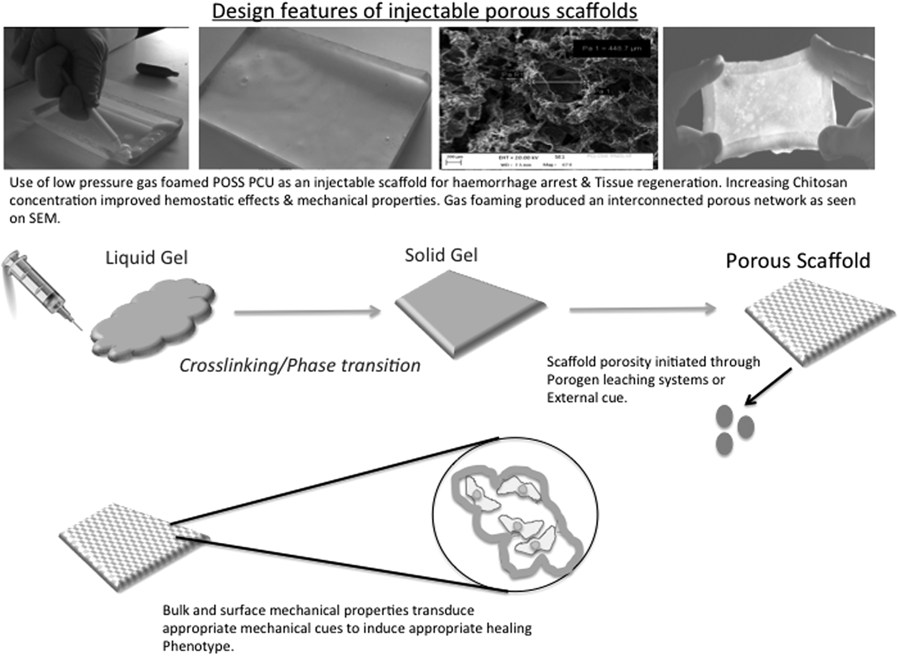
Design features of injectable porous scaffolds. The design of future injectable porous materials for complex wounds requires the engineering of several key components. Primarily, the material needs to undergo a phase transition from a viscous liquid to a more mechanically robust gel or solid. This must be through a biorthogonal chemistry system. Second, and synergistic to this phase transition, is the initiation of a porogen within the material system to build an interconnected macroporous structure. Finally, the nano and macromechanical properties must be tightly controlled to deliver appropriate mechanical cues to the cellular environment. The system therefore is a highly sophisticated and tightly controlled equilibrium. Utilizing a low-pressure gas porogen, nitrous oxide, in our laboratories, we have attempted to create a nanofunctionalized polymer system and synthesize it into a macroporous foam. Utilizing polysaccharide and urethane polymer mixtures under low gas pressures, mechanically robust porous foams could be synthesized for potential bioengineering applications in complex wounds.
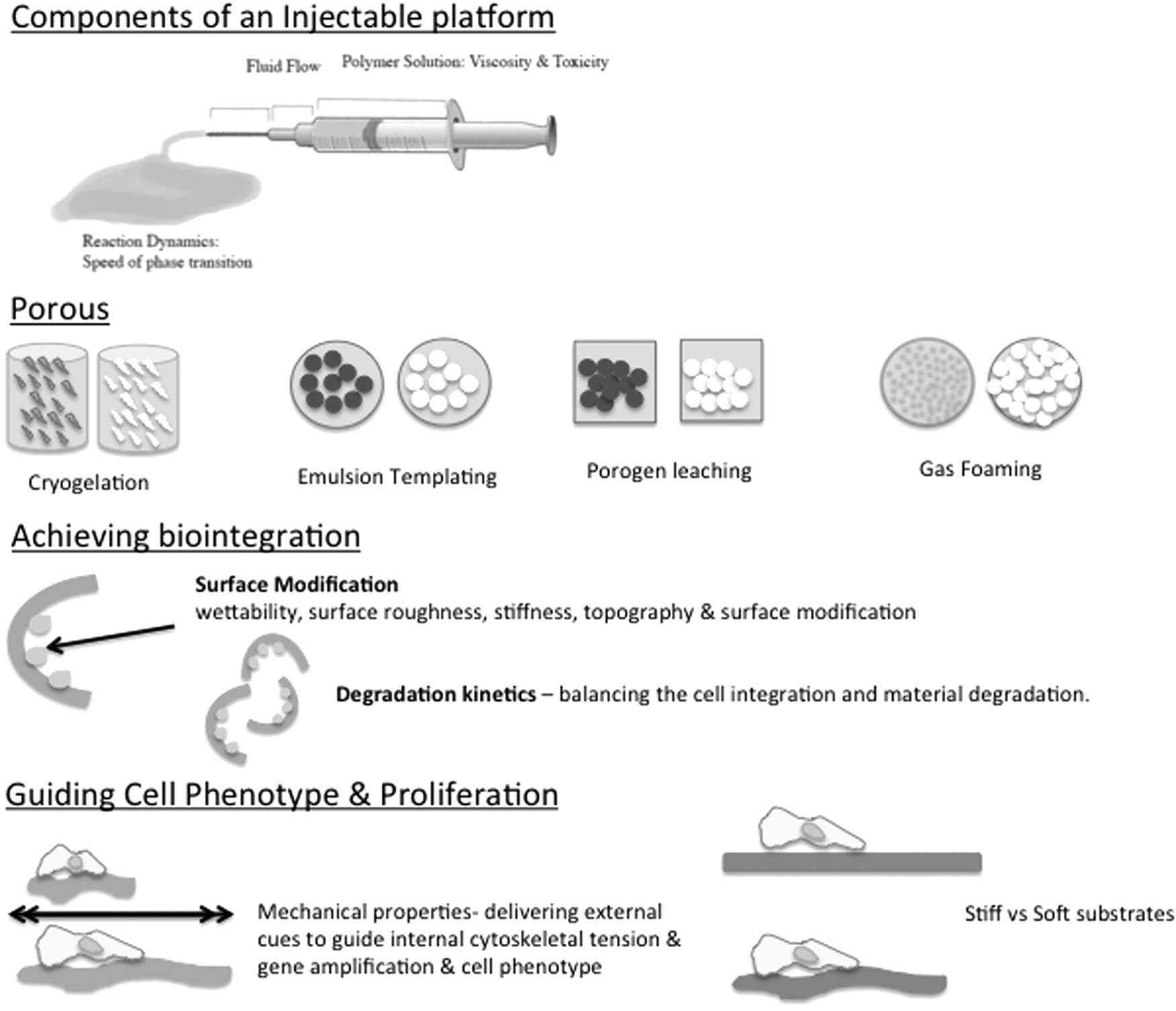
Components of injectable scaffold. Injectable hydrogels require several features to be considered in their engineering. The chemistry and cross-linking of the polymer will determine the level of control for the bulk mechanical properties. The viscosity and toxicity of the polymer solution must also be tested as well as weighed against the reaction dynamics such as the speed of cross-linking and relative reaction chemistry. The system for engineering pores within the system is crucial. Techniques such as cryogelation have been postulated; however, stimulus-controlled porogens or emulsion systems may prove more tunable. The ability to tightly control the surface modification to enable cellular integration, whether through stiffness, protein delivery, or direction chemical modification, will be a major factor in determining the success of the material to evade immune surveillance and promote appropriate cellular phenotypes. There are therefore a multitude of factors to consider in designing such platforms.
Designing a robust mechanical system to achieve a solution gelation change (sol gel) is one of many critical steps in injectable biomaterial design, as is balancing the overall chemical equation to minimize unrequired products or leachable products. Ensuring reaction composition should also provide a material with much greater control over degradation kinetics that will ultimately improve tissue interaction. Isolating polymer material systems that are capable of generating robust large-volume applications in situ is a central translational hurdle in this material design. Choosing relatively appropriate materials that deliver structural integrity is a critical challenge. Particularly, as complex material, systems that function on the in vitro and in vivo microscale may not necessarily perform on a larger scale in vivo or in a human model.
The cost of such materials is also an important consideration. Constructing small-volume, highly functional injectable materials may not necessarily translate to a cost-effective product when upscaled for large-volume wounds. The cohesive material soup of the liquid substrate must also be considered so as to avoid uncontrolled dispersion through tissue planes without compromising the necessary properties of the liquid state.
The transition from a liquid system to a solid is one of the difficulties in designing these systems, particularly as many of the classic chemical processes are potentially toxic. The adverse effects of poor reaction kinetics such as exothermic reactions, toxic by-products, and prolonged cross-linking are potentially deleterious to tissue viability. Use of zeolite powder for control of vessel hemorrhage in trauma produced an exothermic reaction that was not tolerated by patients. 22 Chitosan-based biomaterials for the same application were stable under physiological temperature and pH and generated no thermal properties. 23 Hence, the ability of polymers to create a solid scaffold utilizing in situ homeostatic conditions is necessary.
Although polyurethane-based foam systems have the potential to provide a porous robust structure, the use of isocyanates and surfactants is potentially toxic to a wound bed during polymerization,24,25 precluding their use in material design. Quantifying harmful reactants and their subsequent clearance, necessary for federal drug administration,26–28 remains a challenge.
Alternatively, prolonged cross-linking mechanisms within hydrogel systems such as those seen with click chemistry mechanisms could prevent the construction of a precise porous architecture.25,29 In particular, the external forces of the wound may alter the structural hierarchy of the hydrogel in these circumstances. A further example of adverse reaction systems may be in the synthesis of hydrogen ions that alter local wound pH, resulting in a potential chemical burn to the wound. The key features and drawbacks of potential delivery reactions are discussed in Table 1.
LDI, lysine-di-isocyanate; EDC, 1-ethyl-3-(3-dimethylaminopropyl)carbodiimide.
A central advantage of the injectable hydrogel delivery platform is its ability to orchestrate temporal and spatially dependent materials that coordinate several different complex delivery mechanisms and assembly systems. In essence, material assembly can be orchestrated alongside trophic factor or ligand delivery. Biocompatible and bioabsorbable, supramolecular, self-assembly injectable hydrogels were developed from alpha-cyclodextrins and polyethylene oxide. 30 These hydrogels were shown to be thixotropic and reversible, permitting injection and therefore highlighting the flexibility in injectable platforms.
Alternatively, alginate-VEGF hydrogels were injected in rat calvariae and capable of initiating significant increases in bone regeneration at 8 weeks and improved vasculogenesis. 31 Furthermore, pore-forming injectable alginate gels were capable of controlled release kinetics of granulocyte–macrophage colony-stimulating factor (GM-CSF) through conjugation with gold nanoparticles. 32 These examples highlight the overall potential within hydrogel systems to deliver several cohabiting physicochemical mechanisms and the tenability within the elemental constituents of the hydrogel system (cross-linker density, polymer structure, nanoparticle surfaces). Such flexibility strengthens the argument for a hydrogel-based self-assembly system for future injectable materials for soft tissue regeneration.
Hydrogels are often criticized for their lack of inherent strength and their susceptibility to mechanical fracture. The mechanical strength of hydrogels can be altered through the polymer or cross-linker characteristics, the gelling conditions (such as pH). Primarily, it is the polymer and cross-linker characteristics that are able to deliver the inherent material cohesion that is required for durable clinical applications such as a dermal replacement. The utilization of ionic and covalent cross-links is one strategy capable of improving mechanical durability and malleability. 33
Additionally, click reactions have enabled the synthesis of versatile biocompatible hydrogels with durable mechanical properties.29,34 Recently, the production of tough gels has shown the ability of hydrogels to be versatile adhesives with anisotropic mechanical properties. These gels are able to adhere to a variety of surfaces and withstand significant shear and tensile stretch forces without fracture, 35 paving the way for a new generation of mechanically durable and highly viscoelastic hydrogels.
Scaffold Porosity
Scaffold porosity permits cell migration and tissue integration, while nanoscale pores permit fluid diffusion and may have a role in directing cell fate. In ex vivo implantable materials, the generation of pores of preferred sizes is straightforward. Generating in situ pores is complicated by the need to control mean pore size. Pore size is crucial for the depth of penetration of cells within the 3D scaffold architecture. 36 Furthermore, mean pore size has been demonstrated to be crucial for controlling the degree of graft contracture after implantation. 18 Pore sizes between 20 and 125 μm in collagen–glycosaminoglycan (GAG) dermal matrices were key to controlling contraction inhibition. The microscale features of individual pores as well as pore clusters are important for controlling the microarchitecture as well as the orientation, aggregation, and function of cells.37,38 Pore sizes smaller than 5 μm are required for neovascularization, 5–15 μm for fibroblast ingrowth, 20–125 μm for adult mammalian cell infiltration, and 100–350 μm for bone. 39 Osteoid ingrowth is successful at 40–100 μm and pore sizes greater than 500 μm are required for fibrovascular tissue growth. 40 This is therefore an important characteristic that needs precise control to initiate appropriate material tissue interaction. 41
Techniques such as porogen leaching or lyophilization, as used in generating porosity ex vivo, may be inadequate. While mixing appropriately selected porogens within the mixture controls pore size, generating uniform porogen dispersion is a challenge. Poor diffusion of fluid through the scaffold may leave areas of the scaffold unleached, leaving the scaffold ineffective.
An alternative approach is the control of cross-linking within the material. Poly(N-isopropyl acrylamide) copolymerized with acrylic acid hydrogels was synthesized with varying degrees of cross-linking using N,N′-methylenebisacrylamide. The pore morphology and pore orientation may be altered through tight manipulation of the cross-linker concentration, ensuring uniform pore distribution control. 42 Techniques such as emulsion templating43–45 or cryogelation also ensure uniform pore formation throughout the scaffold.
In one example, low-pressure gas foaming was utilized to generate porous POSS PCU, polyhedral oligomeric silsesquioxane poly(carbonate-urea) urethane, and POSS PCL, poly(ɛ-caprolactone)/polyhedral oligomeric silsesquioxane-based bioactive foams. 46 Liquid polymer with Tween 20(surfactant) and dimethylacetamide solvent was degassed into a container before leaching in deionized water. 46 This system created an interconnected porous scaffold; however, the system was not clinically translatable due to the toxic surfactants and solvents.
Nevertheless, these techniques show promise. Gas foaming is an inexpensive mechanism for introducing porosity within a biomaterial scaffold as the blowing agents are cell friendly and used without an organic solvent.47–49 However, to preserve the gas pores, surfactants and foam stabilizers are required, which may introduce another toxic component. The use of dense or supercritical gas is an inert, nontoxic, and inexpensive alternative. Dense gas is a fluid above or close to its critical temperature and pressure that exhibits physical properties between those of a gas or liquid. This option eliminates the need for surfactant or foam stabilizers required in standard gas foaming and avoids the necessary calibration period of the reaction. However, dense gases have low solubility in hydrophilic polymers and therefore previous examples are limited to predominantly poly lactic acid, poly lactic glycolic acid, and poly (ɛ-caprolactone) (PCL) systems 50 chemistry.51–53 CO2 water emulsion templating or cosolvent systems 54 have been investigated to improve gas interaction with hydrophilic polymers.55,56 However, the introduction of a solvent is another potentially cytotoxic agent within the mixture. Furthermore, dense gas foaming is yet to be harnessed into an injectable capacity or into a hydrogel platform. In the latter application, the introduction of gas pressure may disrupt the homogeneity of the hydrogel itself.
On the other hand, cryogelation of injectable hydrogels demonstrated a cytocompatible method of delivering injectable in situ porosity. Cryogelation of injectable hydrogels showed shape memory properties and the ice crystals were biocompatible bioinert porogens that leached out during polymerization. 57 While promising, this solution is limited by the necessity for the reactants to be kept in the semifrozen state before polymerization. Alternatively, this technique could be harnessed to deliver micro or nanoporous cryogels as part of a larger self-assembly system. This would circumvent the need for a porogen within the gel system, potentially using the liquid state to house a cross-linking agent. Another drawback is ensuring stable reaction kinetics in all conditions. This may be a problem particularly with cryogelation and could be a potential translational hurdle. 8
More recently, void-forming hydrogels have been developed and tuned to direct mesenchymal stem cell (MSC) fate. 58 In this example, oxidized, hydrolytically labile, alginate solid-phase porogens were encapsulated within high-molecular-weight bulk alginate hydrogel. 58 Hydrolysis of the solid-phase porogen was controlled by physiological conditions and could decouple the elasticity and cell confinement from the degrading bulk material. Hence, cells could be encapsulated within the porogen phase and released during gradual hydrolysis. This also enables control of the spatial ingrowth of cells into the gradually macroporous bulk scaffold. These hydrogels overcome the development of in situ porogens by manipulating chemical degradation of a predesigned porogen. The accompanying in vivo model highlights the suitability of this platform as an injectable porous material system.
Strategies to Achieve Biointegration
Three broad strategies may achieve biointegration with an injectable hydrogel material. First, immediate control over the initial material, fluid, and protein interactions is required to avoid inducing an immune response. Immune sensitivity to biomaterials can be divided into the fibrous capsule formation and foreign body response. The former is a polymorphonuclear cell-driven reaction, while the latter is a macrophage-controlled response. The immediate interaction is therefore governed by the nonspecific blood and tissue fluid interaction with the material's surface. Therefore, control over its hydrophobicity and protein binding, manipulating the Vroman effect, will drive potential cellular interaction. Factors that control this protein surface adsorption include the wettability, surface characteristics, topography, stiffness, and surface roughness.
Hydrogel surface modification has been enabled through surface modification with heparin, poly(ethyleneglycol) (PEG), or albumin to reduce thrombogenicity. Alternatively, modified surface roughness has been investigated. 59 More recently, gold microarray has allowed surfaced patterning of PEG hydrogels to enable surface functionalization to increase adsorption. To direct specific cell material, interaction molecules such as heparin have been methacrylated and copolymerized to dimethacrylated PEG gels to increase fibronectin (FN), collagen, and MSC adherence. 60
Recently, injectable hydrogels have been utilized to directly control immune response. An injectable, pore-forming alginate hydrogel was capable of controlled release of GM-CSF, which resulted in the infiltration of large numbers of immature dendritic cells. 32 In a further development of this model, these hydrogels were encapsulated with cyctosine–phosphodiester–guanine oligodeoxynucleotide as a dendritic cell activator. After injection into an in vivo melanoma model, the result was a strong specific antitumor T cell response. 61 This hydrogel model points to a promising role for future injectable hydrogels to manipulate local host reactions to materials, thereby enabling integration.62,63
The degradation kinetics represents another challenge in material design as degradation requires a precise orchestrated equilibrium. Material deconstruction in a precise space and time must control release of particular biosignaling agents to allow functional regeneration of tissue. 64 Polylactic or polyglycolic synthetic materials have been investigated for their potential biodegradation in vivo. However, the production of bioabsorbable lactic and glycolic acid produces local changes in pH attributed to an immune response. 64 The development of injectable hydrogels from these materials has been sparse, potentially due to the superiority of polysaccharide-based systems (such as gelatin or alginate).
Consequently, there has been a shift toward more precise degradation. Poly aldehyde guluronate hydrogels were covalently cross-linked and shown to have controllable degradation that maintained robust mechanical properties. Traditionally, the loss of cross-link density directly affects mechanical stability. However, in the aforementioned example, the use of partially bound cross-linking molecules was key in being used as a reversible cross-linker to form the hydrogel, thereby overcoming loss in mechanical strength during degradation.65,66
Guiding Cellular Adhesion and Mechanical Cues
The second strategy to improve material integration is the functionalization of the material surface to encourage cell material bonding as well as extracellular matrix (ECM) deposition and material remodeling. This may be produced by the grafting of known adhesion motifs or by building mechanical and tensile cues to direct cell fate. A crucial problem in the former technique is ensuring the viability of such motifs throughout the delivery mechanism and avoiding denaturation of key proteins. Hence, temperature and pH sensitivity are important to material design.
Several techniques have been used to circumvent these problems and protect the inherent functionality of the polymer. Thermoresponsive polymers can be used to protect the adhesion motifs within grafted cages. 67 Water-soluble, thermoresponsive, chitosan pluronic hydrogels were capable of delivering grafted RGD peptides to improve cell adhesion in cartilage in vitro models.68,69 Alternatively, UV photoinitiation has also been utilized in protein peptide photocage models. In these scaffolds, the UV initiator controls protein unfolding and hydrogel gelation. These initiators may be able to protect key functionalized peptides through the solidification process. UV initiation was utilized to unmask caged RGD peptides within polyethylene glycol diacrylate hydrogels. 70
Another strategy for delivering the required motifs is manipulating physiological pH. Chitosan is one of the few natural polymers sensitive to fluctuations in physiological ph. Chitosan is positively charged at pH below 7.4 due to protonation of primary amine groups. Variations in pH between 6.99 and 7.20 have been able to improve cell–chitosan adhesion due to increased FN protein absorption.71,72 This pH-responsive material has been used to reduce cell adhesion with increases in pH, particularly with 3T3 fibroblasts, human cervical carcinoma cell line (HeLa), and human keratinocytes (HeCaT). 73
A global strategy to direct cellular fate and proliferation can be harnessed through mechanotransduction signal pathways. Mechanotransduction and cellular tensegrity are the central pathways for cells to internally and externally control their microenvironment. External mechanical cues relayed through integrin (cell–material surface) or cadherin (cell–cell) cell surface receptors are able to transduce force on the nuclear envelope. Such communication can tune cytoskeletal organization and tension through myosin light chain and actin interactions. However, it can also precisely regulate gene transcription and protein unfolding. In essence, there is a direct tangible connection between external force stimuli and the contents of the cell nucleus. The central role of mechanosensory pathways in cancer and skeletal muscle repair, as well as scar formation, has begun to be elucidated. 74 Harnessing the stimulation of mechanical cues to control cellular phenotype and proliferation is a fundamental consideration for future injectable biomaterial design. 75
Hydrogels present themselves as an obvious candidate material due to their tunable viscoelastic properties, which is a unique property of dermal tissue. Revealing the mechanical pathways in wound healing has quantified some of the potential mechanical requirements for such materials. However, the next challenge is to physically quantify the stimulation of these pathways into a numerical value that can be reflected in material design. Translating this into a potential structure that actively receives and translates these mechanical cues to and from the cell is the next step.
Ideally, such a material would allow surrounding temporal and spatial tissue cues, stiffness, and strain to precisely control stromal and stem cell phenotype,76–78 signaling, 79 and proliferation.80–85 Furthermore, such mechanical signals may attenuate the local inflammatory response seen in wound healing that has been identified to be controlled through similar mechanical pathways.86–89 In vitro studies using MSCs have revealed that MSC phenotype can be acutely controlled by substrate stiffness through mechanical signaling 90 and MSCs can be differentiated into osteoid lineage with the application of subatmospheric pressure biomaterial systems,91–93 thus indicating a potential role for these systems in guiding epidermal stem cell niches to induce wound repair through appropriate material cues.
Substrate stiffness has been demonstrated to be able to control stem cell fate,76,77 in particular neuronal, 94 chondrocyte, 95 cardiomyocyte, 96 dermal, 97 and limbal stem cells. 98 Recently, it has been shown that substrates of varying stiffness have different concentrations of protein anchorage sites that control stem cell fate through mechanical signals from cells attached to the anchorage proteins. 90 Substrate stiffness is controlled by material concentration and cross-link density. Therefore, the ideal injectable material for this application would dynamically reorganize cross-link densities to match the material stiffness to time-dependent phases of wound healing. This would tightly control the cell anchorage densities and the intracellular cytoskeletal tension, as well as gene proliferation.
Directing Cell Fate and Proliferation
The final route to achieving biological integration involves stimulating cell proliferation after cell adhesion. This has been achieved through various techniques such as enzyme-controlled release of proteases, 99 hyaluronidases, 100 and control of ligand density.63,101 In the latter technique, the temporal control of enzyme-degradable RGD sequences on polyethylene glycol hydrogels increased human mesenchymal stem cell (hMSC) chondrogenesis. 102 There was also a 10-fold increase in GAG deposition compared with controls. This technique simulated the natural occurrence in chondrogenesis where hMSCs initially upregulate FN production to direct cell fate through increased cell signaling and cell adhesion. 102
Another technique to determine cell fate and proliferation, particularly with hMSCs, has been through photolight enzymatic control. In this example, hMSC proliferation and 3D infiltration of hydrogels could be controlled by micropatterning the gel with photomasked transglutaminase factor XIIIa. 103 Transglutaminase factor XIIIa is a potent natural ECM cross-linking enzyme and, by blocking its active site with a photolabile cage group, it may become photosensitive.
Furthermore, the acute inflammatory response to wounding could be controlled to accelerate healing. In a polyion complex (PIC), protein-loaded, redox-active injectable gel (reactive oxygen species [ROS]), the use of electrostatic interactions between protein and PIC could deliver sustained protein release. When interleukin-12 was attached to the ROS gel and injected in tumors in vivo, there was significant tumor growth inhibition compared with ROS gel alone. Hence, hydrogel platforms can be utilized to actively control local cell interactions. 104
Gold nanoparticles coated onto polyethyleneimine (PEI) surfaces were able to stimulate neurite outgrowth with alternating electrical stimulation. In another example, gold nanoparticles functionalized with FN and PEI could control hMSC differentiation through electrical stimulation to an osteoid lineage, proven with increased proteins such as core binding α1 and collagen type 1.105–108 Gold nanoparticles may be harnessed by hydrogels to precisely direct cell growth and fate. Hence, functioning hydrogels functioned with nanoparticles are a future avenue for injectable hydrogels to ensure acceleration of cellular regeneration.106,109
Mechanics of Wound Healing
The mechanical properties of skin, namely dermis and epidermis, have been the subject of several investigations. Broadly, skin is an anisotropic, nonhomogeneous, nonlinear viscoelastic organ with an ability to alter its tension and modulus depending on the applied load. Resting tension in skin has been reported as between 12 and 36 nm that can increase up to five times during flexion. Prestress values have been reported ranging from 12 to 36 nm or as high as 28–96 kPa. The elastic properties of skin are an adaptation to reduce the overall mechanical tension within its bulk mass. This can be seen when plastic surgeons utilize expansion devices to stretch skin for surgical reconstruction. In this example, the thickness, mass, and surface area of the skin expand to reduce overall tensile stress.
Nevertheless, there are little data to objectively quantify what stress and strain values are required to initiate these pathways of tissue growth. In one experimental study, a 10% threshold of stress strain was utilized to investigate this phenomenon. Several factors have been postulated as affecting the mechanical properties of skin, namely skin thickness, surface load, force applied, and the level of hydration. These factors are acutely transferable to the design of hydrogel scaffolds.
To investigate the mechanical properties of the skin and its underlying layers, studies have focused on simplifying the layers to dermis, hypodermis, and smooth muscle. 110 In one study, these layers were investigated in a finite element model analogous to three springs. 111 This revealed the moduli of dermis to be 35 kPa, hypodermis to be 2 kPa, and muscle to be 80 kPa. Furthermore, these values fluctuated under different isometric forces (flexion and extension) as well as venous occlusion. Isometric flexion increased the modulus by 1112% to 446 kPa, while isometric extension raised it by 210% to 651 kPa. Venous occlusion only moderately increased modulus by 21% to 254 kPa. Therefore, the elastic modulus of the dermis and hypodermis is controlled by the active and passive states of underlying muscle as well as the shear forces applied to it. Therefore, hydrogel designs will need to take account of this complex phenomenon to provide a robust substitute.
The adhesion property of skin is another major factor that is often overlooked in material design. 112 Shear modulus values of 13.3 kPa have been observed for skin, which are particularly important for preventing loss of the material–wound interface in clinical use. In clinical practice, the loss of skin grafts due to shear forces is overcome by the use of surface-modified nonshear dressings to prevent graft loss. Injectable highly adhesive hydrogels may circumvent this problem. Highly adhesive tough hydrogels have been developed using acrylate systems that show promising tensile and adhesive properties to nonbiological surfaces 35 ; development of this concept for biological systems would be a promising step in this area.
Wound healing itself is therefore a dynamic process of inflammation, tissue formation, and subsequent remodeling. 113 Fibrin matrix deposition is superseded by fibroblast infiltration, collagen ECM deposition, and remodeling. 114 In vivo and in vitro data demonstrate that increase in tension across a wound results in increased myofibroblast and fibroblast proliferation, 115 which is responsible for granulation tissue and potentially increased scar formation. Stretch further augments mitotic activity as well as upregulates the MAPK and protein C pathways, the latter of which is attenuated by blocking of the integrin B protein. 89 Conversely, Gurtner and colleagues have demonstrated preclinical and clinical data of a stress-shielding polymer device to reduce scar formation in surgical wounds.89,116,117 It is therefore imperative that future hydrogels are able to modulate their inherent mechanical properties to reflect the changing mechanics of the wound environment so as to encourage appropriate cellular integration and proliferation, as well as avoid unnecessary scar formation.
The control of hydrogel mechanical properties has been obtained largely through physicochemical external cues such as temperature, pH, light, oxidation/reduction, or solvent concentration to control the relative cross-link density or release ligand-bound particles. In one example, a PEG-chitosan anisotropic hydrogel was synthesized, the mechanical properties of which were tunable by salt concentration, solute concentration, and pH. 118 Rheology demonstrated that under different conditions, the gels underwent phase transition from solution to gelation. Hence, multistimuli-responsive gels are an important step for constructing future wound dressings, particularly as healing involves a broad range of interactions between inflammatory proteins, mechanical stress cues, and cells.
Mechanically, anisotropic hydrazine cross-linked poly(oligoethylene glycol methacrylate) and cellulose nanocrystal freeze-dried gels were synthesized with a range of mechanical properties. 119 In controlling the cellulose nanocrystals dispersion and freeze temperature, different architectures could be synthesized that yielded different mechanical properties. 119 However, these traditional methods are not transferable to the injectable platform. One clinically relevant example utilized gelatin–hydroxyphenylpropionic acid that used the oxidized hydroxyphenylpropionic acid conjugates as a cross-linker to achieve gelation. Using hydrogen peroxide and horseradish peroxidase as catalysts, the mechanical properties could be tuned by altering the H2O2 concentration during synthesis and before lyophilization. 120
Temperature has been shown to be successfully harnessed to control true injectable gel mechanical properties for vitreous humor replacement. 121 Utilizing poly(methacrylamide-co-methacrylate-co-N′,N′-bis(methylacryloyl-cystamine)) (poly(MAM-co-MAA-co-BMAC) and thiolated gellan, hydrogels were synthesized that underwent cross-linking in a range of physiological and nonphysiological temperatures. This system had two mechanisms. The first initiated gelling through temperature, the range of which could be predetermined. Second, the mechanical properties could be tuned, within broad values, through an externally controlled cross-linker, separate to the temperature gelation. While this example demonstrates the feasibility in an injectable platform, there remains a lack of dynamic control of the material's mechanical properties after gelation.
Greater control can be achieved through the use of a primary cross-linker, such as an addition reaction, followed by controlled secondary system, such as UV polymerization.122–124 In a methacrylated hyaluronic acid system, the mechanical properties may be controlled to a greater degree by the length of UV exposure, which increased the material's modulus. 125 Furthermore, the authors demonstrated that hMSCs were able to adapt lineage as material moduli changed. This demonstrated the feasibility of a dynamic material's ability to control the surrounding cell fate in situ.
This area is one for future focus in the development of mechanically dynamic injectable wound dressings. 126 One example that demonstrates the potential for such dynamic gels is a bioglass–agarose/alginate BG-AA hydrogel. The thermosensitivity between the agarose and alginate polymer chains allowed for immediate gelling of the structure, while the bioglass ions could be utilized to enhance the cross-links subsequently. 127 Modifications of this system to wholly respond to external stimuli to control these functions would be an interesting development. Furthermore, enabling the material stiffness to be dynamically controlled by biological cues inherent in the wound environment would overcome the problems with modulus mismatch in biodegradable dressings and present a viable solution to scarless healing.
Clinical Translation
Hydrogels are material platforms that have achieved clinical utility in several areas. Hydrogels have the ability to deliver fluid to a dry chronic wound bed, facilitating autolytic debridement and thermally insulating a wound.128,129 They have been most successful in the treatment of chronic wounds. Clinical hydrogel dressings are also capable of delivering growth factors to wound beds. 130 Regranex 131 is a platelet-derived growth factor hydrogel dressing for chronic wounds that supports epithelialization. Such translation has cultured a broad array of hydrogel scaffolds capable of delivery growth factors, antibacterial agents, and cell lines. These demonstrate the breadth of capability within the engineering of hydrogel materials and the degree of control that can be exerted on cell, growth factor, or drug delivery.
Several groups have focused on injectable scaffolds for soft tissue regeneration. Preformed silk foam scaffolds were first salt leached into porous discs, then injected using a bespoke injection gun device. 132 Unlike aforementioned techniques that generate the scaffold in situ, in this study, the scaffold was presynthesized before delivery through a needle subcutaneously into a rat hindback wound. The scaffolds integrated fully by day 90 as adipose stem cells migrated through the 3D structure. This injectable scaffold employed a technique for injection based on breast augmentation using a preformed scaffold and injection through a small puncture wound.
Cryopolymerization of gelatin and methacrylate are alternative methods for porous hydrogel scaffold synthesis and have produced a cryoGelMA hydrogel. 133 The hydrogel was injected through a 26G subcutaneously to form in situ, demonstrating proof of concept for soft tissue reconstruction. Furthermore, after incorporation of GM-CSF within the scaffold, the level of cell recruitment in vivo increased 20-fold. Although the cryogel platform is not a viable injectable platform on its own, it may be a useful mechanism for a more complex injectable self-assembly system.
Alternative material platforms may provide mechanisms for future adoption within hydrogel systems. Polyurethane scaffolds with in situ swelling have been demonstrated for nucleus pulposus regeneration. 134 In a true injectable polyurethane with hyaluronic acid or carboxymethyl cellulose scaffold wound healing model, the bioactive scaffolds demonstrated greater cell proliferation than controls. 135 Control wounds decreased Ki67+ proliferating cells by 67% compared with control. In addition, TUNEL staining identified that the level of apoptosis in the treatment groups did not change. Last, treatment scaffolds had more randomly oriented myofibroblasts and collagen fibers; a positive indicator of normal tissue generation. This represents a unique demonstration of the ability of scaffolds to fill large wound defects to accelerate healing.
Several examples demonstrate the evolution and challenges in designing injectable hydrogels for complex wound healing. In one technique, a composite hydrogel of N,O-carboxymethyl chitosan and oxidized alginate hydrogel was synthesized that delivered curcumin to the wound bed.136,137 Curcumin induced TGF B1 production in wounded tissue. This model demonstrated that curcumin-loaded hydrogels were accelerating wound healing against nonloaded hydrogels. However, the wounds produced superficial rather than full-thickness defects and therefore this system will need investigation in a more robust model of complex wounds. Nevertheless, this system proves the utility of injectable hydrogels in wound healing.
An alternative approach was using GAG hydrogels for the controlled release of basic FGF (bFGF). 138 Thiol-modified heparin was used as an analog to the naturally occurring proteoglycan, heparan sulfate. The thiol-modified heparin was cross-linked with thiol-modified heparan or chondroitin sulfate poly(ethylene glycol) diacrylate to control the delivery of the bFGF to the wound bed. In vitro results demonstrated that thiol-modified heparin was capable of tightly controlling bFGF delivery. Furthermore, this multivalent material was capable of significantly improving neovascularization.
A dual-control, drug delivery injectable hydrogel was developed to primarily treat 4T1 breast cancer recurrence; however, it showed that through mechanical offloading at the wound site, it was capable of accelerating incisional wound healing. 139 The PEG-PCL-PEG (PEG-PCL-PEG) hydrogel utilized physiological temperatures to control delivery of local chemotherapeutic agents. These gels demonstrate the versatility in hydrogel design and broad applications to improve wound regeneration. However, these systems fail to utilize the dynamic changes in the mechanics of the wound microenvironment to control the inherent mechanical properties of the hydrogel. Such temporal reorganization of the cross-linking structure would potentially result in more appropriate collagen deposition, reorganization, and surface cues for phenotype direction.
The employment of MSCs within hydrogel dressings has been utilized as a potential trauma bandage for complex wounds. 140 Although not injectable, this system allows for robust storage of the stem cell niche in a variety of temperature conditions that maintain cell viability. It is a promising step for hydrogel cell delivery, allowing such cell lines to be utilized in more austere environments. The use of this system in an injectable delivery mechanism would be a considerable step to transferring regenerative technologies into trauma care.
A group from Los Angeles has used microfluidic fabrication to synthesis hydrogel beads that are capable of cell encapsulation and self-assembly on injection. 141 This system has been shown to produce a structurally organized macroporous hydrogel system capable of rapid cell proliferation within 48 h and cutaneous tissue regeneration in an in vitro model. It reflects the most viable injectable platform for future materials for complex wound healing.
One current design limitation is the ability to chemically modify current hydrogel polymer systems to withstand the mechanical disruption from NPWT, which is an increasingly adopted platform for wound temporization by surgical specialists.15–17,142 Negative pressure therapy delivers a considerable mechanical deformation to materials. This force is a mathematical function of the surface area of the wound and a constant pressure that is applied through the machine.
Injectable Scaffolds in Hemorrhage Control
Hydrogels have demonstrated the capability to reduce the thrombogenicity of material surfaces or increase protein adsorption. A clinically applicable hemostatic hydrogel has yet to emerge.
Alternatively, the ability of polyurethane (PU) scaffolds to prevent uncontrolled hemorrhage is an evolving area of research. In particular, PU scaffolds have been developed for controlling noncompressible life-threatening hepatic hemorrhage. 143 However, further to this, hydrophobic chitosan-modified PU scaffolds were investigated in vitro and in vivo. Blood cells formed tight colonies in adjacent foam bubbles and there was no adverse effect on cell viability. In a hepatectomy rodent model, there was an improvement from 0% to 100% survival in control versus chitosan scaffolds. These results refer to an intra-abdominal hemorrhage model and further evaluation of such scaffolds in an extremity or exsanguinating large vessel model is needed to quantify the effect of foams in open wounds.
The real advantage of adopting a hydrogel platform for these materials is the breadth of chemical flexibility and functional delivery that these materials are capable of compared with other platforms. There is therefore a substantial need for highly functionalized injectable or spray-on hydrogel materials for the arrest of superficial or compressible hemorrhage.
Conclusions and Future Directions
There exists an urgent unmet need to develop injectable scaffolds for use in soft tissue wound regeneration. For trauma surgeons, to utilize such a material with NPWT fulfills the dual role of wound temporization and regeneration. Such a therapy may reduce the surgical burden following major trauma and hasten definitive wound closure.
This review has demonstrated that such a material must be mechanically robust and gelled in situ, capable of becoming pore forming, and pore geometry must be tightly controlled. They must be able to deliver secondary functions to the wound bed as well as undergo structural reorganization in response to altered wound bed mechanics. Last, they must be able to fully integrate with host tissues through cellular degradation and encourage cellular ingrowth and proliferation.
The key hurdles are the creation of a biorthogonol, interconnected, mechanically robust porous scaffold that undergoes rapid phase transition. Although mechanically robust gels can already be synthesized, incorporating micro and macropore hierarchy has been a challenge. Tightly controlling this porous network and ensuring its reproducibility are the future areas of experimental work in this area of research. Further characterizing the role of mechanical cues through materials in wound healing will create a blueprint from which polymer chemists can tune future hydrogel design. Last, the clinical goal should be to integrate these novel materials within existing therapeutic systems to accelerate their bench-to-bedside translation and thus broaden the scope of future biomaterial scaffolds.
Footnotes
Disclosure Statement
No competing financial interests exist.