
Abstract
Three-dimensional (3D) printing technology has been used in industrial worlds for decades. Three-dimensional bioprinting has recently received an increasing attention across the globe among researchers, academicians, students, and even the ordinary people. This emerging technique has a great potential to engineer highly organized functional bioconstructs with complex geometries and tailored components for engineering bioartificial tissues/organs for widespread applications, including transplantation, therapeutic investigation, drug development, bioassay, and disease modeling. Although many specialized 3D printers have been developed and applied to print various types of 3D tissue constructs, bioprinting technologies still have several technical challenges, including high resolution distribution of cells, controlled deposition of bioinks, suitable bioink materials, maturation of cells, and effective vascularization and innervation within engineered complex structures. In this brief review, we discuss about bioprinting approach, current limitations, and possibility of future advancements for producing engineered bioconstructs and bioartificial organs with desired functionalities.
Introduction
O
During the past two decades, numerous conventional techniques, including particulate leaching, solvent casting, gas foaming, phase separation, melt molding, and freeze-drying have been applied to fabricate porous scaffolds.4,5 However, there are still several intrinsic obstacles associated with these methods, including the inability to adequately control the following factors: (1) cell distribution in three-dimensional (3D) structures, (2) control of heterogeneity or the localization of multiple cell types, (3) control of the significantly designed internal structure of the 3D structures, (4) local concentration of growth factors, (5) induction of blood capillaries, (6) selective enhancement of targeted organ cells, and (7) biodegradation of the scaffold material. Owing to all of these shortcomings, it is still an intractable issue to produce complex 3D tissues with multicell types or specialized cellular components and complicated architecture of the important organs, such as heart, lungs, liver, and kidney.
To address the above mentioned issues of traditional tissue engineering, an innovative approach of “Biopatterning, Bioprinting, and Biofabrication” was proposed to construct two-dimensional (2D) and 3D tissue constructs by positioning the desired biological materials or living cells at predefined locations. The first international meeting of “Biopatterning, Bioprinting, and Biofabrication” was held in Manchester (2004), and since then several international meetings/symposiums were held in the United States, Asia-Pacific, and European regions at regular intervals. In 2009, the first International Journal on “Biofabrication” was launched. In 2010, a formal organization, International Society for Biofabrication (ISBF), was setup to provide a platform for promotion of this research field for further technological advancements, broaden the scientific, academic, and industrial community and to maintain the continuity between the biofabrication congresses.
Over the past few years, this interdisciplinary technological field has experienced a continued research growth and innovations. Today, bioprinting (a form of biofabrication approaches) has become one of the most important research areas in tissue engineering and regenerative medicine. Meanwhile, in the industrial sector, a worldwide big boom of 3D printing has surged as a new era of additive manufacturing and digital fabrication. Global 3D printing market has been witnessing strong growth over the past 3–4 years on account of increasing demand in various industries such as defence, architecture, automotive, aerospace, consumer products, and many more. As far as biomedical field is concerned, the applicability of the 3D printer has received a tremendous attention too. Biomedical research applications for 3D bioprinting are expanding rapidly, and more and more researchers are expressing interest in bioprinting methods to bring exciting revolution in healthcare and medicine avenues. The global spread of bioprinting awareness for fabricating realistic millimeter- to centimeter-sized bioconstructs and 3D tissue/organ models by using complex designs has generated a big boom.6–8
These dynamics have extended 3D printing into a new key biofabrication era. In this brief review, we discuss about bioprinting approach for producing therapeutic substitutes for diseased/damaged tissue and organ, current limitations, and possibility of future advancements for engineering sophisticated tissues/organs.
Engineering of Therapeutic Substitutes for Repairing Problematic Tissue/Organ
Present therapeutic means for organ failure
As mentioned before, the intrinsic purpose of tissue engineering is to create therapeutic substitutes for regenerating problematic tissue and organ.1–3 When the organ functions become impaired by various factors and conditions, including disease, injury, aging, and congenital defects, several critical signs and symptoms appear.9–13 Organ impairment may be acute or chronic—both of which can lead to a clinical state of organ failure within single or multiorgan, depending on the underlying circumstances. To treat organ failure, drug administration is the first choice, but it is effective only in the early phase. If intensive drug therapy is not effective, next residual option is organ transplantation or the use of artificial organs as replacement for natural organ in the patient's body. However, the nonavailability of suitable donors has resulted in persistent organ shortage crisis worldwide. There has been a significant increase in the number of patients on transplant waiting lists as well as in the number of patients dying while awaiting transplant operations for years. Artificial organs that fully mimic the function of natural organs can help to alleviate organ failure/replacement issue.
During the last few years, mechanic artificial organs have been developed and used for augmenting or duplicating organ-specific function. 14 However, such systems can substitute only physical or mechanical functions. Consequently, there is critical need for generating bioartificial organs as substitutes possessing complete package of essential biological, biochemical, and metabolic organ functions such as energy generation, hormone, and growth factor secretion, and growth and immunity functions etc.
Although, tissue-engineering approach has the ability to create substitute tissue and bioartificial organ, but none of the available approaches has the capability to produce fully functional complicated organ. Many researchers are engaged to address these challenging issues. There is no doubt that advancements in tissue engineering and regenerative medicine research depend not only on novel ideas/methods and paradigm shift in the way biomaterials are being designed or their biofunctionalities are improved for generating new cell/tissue culture platforms but also to a great extent on development and implementation of advance biofabrication technologies (e.g., bioprinting). To understand the need for advanced technologies for the fabrication of bioactive structures, it is first important to acknowledge the potential limitations of conventional methods.
In this section, some of the concerns with traditional therapeutic approaches, which are utilized for organ failure and treatment/replacement processes, are outlined (Fig. 1). The sketch in Figure 1 shows a schematic drawing of the therapeutic strategies based on cell transplantation, cell spheroid transplantation, cell sheet transplantation, and cell-seeded scaffold constructs transplantation. In general, cell transplantation strategy is considered as significantly effective approach, but the main issue with this approach is that most of the transplanted cells normally disappear, that is, cells do not remain at the injected site. 15 On the contrary, cell spheroids, cell sheets, and cell transplantation with hydrogel approaches are also being widely applied to prevent dispersion of cells and to maintain multiple cells at the transplanted site with high density. 16 Similarly, 3D scaffolding approaches have also been widely used to engineer 3D tissues.

Schematic representation showing therapeutic strategies of organ failure and different tissue-engineering approaches.
In the case of cell-laden scaffold, cells are generally seeded and attached on the scaffold, which can be subsequently transplanted into the body to grow significant tissue structures. 17 However, all these transplantation-based procedures can be used only in the early phase of organ dysfunction, and immediate effects cannot be expected in the completely irreversible organ failure stage. It is believed that the generation and maturation processes of vital structures must be carried out perfectly soon after implantation, and it could take a long time to duplicate/substitute specific organ functions. All these processes are entirely dependent on the cells and the recipient's body, and it is quite difficult or rather impossible to control their maturation process throughout. Therefore, a lot more need to be done to address not only intrinsic obstacles and associated disadvantages of the conventional tissue engineering strategies but also a breakthrough approach is desperately needed to bridge big gaps to produce sophisticated tissues and organs.
Rationales of 3D printing or additive manufacturing to construct complex tissues
Organs are highly complex in nature, and their spatial constraints, cell types, role of physical forces in development, biochemical signals, and growth and remodeling framework differ in each tissue/organ. Each individual organ possesses its own specialized anatomical and morphological structures to perform all essential organ-specific physiological activities. Development of organs and their appropriate functionalities largely depend on the organic integration and interfacing of diverse population of cells, tissue components, growth and differentiation factors, and complex architectures. The coordinated inner structures and intricate physicochemical and spatiotemporal environments regulate the ability of each organ to render quantitatively sufficient physiologic support to keep patients alive to maintain their daily life activities. Therefore, to generate sufficient organ functions as alternative to organ transplantation, both qualitatively and quantitatively, significant biological system with desired size, histological components, and sophisticated structures should be considered, designed, and constructed.
In conventional tissue engineering approaches, which are generally based on cell, spheroid, cell sheet, or 3D scaffolds (fabricated with molding or subtraction) transplantation, the precise structures can be engineered with defined morphology, but the inner structure cannot be controlled at all. Thus, it is highly required to develop novel and effective procedures with the ability to construct 3D structures and to control inner structures, simultaneously. Three-dimensional printing technique which relies on a methodology known as additive manufacturing meets this requirement. In this approach, digital data of a 3D image is translated into an actual physical entity. In brief, a printer layers successive 2D sections to manufacture reproducible 3D constructs with sufficient precision, accuracy, and controllability of inner structures. Same as how color printer is used to create desired documents, biocolor printing is also possible for the construction of distinguish multicomposite bioproducts.
Using additive manufacturing strategies, desired 3D scaffolds with controlled microarchitecture, geometry, and interconnectivity of pores have been fabricated and used.18–21 It has been demonstrated that the positions of cells can be controlled even inside of the 3D structures. In contrast to methods that involve conventional scaffold system where cells are seeded afterward, it is impossible to control the localization and distribution of particular cell type. The emergence of 3D bioprinting technology as one of the most promising and advanced tissue-engineering approaches has enabled the successful fabrication of various bioprinted tissue structures.22–41 During the past few years, development and applications of the three most widely used bioprinting techniques, including inkjet printing, extrusion printing, and laser printing have gained a tremendous interest in the field of biomedical research.42–44
Recent Developments and Issues in Bioprinting
Redefinition of bioprinting and biofabrication
After realizing that bioprinting is gaining continuous big boom beyond expectation, the working definition of the term “biofabrication” (including bioprinting) is recently proposed by the ISBF. According to ISBF, biofabrication can be defined as the automated generation of biologically functional products with structural organization from living cells, bioactive molecules, biomaterials, cell aggregates such as microtissues, or hybrid cell-material constructs through bioprinting or bioassembly and subsequent tissue maturation processes. 45 In brief, bioprinting technology offers a straightforward approach to apply computer-controlled 3D printers for the spatial and temporal dispensing and deposition of biologically relevant materials with accurate geometric distributions of cells for fabricating biologically mimicking 3D tissue constructs and organs in the laboratory or ex vivo. In a typical 3D bioprinting experiment, biologically inspired computer-assisted designs can be used to print tissue constructs and organs for implantation, or tissue models for normal and pathological modeling, diagnostics, and drug discovery and efficacy tests.
Bioinks for 3D bioprinting
In the printing technology, the printing device (printer) and the printing material (ink) are the two main key elements. As discussed above, industrial 3D printers were initially developed for printing of nonbiological and nonbiocompatible materials. In general industrial printing processes, 3D printers often utilize toxic organic solvents, high temperature or harsh environmental conditions. However, hazardous/toxic inks or biologically intolerable environments are unsuitable for printing of living cells, biocomponents, or associated biomaterials. Hence, selection of appropriate materials for designing and implementation of an ideal bioink is very essential for establishing biofabrication (especially bioprinting) technologies as clinically relevant techniques.
In the keynote lecture at the Biofabrication 2014 (The 4th annual conference of ISBF) held in El Paso, Sun W. (The former president of ISBF), proposed a four-level classification of 3D bioprinting for medical application based on printing materials. Level 1 is proposed for 3D printing with nonbiocompatible materials. In this category, 3D models for medical education can be produced based on CT scan data using 3D printer. Level 2 is proposed for 3D printing with biocompatible materials. In this category, an on demand 3D prosthesis can be manufactured using 3D printer based on special design for the recipients. Level 3 and 4 are entirely devoted to tissue engineering and regenerative medicine. In level 3, printing strategy can be applied to generate 3D scaffolds using biodegradable materials, and in level 4, 3D printing can be used to print biologically relevant materials, including living cells and extracellular matrices (ECMs) (real components of biological tissues and organs).
This classification reflects that the ink material for 3D printing process play a key role in fabrication of required constructs. In a broad sense, the material used in 3D bioprinter can be called as bioink. However, from biological point of view, the intrinsic bioink is used only in level 4 printing process. In level 4-type printing, cells are incorporated in the bioink, which implies that the biocompatibility of surrounding medium/materials and manufacturing process is highly mandatory because any undesirable condition may lead to cell death.
Consideration of bioinks for organ fabrication
The fundamental issue in fabricating tissues/organs in a controlled environment outside of a living organism is reproducing/mimicking in vivo tissue environment in terms of chemical, mechanical, and morphological properties. Therefore, bioink material, in which the cells may be cultured, encapsulated, and protected from the external environments during the bioprinting procedure, is one of the most crucial choices to be made. It is well established that each individual bioprinting method has its own requirements. For instance, to print a desired construct using inkjet and laser-based bioprinting methods, the prepared bioinks should have less viscosity (liquid-like). Similarly, an extrusion printer requires bioink formulated from viscous materials (need certain viscosity). This suggests that selection of appropriate biomaterials for designing an ideal bioink should be made based upon the type of printing method.
There are two important factors to consider when designing an ideal bioink. Firstly, the fabricated bioink should possess optimal physicochemical properties. Secondly, the prepared bioink should have sufficient cytocompatibility as well as the capability to reciprocate cell-mediated matrix remodeling during tissue growth, maturation, and regeneration stages. 46 As far as the fabrication of bioink is concerned, several factors concerning to printing process (e.g., viscosity, viscoelasticity, gelation kinetics, hydration degree, and biodegradation etc.) should be well assessed. To print a desired bioconstruct for transplantation purpose, it is highly recommended that the bioink should be formulated using real raw materials (components of organs), including ECMs, biomolecules, and living elements (cells).
When working with cells encapsulated bioinks, it is mandatory to ensure biosafety and cell handling considerations because cells must remain viable for the desired duration. However, the main constraint with cell-incorporated bioink is that cells or organ-derived materials are usually suspended in defined culture media, which makes it quite difficult to print and control spatial geometry or 3D shape using low viscous fluids or liquid medium only. Therefore, supplementary bioingredients with excellent gelation properties (for making desired bioink compositions, maintain printing environments, or holding times) are needed for systematically improving rheological properties of ideal bioinks. Furthermore, to address structural and mechanical framework-related issues, it is better to use hydrogel precursors that can be easily transformed to semisolid hydrogels via phase transition processes. Previous studies have also demonstrated that the solidified hydrogel has great potential not only in providing 3D structural integrity and rigidity but also in facilitating favorable aqua-environmental conditions and enable nutrient uptake and oxygen transportation, all of which are fundamental requirements for the proper growth and maintenance of cells/tissues.47,48
Another important consideration for bioink preparation is the printability, which depends on several rheological factors (viscosity, surface tension, and thixotropy, etc.). During bioprinting process, it is necessary to be able to eject the bioink, deposit, solidify, and retain spatial resolution of the material to control and generate desired high-quality 3D construct with accurate geometry. Thus, viscosity of the bioink plays a vital role in biomanufacturing and determining the flexibility of freestanding bioconstructs and preserving their structural integrity during/post printing process. It is believed that cells or biomolecules may experience shear stress, local rheologic forces, or other external physical forces during printing process, which may influence cell responses in several contexts. Therefore, to obtain viable bioconstructs, it is very essential to have a comprehensive understanding of how all the bioprinting parameters impact on the cellular processes at different time points.
Biocompatibility is another major concern of bioink for the tissue/organ regeneration and should be addressed properly. Ideally, bioink should allow cells and ECMs to interact in a 3D microenvironment and to regulate a variety of surrounding biophysical and biochemical cues. It is widely believed that the bioprinting of functional tissue/organ is directly dependent on biocompatibility of bioinks. In recent years many efforts have been made to develop bioinks with improved biocompatibilities, but most of the reported bioinks are subjected to grave shortcomings since properties favorable for cells viability, adhesion, proliferation, migration, and differentiation are at odds with those that facilitate printing of desired 3D bioconstructs. 49 Therefore, a careful consideration has to be given to the printing parameters as well as to the cytotoxic impacts that the bioink may have on cell viability at pre-, during, and postfabrication processes.50–54
Natural polymers versus synthetic polymers for bioinks
The predominant polymer materials that are being used for the development of bioink include both natural as well as synthetic polymers. The important natural polymers that are frequently used in tissue engineering and regenerative medicine research include alginate, fibrin, hyaluronic acid, collagen, laminin, gelatin, fibronectin, chitosan, and silk-derived materials.55–57 More recently, it is proposed that decellularized ECMs derived from organs could be used to fabricate organ-specific bioinks for bioprinting purposes.58,59 Synthetic polymers are generally synthesized through chemical reactions and also biological recombinant procedures. While natural polymers are familiar in the natural tissues, synthetic polymers are not familiar and need to be tested for their possible effects on cells behaviors. Although the biocompatibility of natural polymers in a living body has already been proved historically, natural polymers are generally composed of several subtypes of natural polymers, and their actual composition rates are usually unknown and could also vary from organ to organ. It is highly possible that some unknown biological materials might link with known ones. Hence, use of natural polymers without proper purifications may cause some undesirable side effects to cells.
On the contrary, synthetic polymer materials are generally synthesized through different chemical reactions and are considered to be more controllable in terms of their chemical and mechanical characteristics. A synthetic polymer means that the material is new and pure which has never been used, it is thus, possible to design new materials by chemical and biological synthesis procedures containing excellent biofunctions and mechanical properties. To synthesize and understand the biocompatibility of novel synthetic polymers in greater detail, a lot of research and analysis is required. Bioink preparations using biopolymers are considered to be more effective and promising direction, but merits and demerits should be evaluated deeply.60–62
Bioparts for bioassembly
Our body system is truly a marvel of engineering. It comprises an amalgamation of different organs, and each individual organ is made up of a variety of interrelated bioparts that work together to perform numerous essential biological functions. Therefore, to biofabricate a fully functional organ, we need not only the small components of organ, that is, various cell types and ECMs alone but also several types of small tissue components such as blood vessels and myofibers are also of primary necessity. For this reason, it is necessary to fabricate excellent bioparts or small tissue components, which can be efficiently used for bioassembly purposes. It is well known that cultured cells first undergo through adhesion, proliferation, and differentiation phases, and then actual development of specific tissues takes place. Finally, precultured cell constructs show maturation and become pretissue components, which can be used as promising spare parts for bioassembly. Use of such bioparts may be helpful to fabricate organ substitutes at a rapid rate.
Concept of in-factory tissue/organ engineering
In biofabrication, several bioassembly techniques have already been proposed such as rolling, lapping, folding, piling up, building, and so on. All conventional approaches are time-consuming and labor intensive. Therefore, computer-controlled automated technologies are urgently needed for efficient bioassembly processes. Although bioprinting and bioassembly process can be helpful for the fabrication of 3D tissue constructs, lack of suitable bioreactors may create cell culture, maturation, and maintenance problems. Therefore, development of bioreactors as ambient cell-culture system is also needed for the successful manufacturing of large-scale bioproducts.
Since it is necessary for bioproducts to pass through many processes to become physiologically active tissue and organ substitutes, we specifically propose a new concept of in-factory tissue engineering for carrying out multipurpose industrial-scale production of sophisticated engineered tissues and organs with desired functionalities. The ultimate target of in-factory tissue/organ-engineering research is to design and produce complex therapeutic micro- and macroscale tissues/organs substitutes for both bioindustrial as well as clinical purposes. To promote in-factory tissue/organ-engineering field, advanced biofabrication and bioprocessing methods, and integrated devices with automated systems are greatly needed.
Recent achievements using bioprinting approaches
Bioprinting technology is considered as the most promising approach for the biofabrication of spare parts of human body due to its potential to print bioinks composed of cell aggregates, cells encapsulated with natural or synthetic polymeric hydrogels, cellular subunits, viscous fluids, cell-encapsulated micro/nanocarriers or polymers without cells that act as structural and mechanical framework for generating heterogeneous and clinical relevant bioconstructs.63,64 Due to these advantages, this technology has been receiving a tremendous attention, popularity, and accessibility in the field of tissue engineering and regenerative medicine research. During the past few years, various research groups have demonstrated three major bioprinting techniques, including inkjet printers,28,65–72 extrusion printers,73–91 and laser-assisted printers,92–97 and several pioneering results have been obtained for the fabrication of physiologically active tissue substitutes using a variety of cell types encapsulated within appropriate bioinks (Figs. 2–4).98–105
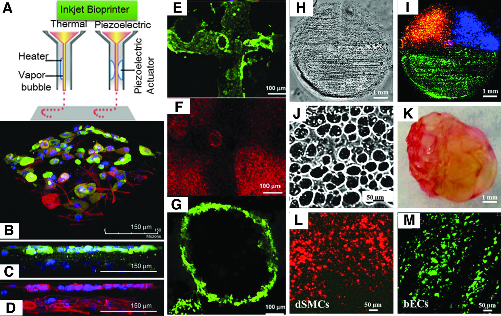
Inkjet-based bioprinting.
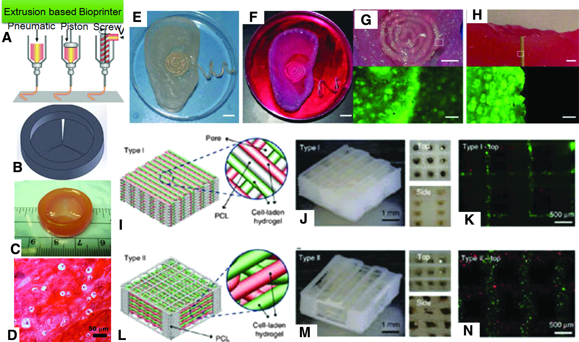
Extrusion-based bioprinting method.

Laser-based bioprinting method:
Recent advances in the development (mechanical platforms) and improvement (software's) of bioprinting techniques have significantly enhanced their applications and have opened up exciting opportunities suggesting that fully functional 3D biomimetic and disease-specific tissue constructs can now be fabricated to repair/restore functionality of a diseased/damaged tissue. Currently, bioprinting technology is being widely applied to print small-scale tissue structures to mimic native tissues (such as skin, nerve, cardiac, liver bone, and cartilage), models for cellular studies, drug discovery, toxicological effect assessments, tissue pharmacodynamics, and so on. However, 3D printing-based research is still at its preliminary stage, requiring comprehensive levels of advanced and integrated research efforts. Table 1 highlights the application of inkjet, extrusion, and laser-based bioprinting techniques for the biofabrication of various tissue constructs by using several cell types and bioink materials.
hiPSC, human induced pluripotent cell; hMSC, human mesenchymal stem cells; HUVEC, human umbilical vein endothelial cells; HUVSMC, human umbilical vein smooth muscle cells; LIFT, laser induced forward transfer; PEGDMA, poly(ethylene glycol) dimethacrylate; PU, polyurethane.
Limitations of the Existing 3D Printers and Bioinks
As discussed, there has been intensive research with regard to bioprinting technology. Among the different additive manufacturing techniques, inkjet, extrusion, and laser-based bioprinting are the three major printing technologies, which have the capability to print cell-laden bioconstructs under physiological conditions. All the above-mentioned technologies apply different printing principles, and each individual bioprinting method has its own intrinsic advantages and associated disadvantages with respect to its resolution, compactness, automation, printing capabilities, precision, and accuracy in ejection and deposition, scalability, and availability of ideal bioinks. Currently, a number of improved versions of 3D printers have been launched in the market for bioprinting purposes. Although various advance features of the latest bioprinters provide the opportunity for the biofabrication of various types of desired tissue structures, the development of a unified printing approach that shows compatibility with library of materials for the fabrication of different 3D tissue constructs or organs is still a great challenge. The fundamental issue with the typical bioprinting approach is that these methods perform operation based on a layer-by-layer criterion, which does not guarantee the development of highly complex, hollow, or fully functional and durable 3D complex bioproducts. In addition, there are numerous technological challenges in front of bioprinting technology, including development of novel high-resolution digital bioprinters supplemented with excellent nozzle and cartridge that allow loading and unloading of all types of bioink materials during printing processes, development of an efficient technique for the robotic delivery of target biocomponents, and development of a reliable technique to print prevascularized 3D constructs
Furthermore, selection of appropriate biocompatible materials for the preparation of bioinks is key step toward designing and facilitation of printing process and implementation of bioprinting technique for the fabrication of 3D-complex structures.106–112 Although various types of bioinks have been designed and implemented for bioprinting purposes, development of biofunctional printable bioink materials with excellent chemical, mechanical, rheological, and cytocompatible properties for fabricating desired bioproducts for transplantation is still a serious issue. Thus, there is an increasing demand for the development of tunable bioinks with a wide range of properties that can be easily manipulated by the bioprinting system. Bioprinting technology is considered as a technology in its infancy but it is growing up fast. To enhance progress in bioprinting approach for designing and manufacturing physiologically active bioparts for clinical applications, all the aforementioned technical as well as bioink material-related challenging issues need to be resolved.
Conclusions and Future Prospects
Bioprinting is one of the advanced fabrication techniques for rapid prototyping of cell-laden hydrogels to develop 3D tissue constructs. Early trends of bioprinting research have shown its promising potential in biofabrication of small-scale tissue structures. This technique offers unique advantages compared with conventional microfabrication methods such as microscale, high-throughput manufacturing, and ability of precise dispensing of bioink with high spatial and temporal resolution. All these features permit the efficient printing of biomolecules, living cells, and biopolymeric materials and control their precise localization in the 3D construct with excellent geometrical distributions. To further improve the potential of bioprinting approach, the printing speed and accuracy, improvements in the mechanical and rheological shortcomings of bioinks, and other technological and biological limitations need to be addressed. Although there is a wide room for further advancement and innovation in bioprinting technology, this technique indicates at its future ability to address some of the most daunting issues related to tissue engineering and may help in-factory large-scale production of effective and highly sophisticated therapeutic substitutes for clinical and bioindustrial applications. Bioprinting technology has also great potentials not only in production therapeutic bioproducts but could also be very useful for the fabrication of disease models, models for drug development and bioassays, and so on. In conclusion, the era of bioprinting has come to resolve partial organ dysfunction or complete organ failure issues.
Footnotes
Disclosure Statement
No competing financial interests exist.