
Abstract
The use of platelet-rich plasma and mesenchymal stem cells has garnered much attention in orthopedic medicine, focusing on the biological aspects of cell function. However, shortly after systemic delivery, or even a local injection, few of the transplanted stem cells or platelets remain at the target site. Improvement in delivery, and the ability to track and monitor injected cells, would greatly improve clinical translation. Nanoparticles can effectively and quickly label most cells in vitro, and evidence to date suggests such labeling does not compromise the proliferation or differentiation of cells. A specific type of nanoparticle, the superparamagnetic iron oxide nanoparticle (SPION), is already employed as a magnetic resonance imaging (MRI) contrast agent. SPIONs can be coupled with cells or bioactive molecules (antibodies, proteins, drugs, etc.) to form an injectable complex for in vivo use. The biocompatibility, magnetic properties, small size, and custom-made surface coatings also enable SPIONs to be used for delivering and monitoring of small molecules, drugs, and cells, specifically to muscle, bone, or cartilage. Because SPIONs consist of cores made of iron oxides, targeting of SPIONs to a specific muscle, bone, or joint in the body can be enhanced with the help of applied gradient magnetic fields. Moreover, MRI has a high sensitivity to SPIONs and can be used for noninvasive determination of successful delivery and monitoring distribution in vivo. Gaps remain in understanding how the physical and chemical properties of nanomaterials affect biological systems. Nonetheless, SPIONs hold great promise for regenerative medicine, and progress is being made rapidly toward clinical applications in orthopedic medicine.
Introduction
T
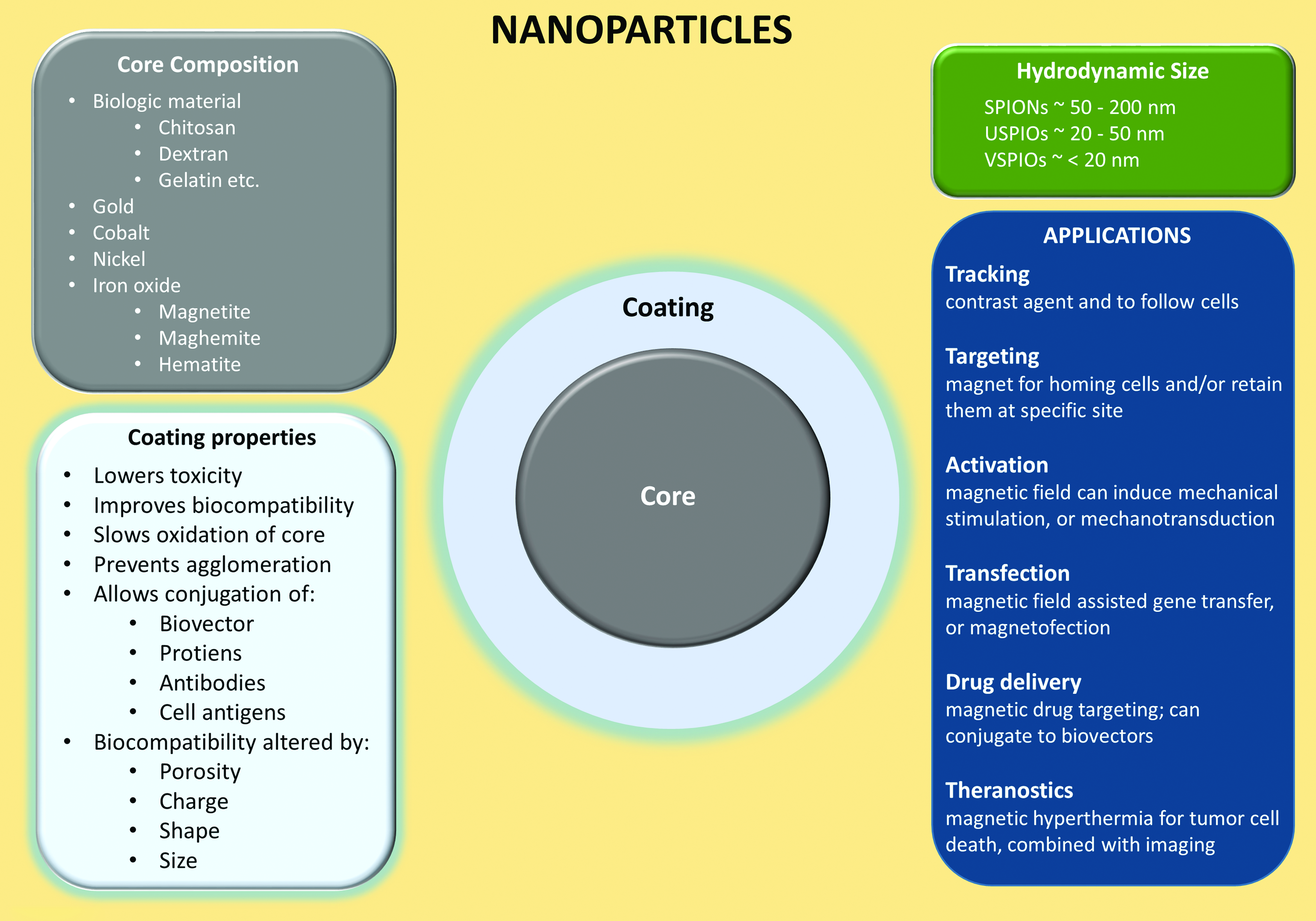
Simplified schematic of nanoparticles. SPIONs are nanoparticles with an iron oxide core (maghemite, magnetite, or hematite core) and are categorized by their hydrodynamic size. Transitional metal oxides (copper, cobalt, nickel, and manganese) mixed with iron oxide also exhibit superparamagnetic properties and are considered members of the SPION family. SPIONs are one of the most employed contrast agents for the labeling of cells, and serve a variety of applications such as imaging, drug delivery, magnetic hyperthermia, and others. Bare SPIONs are cytotoxic, tend to aggregate/agglomerate, and undergo further oxidation, which makes an appropriate coating crucial. Various materials are used for coating, but most SPIONs intended for medical applications are coated with biocompatible derivatives of dextran. SPION, superparamagnetic iron oxide nanoparticle. Color images available online at www.liebertpub.com/teb
Nanoparticle Structure
Core
Ferromagnetic (i.e., iron) and ferrimagnetic (i.e., iron oxide) materials exhibit high magnetization with a low applied magnetic field and have a remnant magnetization with the elimination of the applied magnetic field. 12 Small ferro- and ferrimagnetic materials (<20 nm diameter) exhibit superparamagnetism, in which the nanoparticles saturate with relatively high magnetization with a low applied magnetic field, but have no net magnetization with the removal of an applied magnetic field. While metals such as nickel and cobalt also exhibit superparamagnetism with small diameters, they are highly toxic. 13 Iron oxide (usually magnetite and its oxidized forms, maghemite or hematite) is preferred for biological application because it is a naturally occurring metal in humans (e.g., ferritin in myoglobin and hemoglobin), allowing preexisting metabolic pathways to process the remaining iron from nanoparticles.
Proteins such as deoxymyoglobin and deoxyhemoglobin are paramagnetic and have some magnetization with a low applied magnetic field, although the magnetization is not at the high saturation level of superparamagnetic materials (∼50–60 emu gFe−1). They also have no remnant magnetization with the removal of the applied magnetic field. The high magnetization in presence of an applied magnetic field is crucial for SPIONs as relaxation-darkening (negative) magnetic resonance imaging (MRI) contrast agents, because this property eliminates background effects of biological paramagnetic materials (i.e., deoxymyoglobin, deoxyhemoglobin), allowing for a greatly reduced T2 signal.
SPIONs are composed of an iron oxide core, which is enveloped by a polymeric or polysaccharide coating, and categorized by their hydrodynamic diameters. 12 Typically, standard SPIONs have a hydrodynamic diameter between 50 and 200 nm (Fig. 1), and can contain more than one iron oxide core per particle. Ultrasmall SPIONs (USPIOs) have a hydrodynamic diameter between 20 and 50 nm, and very small SPIONs (VSPIOs) have a hydrodynamic diameter less than 20 nm. Both USPIOs and VSPIOs behave as ferrofluids when suspended in solution (i.e., by not separating from the solution in the presence of a magnetic field). SPIONs can be targeted to a specific tissue area (e.g., delivering platelet-rich plasma [PRP] or stem cells to injured muscle, phagocytic cells to tumors, etc.) using a gradient magnetic field.14,15 Magnetic mediated hyperthermia in target regions can also be induced using an alternating magnetic field as well.16,17
Coating
The coatings of SPIONs serve many important roles, such as reducing iron oxide oxidation, preventing aggregation and agglomeration of extracellular SPIONs, increasing biocompatibility, improving targeting, increasing tracking duration, limiting nonspecific cell interactions, and improving localization by providing a chemical handle for conjugation of targeting ligands and drug molecules. 18 The coating of SPIONs can change its size, shape, porosity, and surface charges, and it can be composed of polyethylene glycol, dextran, citrate, chitosan, polyethyleneimine, phosopholipids, or copolymers. SPION size (core plus coating) can affect their passage into tissues and cells, with most endothelial barriers allowing SPIONs <150 nm to pass. 18 SPION size can also affect the rate of cellular uptake, with diameters between ∼30 and 150 nm having longer blood circulation duration, as they are not phagocytized as readily. 19 When shape is altered such that the nanoparticle is rod-like and not spherical, the coating is anisotropic, resulting in increased in vivo blood circulation time. 20 SPIONs can also be conjugated to a peptide to specifically target a ligand, or be conjugated to drug molecules, such as bisphosphonates for osteoporosis 21 or Bcl2 (B cell lymphoma 2) to inhibit apoptosis and enhance bone regeneration, 22 allowing for magnetic mediated drug delivery to the targeted tissue.14,23 Thus, careful consideration of coating parameters must be ensured for the specific therapeutic method. 18
SPIONs as a contrast agent
At present, the most commonly used MRI contrast agents utilize paramagnetic gadolinium ions, which have a typical elimination half-life of ∼1.6 h. 24 SPIONs have also been employed clinically as contrast agents for hepatic imaging.1–4 SPIONs have a unique advantage over previously developed contrast agents in that they can be tracked for a much longer duration. Human muscle progenitor cells (labeled with dextran-coated SPIONs and labeled with poly-L-lysine-coated SPIONs) can be tracked for over 4 weeks.25,26 The magnetic force experienced by a magnetic particle in a magnetic field is directly proportional to the magnetization of the particle, the gradient of the magnetic field, the volume of the particle, and the particle density. 27 The magnetization of a SPION becomes saturated with a low applied magnetic field; thus the magnetic force is not dependent on the magnetization of the particle. During MRI, the magnetic force experienced by these nanoparticles is much less due to their small size (proportional to volume, in the order of ∼r 3 ). The drag (proportional to r) experienced by SPIONs, 27 together with the short time period of the applied gradient magnetic field, 28 results in no movement during MRI. However, MRI systems have inherent magnetic field gradient coils that can, if desired, generate gradients for magnetic resonance targeting of SPIONs to target tissue. 7
SPIONs in Musculoskeletal Therapies
SPIONs are absorbed by a variety of cells through spontaneous endocytosis or phagocytosis 29 with no signs of toxicity at trackable loads of iron oxide in cells.25,30,31 Labeling of cells with these nanoparticles is simple, typically requiring no more than 1 h of incubation of nanoparticles with the cells. Some labeled cells can be frozen for storage, and replated after thawing, with viability identical to unlabeled cells. 31 However, for nonphagocytic and slowly dividing cells, SPION uptake by cells requires transfection agents, which can impact cells negatively. 32
Tracking and targeting platelets
Platelets, which are small, anuclear, disk-shaped fragments are released into the vasculature from large bone marrow cells called megakaryocytes. A sizeable volume of new platelets (∼1 × 10¹¹) is produced each day, 33 and this yields a turnover of circulating platelets roughly every 10 days. Platelets contain α-granules, which are secretory vesicles that contain a range of growth factors34–36 associated with improved repair of damaged tissue, including cartilage, tendon, muscle, and bone.35,37–41 Plasma with a concentration of platelets above the concentration in whole blood is termed PRP (Fig. 2). 42 PRP can be isolated using either centrifugation protocols, or a commercial system. PRP can have as high as an eightfold increase in the concentration of platelets found in whole blood. 34 The resulting increase of growth factors, still present in physiological proportions to each other, is presumably an advantage compared to the use of isolated growth factors.

Isolation and in vivo magnetic cell targeting of PRP and MSCs. SPIONs are easily taken up by a variety of cells, reaching levels suitable for tracking, with labeled cells showing no signs of toxicity. They are internalized through spontaneous endocytosis or phagocytosis, and cell labeling is simple, chemically safe, and typically requires no more than 1 h of laboratory contact time. Top: Platelets were isolated from whole blood using a commercial system. Photographs show the whole blood and PRP (platelets) obtained after separation by centrifugation. Transmission electron microscopy shows example of SPIONs inside a platelet. The iron oxide core of the SPIONs is present as small dark spheres. Bottom: MSCs can be isolated from bone marrow and cultured. SPIONS can be tagged with a fluorophore, as in this example, for later detection during histological analysis. Red arrows: an internal magnet, external magnet (pictured here: red region on schematic of rat leg), or even a clinical MR scanner can be used to localize labeled platelets or MSCs at target locations and MRI can be used to track the SPION-containing platelets or MSCs in vivo (MRI shows rat leg with SPION-labeled cells targeted to tibialis anterior muscle). EM, electron microscopy; IF, immunofluorescent microscopy; MRI, magnetic resonance imaging; MSCs, mesenchymal stem cells; PRP, platelet-rich plasma. Color images available online at www.liebertpub.com/teb
A major challenge is sustaining PRP at the tissue damage site in vivo for the platelet lifespan (∼10 days), as the leakage of PRP from the region of tissue damage will likely limit its value. Alternatively, biosynthetic scaffolds can be employed for local activation of PRP over a prolonged period of time, but this has drawbacks as well. 43 From a practical and comfort standpoint, recurring injections are unfavorable and are more likely to lead to undesirable systemic effects.42,44 Furthermore, storage of platelets from a single blood draw is challenging, with potential premature activation of platelets. PRP appears to be safe, but effectiveness remains to be proven.
PRP is currently applied to many musculoskeletal disorders,35,37,39,41,45 but with questionable efficacy as described above. A novel method has been described for injecting muscle with platelets containing SPIONs, which can be imaged in vivo by MRI and in vitro by fluorescence microscopy. 15 Platelets endocytose SPIONs (without linkers or binding agents) with ∼98% efficiency. 46 Use of an external magnet can be employed to target and retain the location of PRP with SPIONs.15,47 If translated from a preclinical notion to successful clinical studies, this technique allows for targeting of PRP to a desired site. It could also arrest premature loss of the platelets at damaged muscle, cartilage, tendon, or bone (Fig. 2).
Homing and monitoring stem cells
Regenerative medicine aims to repair or replace damaged human cells, tissues, or organs to restore normal function via stimulation of the body's own repair mechanisms. 48 To translate stem cell therapies into clinical use, the long-term distribution, engraftment, and fate of stem cells must be monitored using a reliable and noninvasive tracking method. Mesenchymal stem cells (MSCs) are a cell population of undifferentiated cells isolated from adult tissue (Fig. 2). With the application of specific growth factors or bioactive molecules in vitro, they have the capacity to differentiate into mesodermal lineages, such as bone, cartilage, fat, muscle, and other tissues (Fig. 3).49,50 MSCs have the ability to respond to the local environment in vivo and have been used clinically in several fields to repair dysfunctional tissue.50–54 Although MSCs can be forced to differentiate into various cell types in vitro, 55 when used in vivo, exogenous MSCs most likely do not directly incorporate, differentiate, and repair tissues. 56 Instead, it is now evident their predominant mode of action is indirect via the secretion of “trophic” factors into the tissue microenvironment that permit the host tissue to regenerate and repair. 57
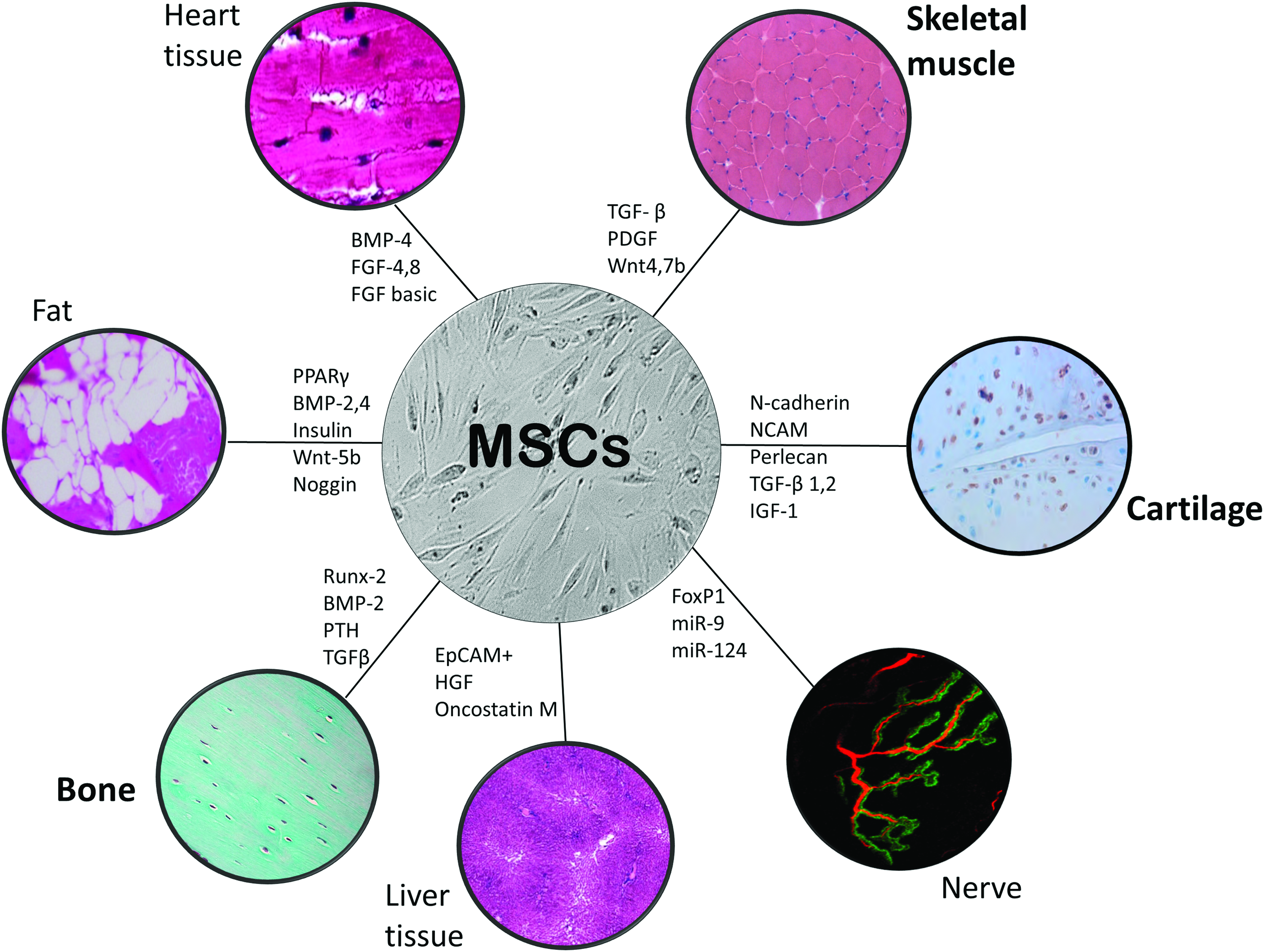
Using various signaling conditions, MSCs can differentiate into different types of cells in vitro, or support endogenous cells in vivo. MSCs derived from bone marrow and can be isolated from most adults with the potential of autologous transplantation, not requiring immunosuppressive agents. They are easy to isolate, expand, and can be differentiated with various growth factors (transcription factors and signaling molecules participating in regulation of MSC differentiation; examples of molecules and factors participating in regulation of MSC differentiation are given, but by no means a complete list). MSCs can support tissue repair and regeneration either directly (direct differentiation) or indirectly (trophic factor secretion permitting endogenous progenitor cells), but with in vivo delivery the evidence points to the latter. MSCs can be successfully labeled with SPIONs, which can be conjugated with a chemical handle for differentiation. SPION-labeled MSCs can also be localized to target tissues with the use of a gradient magnetic field. Color images available online at www.liebertpub.com/teb
Transplantation of exogenous MSCs at the site of skeletal muscle injury can enhance regeneration58–60 and accelerate skeletal muscle repair.59,61 However, effective retention of transplanted MSCs following injection of MSCs into the area of injury has not been demonstrated. Articular cartilage has very little regeneration and healing (scar formation) is often inadequate, which could be augmented by the use of locally applied PRP39,40,43 or transplanted MSCs.51,62 Tissue-engineering has made great progress toward transplantation of ex vivo tissue-engineered cartilage growth, but implementing such methods involves multiple surgeries to harvest cartilage cells and then implant the newly grown autologous graft into the defect. Bone has regenerative capacity, but there are still conditions where MSC implantation could be useful, such as non-unions, bone grafts, bone disease, and potentially with osteoporosis.63–66 While, already there are methods to surgically address traumatic and degenerative conditions in bone and cartilage, the invasive nature of surgery, implant failure, immune rejection, infection, donor site morbidity, and others limit successful regeneration of these tissues. Noninvasive magnetic targeting of MSCs offers the possibility of regeneration without many of these issues, and the potential to track delivered cells over time.
Magnetic cell targeting is the use of a magnetic gradient with the goal of accumulating transplanted SPION-containing cells to a specific site (Fig. 2). Magnetic cell targeting with use of an external magnet provides a noninvasive means of enhancing tissue regeneration and has now been described for both platelets 15 and MSCs 67 (and a series of nonmusculoskeletal conditions). 68 In preclinical studies, magnetic cell targeting has been employed to accumulate MSCs in bone and cartilage tissue,69–71 or targeting growth-promoting factors to such tissues. 22 A localized magnetic field gradient could be achieved in deeper tissues using fixed internal magnets,72,73 but this necessitates invasive surgical implantation. Recent reports indicate that clinical MRI scanners can not only track the location of magnetically labeled cells, but also guide them into tissues that are inaccessible using an external magnet. 7
The iron-positive signal in MRI can persist for as long as 2 months, however, this long lasting signal intensity can be the result of macrophages that have consumed the SPION bound to dead cells 6 or generated by extracellular, instead of intracellular, iron particles. 74 Thus, monitoring of platelet/cell viability, which is thought to correlate with the strength of the SPION signal, can present some challenges. Another major obstacle surrounding the SPION-labeling strategy is leakage of SPIONs into adjacent cells 75 and, in the case of stem cells, the potential for dilution after cell division.76,77 Thus, one of the current drawbacks associated with SPION is its inability to distinguish between viable and nonviable cells, and long-term monitoring may provide an overestimation of cell survival and a false positive signal. 25
SPIONs in muscular dystrophy/gene therapy
There are now a series of studies developing applications for nanoparticles in musculoskeletal tissues,21,78–86 but their use is not limited to orthopedic acute conditions. There are several preclinical studies describing the use of nanoparticles for diseases, for example, muscular dystrophy.87–89 Muscular dystrophies are a heterogeneous group of genetic disorders with progressive skeletal muscle weakness and degeneration. Duchenne muscular dystrophy (DMD), the most common form of muscular dystrophy, an X-linked disorder, was first described a century ago 90 and is caused by the lack of dystrophin at the membrane of muscle fibers. Approximately 1 in 3500 newborn males worldwide are affected with DMD91–93 and patients develop progressive wasting of muscles and ultimately death, usually occurring by the ages of 20–30 due to cardiac or respiratory decline. 94 Muscle regeneration, typically occurring after damage in healthy skeletal muscle, is lost in patients with DMD.95–97 Thus, magnetic cell targeting of MSCs could have potential use in treating muscular dystrophies. However, the limitation of SPIONs in long-term monitoring due to dilution with cell division, leakage to adjacent cells, and macrophage uptake, pose a challenge in successful use of SPIONs to track MSCs survival in DMD patients.
In DMD and in mdx mice, the murine homolog of DMD (mdx also lacks dystrophin), an ideal treatment would be to restore dystrophin. To date, the efforts to treat dystrophies have focused on gene therapy, however, delivery of such a large gene, and to all muscles throughout body, presents challenges. The dystrophin gene is 2.4 Mb in size and is not easily inserted into available vectors. Dystrophin can retain a large part of its function even when missing much of its middle region, and so use of “mini-dystrophin,” containing the N-terminal and C-terminal sequences responsible for binding, has shown promising results.98–101 Other therapeutic options include nonviral carriers (e.g., polymers) to deliver the dystrophin cDNA to muscle. 102 A potential therapy could involve stem cell therapy. MSCs injected intravenously in the mdx mouse can move into muscle, differentiate, and result in partial, although transient, restoration of dystrophin.103,104 The potential of MSCs as an antiapoptotic agent 105 and inhibitor of inflammation 106 is enticing, yet even though animal studies yield successful outcomes, clinical trials have failed to yield significant benefits for patients with DMD, and the problem of delivery efficiency remains a challenge.
SPION containing MSCs appear to differentiate and mature into muscle cells, and studies in mdx mice show the ability to track SPION-labeled MSCs in muscle noninvasively with MRI. 89 SPION-labeled mesangioblasts and magnetodendrimers-labeled MSCs also exhibit normal differentiation and growth and have been tracked after implantation in mdx mice.87–89 Such studies hold great promise for imaging and tracking stem cells in DMD.
Magnetofection is the term given to strategically introducing DNA into cells using coated magnetic nanoparticles, coupled with the influence of an external magnetic field.107–109 This magnetic field-assisted gene transfer is especially useful in cells that are difficult to transfect and has been used to efficiently overcome transduction resistance in skeletal muscle cells. 9 There are some obvious biological barriers in terms of delivering nucleic acids into cells, such as membranes that surround the nucleus, cell vesicles, and the cell itself. Methods to overcome such barriers have been tried, such as cell bombardment methods (the “gene gun approach”) and application of an electric field (electroporation), 110 but the efficacy is variable and they can even result in cell damage. 111 With magnetofection, the vector can be coupled to SPIONs and accumulated on target cells by the application of a gradient magnetic field (magnetic cell therapy, but for nucleic acids). Magnetofection has been reported to improve viral and nonviral mediated gene delivery into cells, such as muscle,9,112 and it may provide a reliable method to deliver gene therapy to cells that are resistant to transfection, such a musculoskeletal tissues.
SPION removal
Following their internalization, SPIONs are eventually metabolized by the lysosomal pathway and the iron oxide core is gradually incorporated into the body's iron store.3,113 Thereafter, it is eliminated in the same manner as endogenous iron through the feces. Coating degradation is determined by its composition. For example, dextran and its derivatives are degraded by enzymes and are eliminated by renal clearance. 113 Size and coating of the SPIONs can be further tuned for faster or slower clearance time. Since iron-based agents will not cause nephrogenic systemic fibrosis in patients with compromised renal function, SPIONs are a viable option to gadolinium-based contrast agents. Macrophages/the reticuloendothelial system in the liver, spleen, and lymph nodes, absorb the iron and this pathway could potentially be utilized to image vascular lesions, tumors, and lymph nodes. 114
Limitations for Clinical Use
SPIONs are reported to be highly biocompatible nanomaterials with little to no toxicity, but recent research sheds doubt on the benign nature of these nanoparticles in biology.115,116 Although high SPION uptake is desirable for improving imaging contrast and therapeutic delivery, high SPIONs loads can be cytotoxic.25,117 SPIONs could have a clinical role someday in musculoskeletal medicine for uses such as diagnostics, cell labeling, drug targeting, and gene delivery, but some studies point to adverse effects on cells, including mitochondria damage, oxidative stress, chromosomal and oxidative DNA damage, altered cell cycle regulation, and protein denaturation.118,119 Whether SPIONs can act as a mutagen is unclear, but there is some experimental evidence of nanoparticles having potential mutagenic interactions on human cell lines. 116 Thus, SPION toxicity, although usually reported as low, has not been completely established. 120 Despite the numerous SPION uses being explored, currently available information on their potential toxicity is scarce and controversial data have been reported. Hence, concerns such as toxicity, stability, and resident time still need to be addressed.
Still undetermined variables for using SPIONs include proper selection, dose, and biophysical parameters to enhance binding, specific tissue compatibility, and others. The use of magnetic cell targeting is exciting, but there are many variables that still need to be optimized, such as optimal magnetic field strength, duration of exposure, and obtaining adequate depth, which are all likely dependent on the makeup of the SPION and the type and state of the tissue being targeted. While SPIONs such as Feridex®, GastroMARK,® and Resovist® have received FDA approval as contrast agents, today only Resovist is available in a few countries.1,4 A USPIO, Feraheme™, has received regulatory approval for ameliorating iron deficiency in chronic kidney disease, and potentially could be used as an imaging agent for lymph nodes and hepatocellular carcinomas. Nanotherm® has European Union-wide regulatory approval and is awaiting FDA approval, with phase II clinical trials for use of magnetic hyperthermia for glioblastomas. Thus, many hurdles remain in transitioning the use of SPIONs into orthopedic clinical use.
Summary
There remains a lack of standardization for isolation and delivery of PRP/MSCs, and the efficacy and optimization of such therapies remain unclear, but the use of such therapies to facilitate musculoskeletal tissue healing appears quite promising. SPIONs can be manufactured from a variety of synthetic or biological materials and they effectively label platelets and cells without compromising capacity for cell proliferation or differentiation. Moreover, studies show that cells loaded with SPIONs can be injected locally or systemically and can be attracted to a target tissue by the application of a gradient magnetic field, such as with an external magnet. However, almost all of the work for such musculoskeletal magnetic cell targeting, and/or subsequent monitoring, is preclinical, involving only animal studies (Table 1). SPIONs can have other roles in musculoskeletal tissues besides regenerative medicine, such as coatings for prevention of biofilm formation 121 (not discussed). To effectively use these nanomaterials in clinical applications, the physiochemical properties and the effects of these properties on physiological processes need to be fully characterized. 122
Representative publications using SPIONs in muscle, cartilage, and bone (superior number corresponds to reference number; ‘X’ indicates when performed in vitro or in vivo, or both). Note that, although SPIONs have been used clinically as a hepatic contrast agent, studies in musculoskeletal applications are still in the preclinical stages.
BSA, bovine serum albumin; hMSCs, human mesenchymal stem cells; PLL, poly-L-lysine; MRI, magnetic resonance imaging; SPION, superparamagnetic iron oxide nanoparticle; uCT, micro-computed tomography scan.
Footnotes
Acknowledgments
This work was supported by grants from the National Institutes of Health, including training grant T32 AR-007592 (S.R.I.) and research grant R21-AR067872 (R.M.L.).
Disclosure Statement
No competing financial interests exist.