




Abstract
Hydrogels have been promising candidate scaffolds for cell delivery and tissue engineering due to their tissue-like physical properties and capability for homogeneous cell loading. However, the encapsulated cells are generally entrapped and constrained in the submicron- or nanosized gel networks, seriously limiting cell growth and tissue formation. Meanwhile, the spatially confined settlement inhibits attachment and spreading of anchorage-dependent cells, leading to their apoptosis. In recent years, macroporous hydrogels have attracted increasing attention in use as cell delivery vehicles and tissue engineering scaffolds. The introduction of macropores within gel scaffolds not only improves their permeability for better nutrient transport but also creates space/interface for cell adhesion, proliferation, and extracellular matrix deposition. Herein, we will first review the development of macroporous gel scaffolds and outline the impact of macropores on cell behaviors. In the first part, the advantages and challenges of hydrogels as three-dimensional (3D) cell culture scaffolds will be described. In the second part, the fabrication of various macroporous hydrogels will be presented. Third, the enhancement of cell activities within macroporous gel scaffolds will be discussed. Finally, several crucial factors that are envisaged to propel the improvement of macroporous gel scaffolds are proposed for 3D cell culture and tissue engineering.
Introduction
T
Currently, two types of scaffolds are commonly used as artificial cell culture matrices: prefabricated porous scaffolds and hydrogels. 7 Porous polymer scaffolds can be easily fabricated via various platforms, such as freeze drying, 8 3D printing, 9 electrospinning, 10 gas foaming, 11 salt leaching, 12 phase separation, 13 and stereolithography. 14 These porous scaffolds generally possess high mechanical strength and stiffness. However, several inherent challenges of porous scaffolds require to be addressed and overcome. First, their preparation processes usually involve the conditions that may lead to cell apoptosis, such as toxic solvents, 15 excess salt concentration, 16 overheating, 17 and ultralow pressure. 8 As a result, the seeding of cells into such porous scaffolds could only be performed after their fabrication, which usually causes inhomogeneous cell distribution and neotissue formation. 18 Second, the low porosity is not advantageous for cell proliferation and migration, although it enables porous scaffolds to exhibit initial structural stability. 19 Bioresorbable porous scaffolds, prepared from biodegradable polymers, hold increasing porosity over culture time to a certain extent, 13 however, it is very difficult to tune the degradation profile in accordance with neotissue development.
Hydrogel, water-swollen, 3D hydrophilic polymer networks, is another type of widely used scaffolding material for 3D cell culture and tissue engineering.20,21 Hydrogel is formed from the gelation of polymer precursor solution, and it consists of numerous polymer networks with submicron- or nanosized pores. 22 Hydrogel has gained great attention as a cell delivery vehicle due to its high porosity, tissue-like water content, injectability, tunable permeability, degradability, and mechanical property. 23 In the past decades, a great number of hydrogel scaffolds have been developed and reported. Depending on the type of seed cells, gel microenvironments have been manipulated and optimized by presenting various topographical, 24 biological, 25 and physical cues, 26 for achieving the promotion of desirable cellular behaviors. 27 This review summarizes the advantages and challenges of the hydrogels for 3D cell culture and tissue engineering, and focuses on the establishment of macroporous gel scaffolds and the effect of the creation of macroporous structure on cellular behaviors and neotissue formation.
Hydrogel-Based Scaffolds for 3D Cell Culture—Advantages and Challenges
Cell encapsulation and subsequent tissue engineering within hydrogels generally involve three steps28–30 : First, seed cells are homogeneously suspended in the isotonic solution of gel precursors; second, cell suspension is transferred into appointed molds in vitro or desired sites in vivo, completing the gelation to obtain 3D cell/gel construct in situ; third, 3D cell culture in vitro and followed by transplantation of the engineered constructs target tissues, or direct neotissue development in vivo. This operation process for cell encapsulation and 3D cell culture within hydrogels exhibits several advantages. First of all, compared to prefabricated porous scaffolds, the cells are able to be more abundantly and homogeneously distributed within hydrogels by carrying out a compatible and rapid curing (several seconds to a few minutes) of cell-suspended precursor solution through various initiation strategies (such as thermo-, ion-, redox-, or photo-curing process).29,31–33 Moreover, cell suspension of gel precursor can be filled into any shaped molds in vitro or tissue defects in vivo, ensuring the spatial fit with host tissues. 34 Last but not least, hydrogel-based constructs can be easily fabricated and manipulated from nanogel particles to macroscopic materials to meet practical requirements.35,36 This process promises effective conveyance of living cells or engineered tissue constructs to target sites of tissue degeneration, which then exert therapeutic effects to repair damaged tissues or organs.
Hydrogels may imbibe from 10% to thousands of fold higher than its dry weight in aqueous solution. 37 The high water content and its elasticity make hydrogels closely resemble native tissues. 38 The high water content not only promises high permeability but also contributes to its excellent biocompatibility. Furthermore, since hydrogel is composed of networks of hydrophilic polymer chains and holds massive water, it exhibits minimal tendency to adsorb proteins from culture medium in vitro or body fluids in vivo. 39 Besides, drugs and growth factors can be easily incorporated into hydrogels and their release behaviors can be modulated by adjusting gel properties such as swelling ratio, water content, and degradation profiles. 40
The abovementioned advantages guarantee the feasibility of hydrogels for cell encapsulation and 3D cell culture in engineering of various tissues such as cartilage, 41 cornea, 42 and liver. 43 Among these applications, cartilage tissue engineering should be the most representative example.44–46 Because of low chondrocyte density and poor blood supply, damaged cartilage exhibits limited self-regeneration capacity, and engineered regeneration of cartilage has been in great and urgent demand. 47 Hydrogel can mimic the extracellular matrix (ECM) of native cartilage tissue, and its hydrophilic networks and high water content can retain the native spheroidal morphology of chondrocytes. 22 In addition, significantly, hydrogel microenvironments can be tuned to enhance desirable cellular processes. 48 A series of excellent studies have been carried out to improve the potential of hydrogels as cartilage tissue engineering scaffolds by illuminating the effects of various gel microenvironmental cues 49 (such as stiffness, 50 degradation profiles, 51 crosslink density, 52 and introduction of physical or chemical factors 53 ) on the behaviors of loaded cells. In addition, some groups have investigated the influence of gel microenvironments on the chondrogenesis of stem cells, such as adipose mesenchymal stem cells, 54 bone mesenchymal stem cells, 55 synovial mesenchymal stem cells, 56 embryonic stem cells (ESCs), 57 and induced pluripotent stem cells (iPSCs). 57 Besides, the metabolic activities of encapsulated cells have also been evaluated at the molecular level in hydrogel-based cartilage tissue engineering. 58
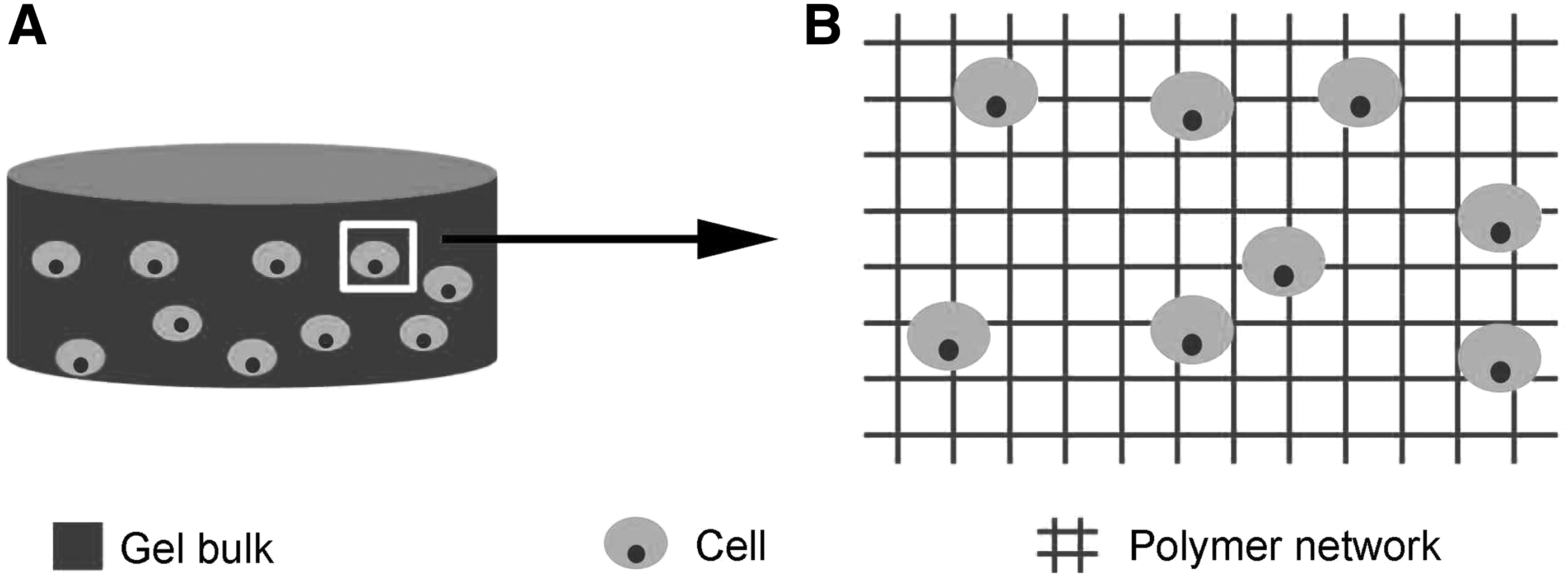
Despite these advantages, a huge challenge remains for the use of conventional hydrogel scaffolds in supporting 3D cell growth and differentiation. The submicron- or nanoscale hydrophilic polymer networks of gel body give rise to serious physical constraints on the encapsulated cells and thus restrict their spreading, proliferation, and migration regardless of cell types (Fig. 1), especially for anchorage-dependent cells (ADCs) (such as fibroblasts, endothelial cells, cardiomyocytes, smooth muscle cells, and neurocytes). 59 ADCs require adhesion to a surface and stretch to maintain their normal morphology, otherwise, they undergo apoptosis gradually. 60 In most cases, the ADCs will die after several days of encapsulation as a result of the failed settlement in the interior of gels. 61 Such disadvantage still has not been overcome even for the “adhesive” gels that are prepared from the precursor containing cell-adhesive domains such as arginine-glycine-aspartic acid (RGD) peptide. 62 The presence of cell-adhesive moieties may obviously enhance focal adhesion of ADCs on gel surfaces. However, the spreading of the cells within the interior of hydrogel will be severely inhibited due to spatial limitations. Similar to ADCs, the proliferation, differentiation, and migration of encapsulated non-ADCs, such as chondrocytes, are also dramatically restricted by the 3D “physical binding” from gel networks. 63 Meanwhile, the limited living spaces within the gel body also depress the secretion and deposition of ECM. 64 Spatial limitation has been proved to be the major bottleneck of gel scaffolds for 3D cell culture and tissue engineering. In recent years, therefore, a series of macroporous hydrogel scaffolds with cell living spaces as well as cell-affinitive interfaces have been established and created for promoting desired cellular behaviors.
Strategies of Preparing Macroporous Gels
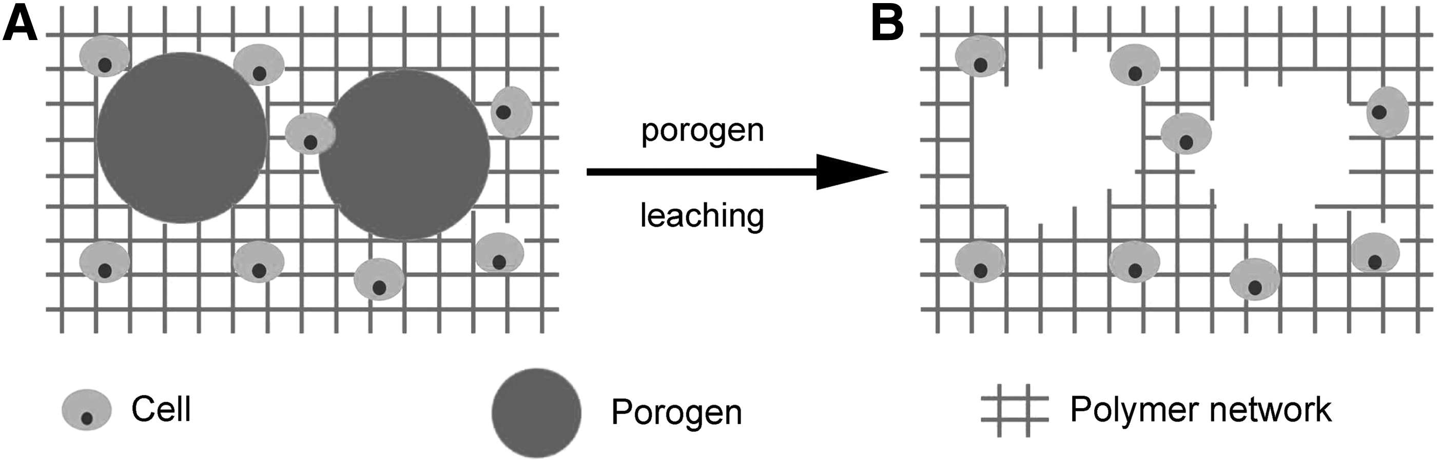
Four types of strategy are currently used to fabricate the macroporous hydrogels that have the ability to achieve in situ 3D cell encapsulation. 65 One of the most common methods is to introduce biodegradable blocks into gel networks, namely, the fabrication of biodegradable gel. 66 In addition to degradation, the macroporous gel scaffolds can be directly developed by means of the fabrication platforms (i.e., 3D bioprinting 67 and electrospinning 68 ). Besides, the macroporous (cavitary) structure can be created in hydrogel via the combining and removal of biocompatible porogens with the size range from tens to hundreds of micrometers in diameter (Fig. 2). 63
Incorporation of bioresorbable blocks
This design of biodegradable hydrogels is based on the hypothesis that the degradation (rupture) of gel networks over incubation time may form cavities that not only regulate hydrogel's physical microenvironments but also provide living spaces for cell proliferation and neotissue formation. 69 For instance, poly(ethylene glycol) (PEG)-based biodegradable hydrogels have obtained extensive studies, and they are generally prepared by incorporating biodegradable parts into PEG networks, such as phosphate groups, 49 poly(lactic acid) (PLA), 70 poly(trimethylene carbonate) (PTMC), 71 poly(epsilon-caprolactone) (PCL), 72 and poly(2,2-dimethyltrimethylene carbonate) (PDTC).47,73 The rupture of these polymer chains is resulted from the hydrolysis or enzymolysis of the ester bonds or carbonate groups. A series of studies have shown that the eroding of such degradable moieties indeed increase the swelling ratio and permeability of hydrogels, and then lead to enhanced cellular activities.74–76 Among these, Anseth et al. have done outstanding works on the poly(lactic acid)-PEG-poly(lactic acid) (PLA-PEG-PLA)-based degradable gels. The results of their studies have shown that the combined use of PEG-dimethacrylate and PLA-PEG-PLA-dimethacrylate precursors and/or the adjustment of polymerization degree of PLA moieties can tune the crosslink density, elastic modulus, and degradation rate of gel scaffolds to modulate cell behaviors. 70 In addition, it is worth mentioning that the aliphatic polycarbonate (such as PTMC, PDTC)-based biodegradable PEG gels can generate carbon dioxide (CO2) and biocompatible diols under degradation, and these degradation products are much less acidic compared to those of poly(lactic acid)-based gels.47,73 Therefore, polycarbonate-based hydrogels are seemingly more beneficial to cell encapsulation and 3D cell culture. Zhang et al. have reported a biodegradable tough hydrogel prepared from the oligoTMC-PEG-oligoTMC-diacrylate precursor for cartilage tissue engineering. 71 Although its degradation facilitates cell proliferation and biosynthetic activity, it is very difficult to exactly tune the timing and degree of cavity generation in accordance with the stages of neotissue development. 77 Besides, the degradation process of gel scaffolds generally become compromised along with the deposition of ECM in the constructs; the ECM is thought to be the “stabilizer” during hydrogel degradation. 71 In recent years, the gel scaffolds that can degrade under stimuli, such as light and enzyme, have attracted much attention.78–80 Anseth et al. devise photodegradable gel scaffolds, and whose properties can be real timely manipulated via light irradiation. This photodegradable gel provides a dynamic cell culture platform to elucidate how cells interact with surrounding microenvironments. 81 Recently, a new cartilage-specific degradable gel scaffold, based on PEG-thiol and aggrecanase cleavable peptide crosslinker, has been developed by Skaalure et al., which is degradable by chondrocytes and enhances the formation of hyaline-like cartilage. 82
3D bioprinting
3D bioprinting, in which the gel materials together with cells can be printed via layer-by-layer fabrication technology,83–85 has been widely exploring as a strategy to produce porous hydrogel scaffolds (with a predetermined internal pore structure and external shape) for various tissue engineering applications such as cartilage, 86 bone, 87 and blood vessels.88,89 Recently, You et al. report a facile bioplotting-based preparation of porous alginate gel scaffold and corresponding cell-laden constructs; the wall thickness between pores, swelling ratio, and compressive modulus of the hydrogel can be adjusted by controlling the concentration of the crosslinker—Ca2+; this fabrication technique can achieve a 3D cell encapsulation with high viability and the porous gel scaffold is capable of promoting the proliferation and chondrogenic differentiation of ATDC5 cells. 86 In addition to pore structure, the macroporous gel scaffolds developed using 3D bioprinting can also be engineered to achieve the establishment of complex architecture by applying rapid prototyping techniques that follow computer-assisted design and manufacturing. Kolesky et al. devised a 3D bioprinting platform that can be used to create delicate heterogeneous macroporous hydrogels on demand; vasculature-like networks and different cell types can be easily placed within such gel scaffolds according to the program. 88 Taken together, the advantages of 3D bioprinting-based macroporous gels include the following: (1) the pore size and gel wall thickness can be facilely controlled through a computer program and (2) complicated tissue construction, such as vasculature and gradient structure, can be accurately introduced to mimic native tissues and organs. However, 3D bioprinting practices, heavily relying on screen printing, are limited to the low resolution in their applications; the resolution of current techniques varies from several to hundreds of micrometers. 87 Furthermore, the paucity of satisfactory biomaterials applicable to bioprinting machines also limits the wide applications of the 3D bioprinting technique in the fabrications of cell-laden gel constructs. 90
Electrospinning
Electrospinning is one straightforward and cost-effective approach that can be applied to produce cell-laden hydrogel fibers using polymer solutions, and these fibers are arranged to form macroporous gel scaffolds.91–93 The electrospinning-based fibrous gel scaffolds have some unique advantages. The gel fibers range from less than 10 nm to several micrometers in diameter, which endows the gel scaffolds with high specific surface area. More significantly, these gel scaffolds can provide topographical cues to induce cell alignment within the constructs. Electrospinning has received growing interest in exploiting its application in producing fibrous hydrogels for 3D cell culture and tissue engineering from a variety of both natural and synthetic polymers, such as PEG 94 and gelatin. 95 Representatively, Han et al. have developed the PEG-methacrylate-based crosslinkable microribbons through electrospinning, which are used as the “precursors” to prepare cell laden macroporous constructs by in situ photoencapsulation of adipose-derived stromal cells. 94 In addition to fibrous constructs prepared from the prefabricated gel fibers, the cells can be directly encapsulated into electrospun fibers during the electrospinning progress and followed by the formation of macroscopic gel constructs. 93 Hu et al. have developed an electrospinning-based approach to fabricate cell laden gel fibers by crosslinking the precursors in cell suspension flowing in the capillary tubes; experimental results show that this fabrication process of cell laden fiber constructs is mild enough to maintain high cell viability.96,97
Incorporation of biocompatible porogen
The application of biocompatible porogens that can be embedded within the hydrogel bulk and subsequently removed in situ is a facile strategy to fabricate macroporous gels. The design inspiration of the porogen-based macroporous gel (referred to as microcavitary gel) is originated from an observation that the chondrocytes proliferate quickly (named as edge flourish, EF) by the edge of cell laden agarose gel constructs compared to the inner chondrocytes.63,98 Gong et al. speculate that the presence of EF phenomenon resulted from the constraints of gel polymer networks on cell growth and the proliferating chondrocytes actively seek living spaces in gel bulk, and therefore, the introduction of cavities within gel constructs by combining and removal of microsphere porogens may create more cell living spaces and enhance cell proliferation in gel bulk. Accordingly, the authors prepare temperature-cured dissolvable gelatin microspheres (150–180 μm) as porogens, which are then coencapsulated with chondrocytes into agarose and alginate hydrogel, respectively. 63 As expected, the EF phenomenon is successfully introduced into the bulk of gel constructs; the cells grow vigorously in gel bulk and proliferate into the cavities formed by the dissolution of gelatin microspheres. 63 Almost simultaneously, Hwang et al. also fabricate similar macroporous hydrogel scaffolds for the encapsulation and 3D culture of hepatocarcinoma cells, and the results again demonstrate the enhancement of the cavities on cell proliferation and biosynthetic activity. 64 The establishment of macroporous structure within hydrogel bulk provides free space for cellular activities as well as tunes the physical properties of gel scaffolds. For instance, the introduction of macropores by using gelatin microspheres (150–200 μm, 0.35 g/mL precursor solution) within hyaluronic acid (HA) hydrogels leads to a significant increase in permeability (more than 2.5-fold elevation), in other words, the diffusion efficiency of nutrients and cellular wastes into and out of gel constructs is distinctly improved, respectively, due to the creation of macroporous structure. 99 Furthermore, interestingly, the presence of macropores is able to regulate the degradation kinetics of degradable gel scaffolds, 77 which may open an avenue to overcome the stabilizing effect of ECM deposition on the degradation of gel scaffolds as well as temporally and spatially control the matching between the formation of living space and neotissue formation. In addition to gelatin microspheres,100,101 other porogens, such as microbubbles and ionically crosslinked alginate microbeads, are also developed for the establishment of macroporous gel scaffolds.102–106 The alginate microbeads can be dissolved via treatment with the solution of chelating agents, such as disodium ethylenediaminetetraacetic acid (EDTA) or sodium citrate.107,108 Kim et al. fabricate macroporous gel scaffolds by using alginate microbeads with 800 μm diameter (that are crosslinked with Ca2+) and photocrosslinkable gelatin methacrylate as porogens and precursor, respectively. 109 Furthermore, one dynamic macroporous gel has been developed by Han et al., in which the three stimuli-responsive polymer microspheres (prepared from gelatin, alginate, and HA, respectively) are used as porogens to create macropores sequentially at predetermined time points through exposure to specific stimuli, namely 37°C, EDTA solution, and hyaluronidase solution, respectively. 62 This process is capable of stepwisely creating cavities within gel scaffold as well as adjusts gel permeability, providing dynamic niches for cell growth. In addition to serving as porogens, polymer microspheres can also be used as vehicles to deliver cells into gel scaffolds. Leong et al. have developed gelatin microspheres (diameter 50–125 μm) as cell carriers for chondrocyte delivery into alginate gels, in which the cell-laden microspheres take on both roles as chondrocyte delivery carriers and temperature responsive porogens to produce macropores within alginate gel constructs. 110 Recently, Fan et al. explore the potential use of Ca2+ crosslinked alginate beads (diameter 1.3 ± 0.1 mm) as both chondrocyte delivery vehicles and porogens in chondroitin sulfate (CS) hydrogels. 114 The cell-laden alginate beads, prepared via the needle extrusion method, are first placed into a cylindrical mold, and the CS precursor solution suspended with chondrocytes is transferred into the mold and then exposed to 365 nm ultraviolet light to obtain cell-encapsulated CS alginate bead composite gels. The alginate beads within composite gel can be dissolved and removed stepwisely by twice incubating the hydrogels within EDTA solution at different time points, forming macroporous gel scaffold. The macroporous gel scaffolds fabricated by using cell-laden microspheres as porogens can initially provide free living spaces for cell growth after porogen leaching, which is more beneficial to cell proliferation and the development of neotissues. Compared to other approaches mentioned above, porogen-based establishment of macroporous structure within gel scaffolds possesses several advantages: (1) the macroporous structure (such as pore size and density) can easily be controlled by adjusting the size and dose of porogens; (2) physical properties and permeability of macroporous gels can be tuned with porogens; (3) the simple fabrication process favors cell encapsulation; and (4) the leaching process of porogens can be modulated to develop a dynamic process for enhancing cell growth and neotissue formation. Therefore, the macroporous gel scaffolds from biocompatible porogens, mainly including gelatin microsphere and alginate beads, have gained most extensive research over the past 6 years.
Macroporous Structures Regulate Cell Fate and Tissue Development
For gel scaffolds, the physical constraints of gel polymer chains on the encapsulated cells heavily hinder their settlements and functions. 59 For instance, cell migration and cell–cell interaction play an important role in regulating cell proliferation, differentiation, and ECM secretion; in addition, cellular aggregation achieved in living space can obviously enhance the organization of some engineered tissues (e.g., cartilage and liver).59,112 However, unfortunately, these cellular activities will be severely depressed when cells are encapsulated into gel scaffolds since they are entrapped in the 3D polymer networks (Fig. 1). Consequently, the creation of cell living spaces (larger than tens of microns in diameter) in gel constructs is required for the establishment of cellular functions, such as cell division, proliferation, differentiation, and ECM secretion. Over the recent years, a series of studies have been carried out to explore the effect and efficiency of macroporous structure in mediating cellular behavior and function of anchorage-dependent and non-ADCs within macroporous gel scaffolds.
Non-anchorage-dependent cell
Chondrocyte is the most typical non-ADC type to study the effect of macropores on cellular behaviors and tissue regeneration. For biodegradable gel scaffolds, Zhang et al. have observed that the chondrocytes encapsulated within the oligoTMC-PEG-oligoTMC hydrogels undergo spontaneous aggregation and form cell/ECM clusters. 71 The authors think this phenomenon may be attributed to the formation of voids within the gel bulk through polymer degradation, which facilitate cell migration and fusion. Sridhar et al. develop cellularly degradable PEG gels with matrix metalloproteinase (MMP) degradable peptide crosslinkers. Compared with nondegradable PEG constructs, the MMP degradable counterparts display obviously enhanced deposition of glycosaminoglycan and collagen. 113
The formation of aggregation/cluster of chondrocytes has also been achieved in the interior of porogen-based macroporous gel constructs via a phase transfer cell culture strategy, namely the dynamic cultivation of chondrocytes at the pore boundaries. 63 Chondrocytes proliferate rapidly at the gel–liquid interfaces and grow into the macropores like the EF phenomenon by gel edges, and the cells acquire extensive living spaces once they infiltrate the cavities. The unrestricted living spaces greatly facilitate the expansion of chondrocytes, and thus, colony-like cell aggregates/clusters are clearly observed within the macropores of gel constructs (Fig. 3).63,99 The formation of cell aggregates not only promote cell proliferation but also significantly enhance the secretion of hyaline cartilage-specific ECM because of the increased cell–cell and cell–ECM interactions. The scaffold-free neocartilage islets form in the macropores. 99 The macroporous gel system has been improved for the engineering of scaffold-free hyaline cartilage graft by replacing agarose with alginate for the gel body. 98 The alginate gel scaffold, prepared via ionic crosslinking, can be completely removed in a sodium citrate or EDTA solution. As the culture time goes by, the extensive and dense ECM are deposited in the macropores, and they are interacted and connected with each other to form the intricate interpenetrating networks across the whole gel constructs. Despite the removal of gel scaffold, the structural integrity of the tissue constructs keeps intact; therefore, the pure, scaffold-free living hyaline cartilage graft (LhCG) is created. 98 This removable macroporous gel scaffolding system for cartilage tissue engineering is further improved by Leong et al. through fabricating and using gelatin microspheres as both cell delivery vehicles and pore making agents in the alginate gel constructs. 110 Along with the dissolution of the gelatin microspheres by 2 days of encapsulation, the loaded chondrocytes are released and directly reside in the macropores. The cells, suspended in the macroporous structure, can expand without spatial limitation to form cell clusters, while the cells in alginate gel can also grow into the pores; they both merge together to form an integrated whole. Subsequently, the pure neocartilage consisting of cells and ECM is obtained by removing the alginate gel through the treatment of sodium citrate solution. When the macroporous gel scaffold is combined with a cell-laden porogen, the cell proliferation rate and hyaline cartilage-specific ECM production are significantly heightened, leading to the accelerated formation of scaffold-free neocartilage. 110 Subsequently, the LhCG is used to treat cartilage lesions of 6-month mini-pigs, and the inspiring repair ability suggests that the LhCG engraftment might be a viable approach for cartilage damage in future. 111 Besides, more easily prepared alginate beads have also been explored as chondrocyte delivery vehicles and gradually dissolving porogens in CS gel constructs for cartilage tissue regeneration, and whose dynamic removal by twice incubating the constructs into EDTA solution at predetermined time points, facilitate the survival and proliferation of encapsulated chondrocytes as well as hyaline cartilage-specific gene expressions and ECM deposition. 114
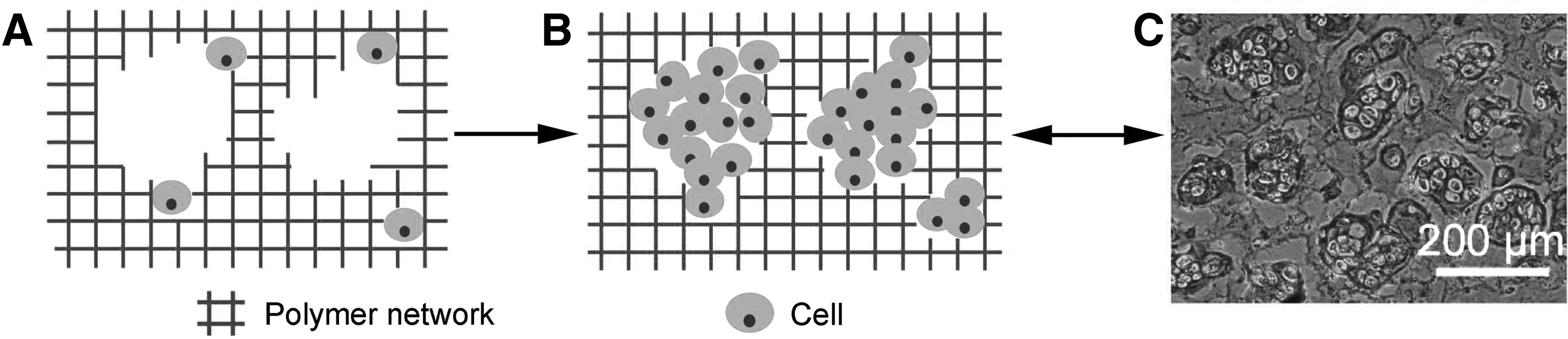
To evaluate how the macroporous structure directs cell fate and neotissue development, Fan and Wang have fabricated three model gel constructs by using photocrosslinkable HA and gelatin microsphere (diameters 150–200 μm) as the precursor and porogen, respectively. 99 The increased permeability of conventional hydrogels achieved by decreasing crosslink density supports better cell viability and rapid proliferation, which results in enhanced secretion of cartilaginous ECM. However, the inhibition of cell amplification and ECM expression by gel polymer networks has been obviously observed with the prolonged in vitro culture. The introduction of macroporous structure elevates the permeability of gel scaffold, promoting the diffusion of nutrients and cellular wastes across constructs; and more importantly, macropores provide living spaces for the proliferation of chondrocytes, inducing the formation of larger cell clusters and scaffold-free neocartilage islets. This study directly demonstrates that the creation of macropores in gel constructs can significantly enhance cell activities and corresponding tissue formation through improving gel permeability and providing cell living space.
The effect of pore size on the proliferation and function of chondrocytes has been investigated by Zeng et al. through adjusting the size range of gelatin microsphere porogens during the fabrication of macroporous alginate gel constructs. 115 Three size ranges of gelatin microspheres from small size (diameters 80–120 μm) to middle size (diameters 150–200 μm) and large size (diameters 250–300 μm) are fabricated by tuning the stirring rate of emulsion solution. Among them, the chondrocytes cultivated in the macroporous gel constructs with 80–120 μm of pore size exhibit better cell proliferation and ECM secretion. This study demonstrates that the enhanced cell proliferation in macroporous alginate gels is resulted from the activation of the Erk1/2-MAPK pathway in the chondrocytes. Han et al. explore chondrocyte behaviors within the macroporous gelatin gels with dynamic pore development, which is achieved by using three microspherical porogens prepared from gelatin (diameters150–250 μm), alginate (diameters 200–300 μm), and HA (diameters 100–200 μm), respectively. 62 The results of this study show that the process of multiple pore formation significantly enhances the viability, proliferation, and ECM production of encapsulated chondrocytes.
Besides chondrocytes, the superiority of macroporous hydrogel scaffolds in promoting non-ADC proliferation and differentiation has also been proved by using ESCs and iPSCs.57,116 Lau et al. have evaluated the potential of macroporous alginate gels to conduct cell colony/embryoid body (EB) formation and subsequent hepatic lineage differentiation of ESCs and iPSCs. 116 The EBs (100–200 μm in diameter) are formed spontaneously in macroporous gel constructs, however, as expected, which are not observed in conventional gel constructs. The upregulation of endoderm markers and hepatic markers as well as higher secretion of urea and albumin in macroporous gel constructs than that in monolayer culture, respectively, has been observed in both iPSCs and ESCs. These results demonstrate the beneficial effects of the macroporous gel scaffolds for directing the proliferation and differentiation of iPSCs and ESCs toward endodermal lineage and following hepatic lineage. This study suggests that the macroporous gels may serve as a continuous scaffolding system to permit iPSC and ESC differentiation and following tissue regeneration, which has been also corroborated through the work performed by He et al. 57 Aiming to prepare tissue-engineered scaffold-free cartilage constructs, they carry out the consecutive chondrogenesis of iPSCs and ESCs in macroporous alginate gel scaffold via the mesoderm differentiation, chondrogenic differentiation, and cultivation, and finally, the alginate gel scaffold is removed to obtain the cartilage graft consisting of induced chondrocytic cells and cartilaginous ECM. Both studies provide a foundation in understanding the differentiation of ESCs and iPSCs in a macroporous gel environment and even aid to realize the applications of iPSC technology in the engineering of autologous tissues. 57 Taken together, the creation of macroporous structure in gel scaffolds not only results in improved diffusion efficiency of nutrients and oxygen as well as removal of cellular waste but more importantly provides free living space for the proliferation and functions of non-ADCs. The formation of non-ADC colonies/clusters, resulted from cellular migration or rapid proliferation, obviously enhances directed cell differentiation and ECM deposition as well as subsequent development of engineered tissues.
Anchorage-dependent cells
The enhancement of macropores on the growth and function of ADCs in hydrogels has also been evaluated in various macroporous gel constructs. Han et al. prepared photocrosslinkable gelatin microribbon precursors via wet-spinning for the fabrication of cell-loaded macroporous gel constructs. 95 The encapsulated adipose-derived stromal cells adhere to the microribbons and spread after 3 h of cell seeding, in addition, continuously proliferate within the 20-day culture period. The biodegradable HA gel scaffolds are developed by Jha et al. using three MMP cleavable peptide crosslinkers (i.e., QPQGLAK, GPLGMHGK, and GPLGLSLGK), respectively. 117 Among them, the gel scaffolds, prepared from the QPQGLAK, support the highest adhesion, spreading, and proliferation of cardiac progenitor cells and the secretion of MMP-13, VEGF165, and angiogenesis-related proteins. The results of the in vivo studies show the gels crosslinked with QPQGLAK outstandingly promote robust angiogenesis and integration of neovascular structure with the host blood circulation system. Hu et al. report one fiber spinning method to prepare hollow gelatin gel fibers by using enzymatic crosslinking of gelatin precursor solution that is flowing through a capillary tube. 97 Human microvascular endothelial cells and NIH/3T3 fibroblasts are mildly encapsulated into the gel fibers with high viability, and those cells can proliferate to form a monolayer on both the inner and outer surfaces of the hollow gel fibers.
Significantly, the EF-like phenomenon is also observed for the ADCs cultured in the macroporous gels fabricated from cell-adhesive polymers such as gelatin. Leong et al. fabricate macroporous gelatin gel constructs by using gelatin methacrylate and temperature dissolvable gelatin microspheres as the precursor and porogens, respectively. 118 Interestingly, the adhesion, spreading, and proliferation of the encapsulated endothelial progenitor outgrowth cells (EPOCs) are achieved within the bulk of macroporous gelatin gel constructs; the EPOCs are found to grow around the boundaries of cavities. However, a similar observation did not appear in the conventional gelatin gel constructs. The higher proliferation and cell–cell contacts of EPOCs in the macroporous constructs lead to the formation of vascular tube-like structures at the gel body–cavity interfaces. The interconnected vascular networks are formed by elevating the density of microcavities. 119 Summarily, the creation of macropores in gel scaffolds prepared from cell-adhesive precursors can introduce numerous cell living interfaces within the gel constructs, and which favors the adhesion, spreading, proliferation, and endothelial differentiation of EPOCs. In addition, the use of cell-adhesive microspheres as porogens and cell delivery vehicles is another strategy to fabricate macroporous gel construct with enhanced cell adhesion and proliferation. Zhong et al. seed human osteoblast-like cells (MG63) on the surfaces of collagen microspheres, and these cell-loaded collagen microspheres, together with collagenase, are encapsulated to alginate gels. 120 The collagen microcarriers provide cell-adhesive interfaces, ensuring initial cell spreading within the gel constructs. Meanwhile, the collagenase promises the biodegradation of microspheres over culture time and further enhances MG63 proliferation, differentiation, and the expression of osteogenic markers by offering increasing cell living interfaces and spaces. A 3D cell culture platform, prepared from the combination of biodegradable gel scaffolds and porogenic microspheres, has been reported by Sokic et al. 121 The gelatin microparticles with different sizes (diameters 50–100, 100–150, or 150–200 μm) and human umbilical artery smooth muscle cells (HUASMCs) are encapsulated into MMP-sensitive PEG gels under cytocompatible conditions. The macropores rapidly generate along with the dissolution of gelatin microparticles on incubating at a 37°C incubator, offering interfaces and spaces for HUASMC adhesion at the initial stage of tissue culture. Subsequently, the gel bulk is degraded gradually, leading to increasing gel porosity. Therefore, this kind of gel scaffold can continuously provide interfaces and living spaces to stimulate cell spreading, migration, proliferation, and then vascularization. 121 Collectively, the introduction of macroporous structures in hydrogels produces cell-adhesive surfaces and spaces, which facilitate ADC adhesion, spreading, and proliferation as well as increased cell–cell contacts. At the same time, the cycloidal porous surfaces may provide physical, mechanical, and topographical cues for enhancing desirable cellular activities and subsequent neotissue development.
Conclusion and Future Directions
Hydrogels have been widely considered as promising scaffolds for 3D cell culture and tissue engineering because of their tissue-like properties, injectability, and the ability to achieve homogeneous cell encapsulation in situ. 122 However, their submicron- or nanosized polymer meshes (smaller than cell size) have drastically inhibited cell settlement, attachment, and proliferation as well as neotissue formation regardless of cell types, besides even limiting the diffusion of nutrients and cellular wastes across the gel constructs. To overcome such shortcomings, tissue engineers have created macropores in gel constructs by using various approaches, such as incorporation of biodegradable segments into gel networks, electrospinning, 3D bioprinting, and utilization of biocompatible porogens. Among them, the introduction of macropores via the porogen leaching method allows facile control over porous size and porosity as well as pore formation kinetics within gel bulk, which is beneficial to investigate the induction effect of macroporous structure on cellular behaviors and neotissue formation. Except for providing cell living space, the creation of larger cavities within gel bulk also leads to a strong increase in permeability of gel constructs, resulting in the rapid diffusion of nutrients and cellular wastes as well as accelerated gel degradation. Besides, the creation of macropores in gel constructs produces numerous cell living interfaces that are significant for ADC attachment, spreading, and proliferation.
Although the last several years have witnessed great progress of macroporous gel scaffolds for 3D cell culture and tissue engineering, several challenges still remain to be conquered for their future development. Successful applications of fabrication platforms, namely 3D bioprinting and electrospinning, in executing macroporous gel preparation with cell encapsulation mainly lie in the development or selection of appropriate biomaterials. Moreover, cell viability of gel constructs fabricated via 3D bioprinting or electrospinning remains to be improved. For biodegradable gel scaffold, it is expected to modulate its degradation rate and mechanical properties to match cell proliferation and neotissue formation by devising or optimizing biodegradable precursors or crosslinkers. The EF (like) phenomenon has been observed for both non-ADCs and ADCs in porogen-based macroporous gel constructs fabricated from noncell-adhesive and cell-adhesive precursors, respectively, however, the specific environmental factors contributing to enhanced cell proliferation and metabolic activity at the “edges” within gel constructs are not completely clear. In addition, the molecular mechanisms behind the EF phenomenon need to be further studied. Besides, the studies on the differentiation of MSCs in macroporous gels have not been reported to date. A better understanding for the roles of gel–pore interface microenvironments in directing cellular fates will guide us to prepare a better gel scaffolding system to support cell proliferation and neotissue development. Next-generation porogens should be controllably leached out based on the requirements to regulate scaffold properties and improve cell functions. The leaching out of commonly used porogens (i.e., gelatin microsphere and alginate beads) is out of control on exposing to stimuli (namely elevated temperature for gelatin microsphere and chelating agents for alginate beads). And therefore, it is difficult to assess the regulation effects of removal process of the porogens on gel microenvironments and cellular behaviors. Future developments of macroporous gel scaffolds need to concentrate on the challenges and problems as highlighted in this review, and we believe that new or improved strategies to treat these issues will advance the applications of macroporous gel scaffolds in 3D cell culture and tissue engineering.
Footnotes
Acknowledgments
This work is financially supported by AcRF Tier 1RG 30/15, Ministry of Education, Singapore, and the National Natural Science Foundation of China (Grant No. 21604045).
Disclosure Statement
No competing financial interests exist.