




Abstract
There is a paucity of in vivo studies that investigate the safety and efficacy of temporomandibular joint (TMJ) tissue regeneration approaches, in part due to the lack of established animal models. Review of disease models for study of TMJ is presented herein with an attempt to identify relevant preclinical animal models for TMJ tissue engineering, with emphasis on the disc and condyle. Although degenerative joint disease models have been mainly performed on mice, rats, and rabbits, preclinical regeneration approaches must employ larger animal species. There remains controversy regarding the preferred choice of larger animal models between the farm pig, minipig, goat, sheep, and dog. The advantages of the pig and minipig include their well characterized anatomy, physiology, and tissue properties. The advantages of the sheep and goat are their easier surgical access, low cost per animal, and its high tissue availability. The advantage of the dog is that the joint space is confined, so migration of interpositional devices should be less likely. However, each species has limitations as well. For example, the farm pig has continuous growth until about 18 months of age, and difficult surgical access due to the zygomatic arch covering the lateral aspect of joint. The minipig is not widely available and somewhat costly. The sheep and the goat are herbivores, and their TMJs mainly function in translation. The dog is a carnivore, and the TMJ is a hinge joint that can only rotate. Although no species provides the gold standard for all preclinical TMJ tissue engineering approaches, the goat and sheep have emerged as the leading options, with the minipig as the choice when cost is less of a limitation; and with the dog and farm pig serving as acceptable alternatives. Finally, naturally occurring TMJ disorders in domestic species may be harnessed on a preclinical trial basis as a clinically relevant platform for translation.
Introduction
T
TMJ disorders (TMD) are estimated to affect between 10 and 36 million individuals in the United States per year, 1 and include clinical conditions and symptoms ranging from clicking of the joint to chronic intractable pain, limited jaw motion, and chronic degenerative disease. These symptoms can have a significant impact upon quality of life.
A wide variety of therapeutic modalities for TMD are available depending upon the symptoms and severity of the disease, and details can be found elsewhere. 2 Generally, noninvasive treatments are attempted first and include behavioral modifications (rest, hot/cold compresses, biofeedback, and physical therapy), pharmaceutical management (NSAIDs, muscle relaxants), and splint therapy. Specific clinical scenarios (i.e., condyle fracture/dislocation or ankylosis) or failure of conservative treatment modalities are indications for surgical management, which range from minimally invasive procedures (arthrocentesis and arthroscopy) to open arthrotomies. In cases of internal derangement, open surgery often includes repair, repositioning, or removal of the fibrocartilaginous TMJ disc.
Tissue engineering and regenerative medicine (TE/RM) could offer potential solutions for replacing damaged TMJ tissues and restoring function to the joint. A thorough review of patient populations in need of TE/RM therapies can be found elsewhere. 3 The absence of a single, well-established animal model for TMJ disease has been a limiting factor for investigating potential surgical and nonsurgical solutions. Furthermore, as TMD span a spectrum of etiologies, a single model of TMJ disease is unlikely to be adequate for all such studies.
The present review highlights the physiologic, anatomic, and practical considerations that impact the utility of preclinical models for TMJ research. Recommendations for guiding the selection of preclinical animal models for TMJ research are presented with a focus on TE/RM approaches.
Preclinical Models of TMDs
To understand the efficacy of tissue engineering approaches, therapies cannot be tested in only disease-free joints. A disease state must be first established, and then treated. Generally, preclinical models of TMDs have encompassed chemical, mechanical, and surgical methods. Chemical methods tend to focus on pain, mechanical methods emphasize structure and function, and surgical methods are more focused on degenerative changes.
Chemical methods
The most common approach to investigate TMJ inflammation and pain involved injecting chemical irritants into the joint space. The rat has been the animal model of choice for evaluating these conditions and the most commonly used irritants were complete Freund's adjuvant (CFA), mustard oil, and formalin. 4
In the rat model, mechanical allodynia (i.e., pain induced by normally non-noxious stimulus) is often studied by the use of CFA, which contains heat-killed Mycobacterium tuberculosis suspended in an oil and saline emulsion.5–7 Allodynia should not be confused with hyperalgesia, which is an exaggerated pain response to a noxious stimulus. The bacteria in CFA are believed to solicit a macrophage immune response that produces chronic active inflammation. In a study with adult male rats (250–350 g), CFA was injected unilaterally in TMJs. 5 This study showed that CFA induced allodynia for up to 18 days.
Rats have been used to study TMJ inflammation and pain induced by mustard oil (allyl isothiocyanate) as an irritant.8–12 For example, one highly cited study explored the effect of mustard oil injection in the TMJ on the jaw and neck muscle activity in rats. 10 Electromyography was used to assess muscle activity. Sprague-Dawley rats of either sex (250–450 g) were used in this study, and it was found that mustard oil injected in the TMJ had the effect of increased muscle activity, suggesting increased nociceptive response.
In the rat model, formalin or formaldehyde have been used to induce TMJ pain.13–17 These chemicals cross-link proteins, which in turn cause inflammation. For example, in one pivotal study, formalin was injected into 54 male Wistar rats (150–250 g). It was found that both head flinches and orafacial rubbing significantly increased for concentrations of formalin above 1.5% when compared to saline control. 13
The rat is by far the most commonly used animal in the preclinical study of pain and inflammation induced by chemical methods. 18 Two reasons for the use of rats are size and cost, which allow for long periods of behavioral training. Furthermore, investigators have developed methods to detect changes in pain/sensitivity in rats; a major hurdle before moving to larger animal models. Nevertheless, the degeneration process caused by chemical insult does not closely resemble the human condition. As such, mechanical perturbation methods have been investigated to better understand TMD progression.
Mechanical methods
A change in loading (mechanical perturbation) can cause dramatic changes to normal TMJ structure and function.19–22 Altered loading can be induced by bite raise, splinting, induced malocclusion, and perturbed mandibular movement. Regardless of the method used, degeneration has been observed, by histologic methods to be rapidly detectable (i.e., less than 6 weeks) in animal models.19–22 It is important to note, however, that these studies replicate a sudden change in occlusion/loading, which is not representative of the controlled change of occlusion associated with orthodontic treatment.
Various animal models have been used to study malocclusion and TMJ degeneration, but the rat remains the most commonly used model.19–22 One study used a unilateral bite raise to study the effects of altered joint loading on the rat TMJ. 23 A unilateral 1 mm thick bite-raising device was applied to the surface of the right maxillary molars. The authors showed that the unilateral bite raise resulted in increased aggrecan expression in the condylar cartilage and increases in expression of a proteoglycan related to versican in the disc and articular surface of the condyle. While such studies can clearly demonstrate the effects of altered loading on extracellular matrix (ECM) expression, that suggests remodeling of the disc, the clinical relevance of these changes remains unclear.
One of the main reasons the rat has been used so often is the rapid remodeling and degeneration of the TMJ after the loading is perturbed. Major changes can be identified in the joint by as little as 4–6 weeks following insult. A notable limitation of this model, however, is that most studies only evaluate histologic changes. Larger animals are typically required to relate such histologic findings to mechanical properties of the joint tissues and the associated degeneration and pain. The rabbit is a larger animal, and provides large enough tissues for reliable mechanical testing such as compression, tensile, shear, and friction. 24 It is important to note that the altered loading models are designed to understand the impact of a large change in the distribution of loads. These models do not replicate clenching or bruxism, but give insight into the mechanisms by which these behaviors could give rise to degeneration of the joint.
Surgical methods
Surgical methods for modeling TMDs include disc displacement, disc perforation, and condylar fracture. Such studies are often performed on larger species that allow for a more detailed and comprehensive evaluation of the disc, unlike with mice and rats.
The rabbit is often used as a model for anterior disc displacement.25–28 The most cited study of a disc displacement model used adult male New Zealand white rabbits to surgically induce anterior disc displacement. 25 The procedure required an incision above the zygomatic process and fracture of the zygomatic process of the squamous temporal bone. The surgical approach alone could have an impact on the results, and is a limitation of this model. Disc degeneration and condylar resorption were both evident by 6 weeks postsurgery. 25 Mandibular head cartilage showed osteoarthritic changes by this same timepoint. 25
The rabbit is commonly used as a model for disc perforation.29–31 However, there is controversy in the field on whether TE/RM approaches are appropriate for disc perforation. 3 In one of these studies, the investigators exposed the joint 29 in adult male New Zealand white rabbits (2.5–3.5 kg) through a transverse incision along the zygomatic arch followed by a horizontal incision through the lateral capsule exposing the superior aspect of the disc. A 4–6 mm full thickness perforation was made at the posterolateral aspect of the disc. The authors studied proteoglycan synthesis and degradation, and found both to be uprelgulated when compared to normal discs. 29 Again, due to the short duration and limited testing in this study, the clinical relevance of these findings is open for discussion.
The sheep is another large animal that has been used in many in vivo preclinical studies in TMJ surgical domain. Sheep have been used as a model for TMJ ankyloses 32 ; TMJ osteoarhrosis33–35 ; to analyze the effect of condylectomy 36 ; to analyze the surgical options for reconstruction after condylectomy 37 ; and to evaluate minimally invasive techniques such as arthroscopy. 38
One of the main reasons that larger animals, rather than mice and rats, are commonly used for the study of TMJ surgical methods is that the larger animal size allows for good surgical access and procurement of tissue large enough for mechanical testing. As seen in Table 1, the sheep, goat, and dog provide direct access to the TMJ as it is in humans. In terms of size of tissue, the pig, sheep, and goat have been tested in both compression and tension with standard mechanical testing equipment.39,40 Mice and rats present technical challenges that frequently raise questions regarding the translational relevance to the human TMJ.
TMJ, temporomandibular joint.
Anatomy, Physiology, and Mechanical Properties
Historically, the pig has been regarded as the gold standard for a nonprimate, large animal TMJ model based on general similarities to the human anatomy41–46 ; specifically, the size of the articular TMJ structures and the shape of the disc (Figs. 1 and 2). Berg 47 examined pigs, sheep, calves, and dogs, and concluded that the pig was the most suitable experimental model of the human TMJ, specifically noting that the pig would best be suited for preclinical studies for disc replacement. The TMJ disc of a pig at market weight (∼260 lbs) is similar in dimensions to the human disc. 39 Bermejo et al. 48 concluded that the pig was the only suitable animal model after a comparison with dogs, cats, rabbits, rats, cows, sheep, and goats. It was proposed that because the pig is the only omnivore of these candidate mammalian models, the condyle and disc have a similar shape to the human TMJ disc. 48 Strom et al. 49 concluded that the domestic pig may be a useful model in the investigation of the function of the masticatory system. The mechanical properties of the porcine TMJ have also been shown to be similar to human.41–46 For example, it has been shown that the mechanical properties of the pig TMJ disc 39 are on par with human (Table 1). Furthermore, a recent comprehensive study has validated the use of Yucatan minipig as an animal model for TMJ TE/RM studies and showed several similarities between minipig and human TMJ discs minipig. 50 However, a more recent article has suggested differences in multiple components of the pig and human TMJ anatomy. 51 The function of TMJ in the farm pig and minipig are also rotation and translation, just like in humans.

Gross anatomy of the TMJ disc in five different species (adapted from Kalpakci et al. 39 , Copyright © 2011 [Reprinted with permission]). TMJ, temporomandibular joint.
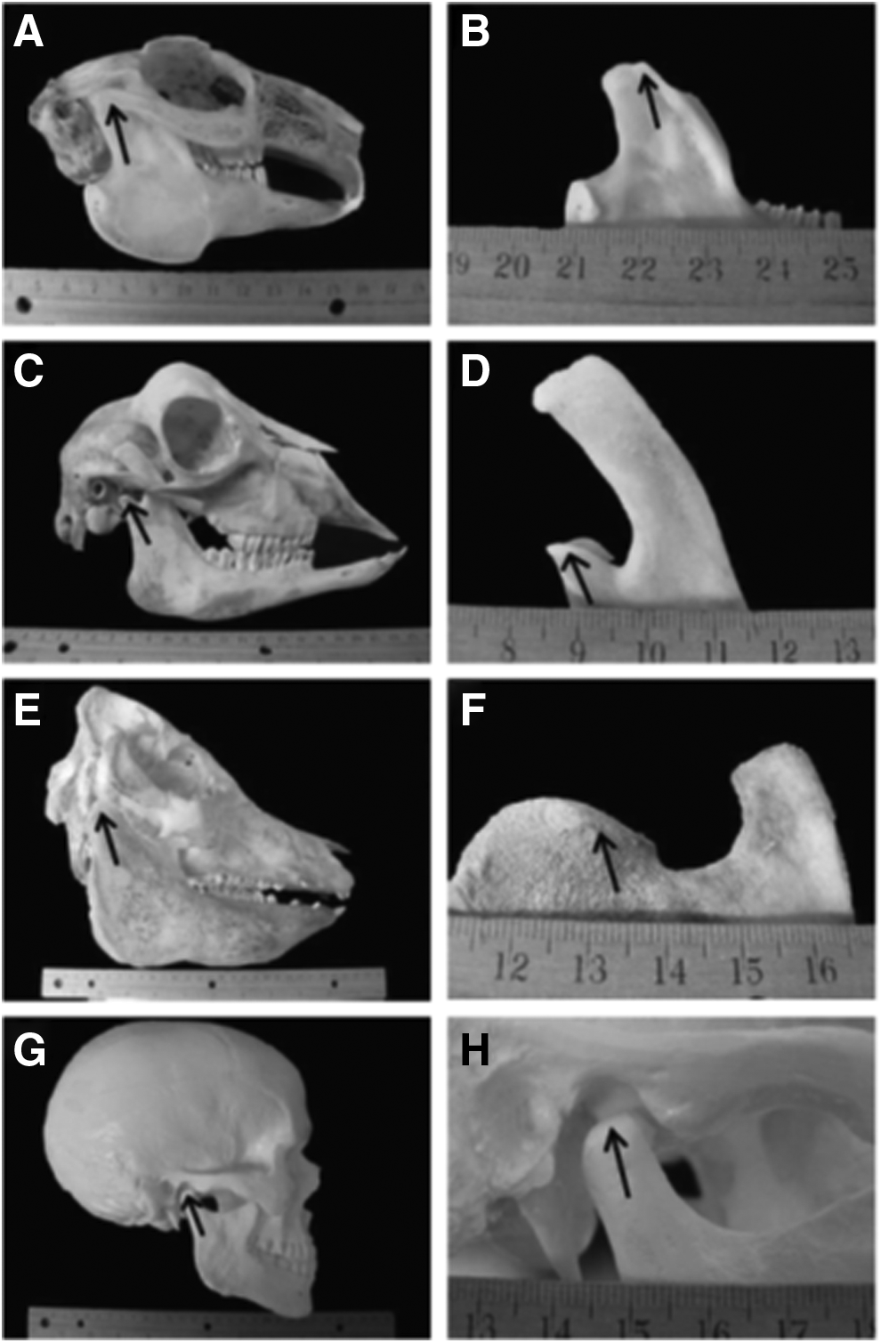
Gross anatomy comparison of the joint space of three species compared to the human.
The TMJ anatomy, histology, and biomechanics of Black Merino sheep have been studied (Table 1). 52 With a preauricular incision and blunt dissection, the TMJ capsule can be easily accessed. The zygomatic arch does not shield the joint space, as is the case in the farm pig. The sheep's condyle is mediolaterally concave with ellipsoidal shape with the longer axis in the mediolateral position. 53 These anatomical similarities to human, associated with the large experience in in vivo TMJ surgical investigation, suggest the sheep as a suitable model to conduct TMJ surgical investigation. 52 It has recently been suggested that the sheep can be used to conduct rigorous preclinical trials in the surgical TMJ domain. 54
No one animal model resembles the human TMJ in all anatomical areas and function. Nevertheless, the farm pig and minipig are a close match in anatomy and physiology. The goat, sheep, and dog are also close to human in terms of anatomy, but their TMJ function is somewhat different, as the goat and sheep mainly function in translation, while the dog mainly functions in rotation.
TE/RM Approaches for TMJ Disc Replacement
TE/RM approaches must be tailored to the TMJ environment, and interpositional devices need to remodel into appropriate tissue analogs.
Two studies using the canine model have established the dog as a potentially useful model in the study of TMJ disc regeneration. In the first study, a prototype device consisting entirely of ECM derived from porcine urinary bladder was evaluated as a bioscaffold for TMJ disc reconstruction.40,55 Implantation of the device was straightforward, as there was direct access into the joint space (Table 1 and Fig. 2). Macroscopically, the implant remodeled very rapidly into newly deposited tissue. Microscopically, remodeling was characterized by robust angiogenesis during the first 2 months and a dense infiltration of predominantly CD68+ mononuclear cells (i.e., macrophages) and smooth muscle actin (SMA)+ large, round cells. During the subsequent months, the bioscaffold remodeled into a structure containing a population of SMA- spindle-shaped cells with a distribution resembling that of the native TMJ disc.
In a second study, 10 dogs were followed for 6 months after implantation of an acellular ECM bioscaffold. 56 The results showed excellent remodeling of the scaffold with articular surface protection while the contralateral, untreated control side showed moderate articular surface remodeling in just a short period of time and no replacement of the TMJ disc tissue.
Sheep have recently been used in a TMJ interpositional material study to evaluate the effect of three different devices in a randomized, blinded preclinical trial 54 ; however, the study is in progress, so the long-term utility of this model is not yet known.
TE/RM Approaches to Condyle Replacement
Like the disc, there have been few attempts to engineer the mandibular condyle in vivo.18,57,58 In two pioneering studies by different groups, Computer design custom-made scaffolds were used for substitution of the condyle in minipigs and sheep. In the minipig study, 57 selective laser sintering method (specific type of solid free-form fabrication) was used to fabricate a polycaprolactone condyle/ramus scaffold. 57 The mandibular head was packed with autologous iliac crest bone marrow, secured to the mandible using miniplates and screws, and evaluated after 1 and 3 months. Compared to controls, there was an increase in regenerated bone volume, and there was evidence of cartilage-like tissue. The authors chose the minipig instead of the domestic farm pig for several reasons, including the fact that the domestic farm pig does not reach skeletal maturity until around 18 months of age. 59 Yet, study animals are usually 3–9 months of age, so they can be housed in normal university facilities. However, the TMJ is still growing in the farm pig at this young age, which may impact results. In contrast, the minipig can be easily handled at 18 months of age, and the size of the TMJ is comparable to humans. In a sheep study, Computer design-customized porous scaffolds of hydroxyapatite were used to replace the condyle. 58 After 4 months, newly formed bone was observed in the scaffold pores. These two studies show the feasibility of both the minipig and sheep as animal models for condyle TE/RM approaches. Future studies should also focus on regeneration of the complex fibrous-cartilage-bone interface of the articular surface of the condyle.
Naturally Occurring Animal Models for TMDs
While previous sections of the article describe the pros and cons of experimental animal models for TMJ research, this section will elaborate on the potential benefits of incorporating naturally occurring TMDs in companion animals as an innovative and clinically relevant model to study TMDs and therapeutics.
A vast number of laboratory animal species have been used as models for TMJ research. These laboratory animals have been proven crucial to provide platforms to study basic disease mechanism and potentially therapeutics. Naturally occurring diseases, such as seen in the TMJ of companion animals, might better reflect the genetic diversity and influence, complex environmental and physiological burden and variation that are present in humans.60–62 Furthermore, the healthcare systems and standards of care in human and companion animals share some similarities and approaches. Therefore, companion animals may serve as a clinically relevant TMJ disease model and provide an opportunity to translate knowledge of safety and efficacy of therapeutics from animals to human in which both the veterinary patient and the human patient benefit from this synergy.60,63
TMDs in dogs and cats
The TMJs of dogs and cats primarily function in a hinge movement.64–66 In dogs, a slight laterotrusion movement is possible. However, in cats the TMJ morphology is more restrictive and a pure hinge motion is more predominant.65,68 The TMJ disc in dogs and cats is thin, which is likely due to the structure-function relationship of the mainly rotary movement of the joint in these species. 66
Examination of the naturally occurring TMDs reveals that TMJ-osteoarthritis (OA) is the most common TMD in dogs and the second most common TMD in cats after fractures. 68 Furthermore, the clinical manifestation of TMJ-OA in dogs and cats is similar to that in humans. It is noteworthy that clinical symptoms may not correlate with the presence and severity of CT findings.68–70 Other naturally occurring disorders of the TMJ in dogs and cats include fractures, ankylosis, luxations, and a variety of neoplasms, which mimic similar disorders in humans.65,71 Similar to the situation with TMJ-OA, these disorders can be harnessed on a clinical trial basis as a clinically relevant platform for translation.
TMDs in horses
Another potential large animal model with naturally occurring TMJ disease is the horse. Horses are similar to other herbivores in that their chewing cycle consists of an opening stroke, closing stroke, and a power stroke. 72 The power stroke in horses is unimodal and there is mediolateral movement of the mandibles. 72 Moreover, previous kinematic work demonstrated that the TMJs of a horse have a lateroventral movement of the working side during the opening stroke and a marked mediodorsal movement of the working side during the power stroke. 73 The TMJ of horses exhibits anatomical variations in the shape and bone density in up to 40% of horses. 74 In addition, similar to humans, horses experience intra-articular disc mineralization (i.e., dystrophic mineralization) as an age-related degeneration. 74 As with dogs and humans, horses can exhibit TMJ fractures, OA and septic arthritis, which may provide a potential platform for translating innovations through naturally occurring animal model.
In summary, naturally occurring TMDs in companion animals provide a future avenue for targets of regenerative therapies. The efficacy of TE/RM approaches must first be validated in repeatable degeneration models in animals of low genetic variance. Many of the species discussed in this review, provide this low variance that is a key controlled parameter when comparing results. Nevertheless, companion animals are the next frontier of these therapies to treat long-term degenerative changes in a high genetic variance population. Furthermore, efficacy in companion animals would provide pivotal data to the Food and Drug Administration (FDA) for clinical translation of potential TMJ technologies.
Conclusions
Preclinical disease models of the TMJ have been performed predominately in small animal species such as rodents and rabbits. These studies have shown that mechanical perturbations to the joint results in degeneration of the joint that more closely resembles TMDs than chemical insult. However, larger animal models are needed to evaluate the safety and efficacy of TE/RM therapies. The pig has often been thought of as the gold standard for TE/RM therapies. However, these assertions are largely based upon postmortem evaluation. It is important to understand the model-specific advantages and limitations that may exist before embarking into tissue engineering preclinical studies. The advantage of the pig and minipig is that anatomy, physiology, and the properties of the tissues have been well characterized. The advantage of sheep and goat is easy surgical access, similar anatomy, the low cost per animal, and the high availability of tissue for practice. The advantage of the dog is that the joint space is very confined, so if attachment of interpositional device is a concern, the device is likely to stay in place. The farm pig has the limitation of continuous growth, therefore the zygomatic arch blocks access into the joint and handling is an issue at skeletal maturity. The minipig is not widely available, thus obtaining cadaveric tissue and enough animals for a large study may be a challenge. Only two farms hold colonies of this breed, and thus access and price can be limitations for investigators. The sheep and the goat are herbivores, and their TMJs mainly function in translation. The dog is a carnivore, and the TMJ is a hinge joint that can only rotate. The type of joint function is not likely to impact the healing potential of the joint. However, it could impact the stability of implant and many future studies want to target animals with joint that both rotate and translate.
Although no species provides the gold standard for all preclinical TMJ TE/RM studies, the goat and sheep have emerged as the leading options, with the minipig as the choice when adequate resources are available, and the dog and farm pig serving as acceptable alternatives. Finally, species in which naturally occurring TMDs occur may be used on a preclinical trial basis as a clinically relevant platform for translation.
Footnotes
Acknowledgments
We gratefully acknowledge support from the National Institutes of Health (National Institute of Dental and Craniofacial Research) with grant R01 DE022055.
Disclosure Statement
No competing financial interests exist.