
Abstract
Herein we review the state-of-the-art in tissue engineering for repair of articular cartilage. First, we describe the molecular, cellular, and histologic structure and function of endogenous cartilage, focusing on chondrocytes, collagens, extracellular matrix, and proteoglycans. We then explore in vitro cell culture on scaffolds, discussing the difficulties involved in maintaining or obtaining a chondrocytic phenotype. Next, we discuss the diverse compounds and designs used for these scaffolds, including natural and synthetic biomaterials and porous, fibrous, and multilayer architectures. We then report on the mechanical properties of different cell-loaded scaffolds, and the success of these scaffolds following in vivo implantation in small animals, in terms of generating tissue that structurally and functionally resembles native tissue. Last, we highlight future trends in this field. We conclude that despite major technical advances made over the past 15 years, and continually improving results in cartilage repair experiments in animals, the development of clinically useful implants for regeneration of articular cartilage remains a challenge.
Graphical abstract
Color images are available online.
Impact Statement
The cartilaginous tissue is very difficult to repair or regenerate due to its lack of vascularization and its small amount of cells. It is a very important biological tissue as it is vital for the lubrication of joints, which promotes a better and more organic movement of the body. This review aims to check the state-of-art of the latest and most innovative trends in the biomaterial field for this tissue, which are the three-dimensional multilayer structures that simulate the behavior under cyclic stresses of the articular cartilage.
Introduction
The main function of cartilage is to keep joints lubricated to ensure a smooth surface, which facilitates transmission of mechanical loads by providing the lowest friction coefficient possible. Unlike most tissue types, cartilage lacks blood vessels and lymphatic vessels and has a low cell population, which chiefly comprises chondrocytes. Consequently, endogenous cartilage recovery is slow and is limited by weak local transport of nutrients. Minor cartilage defects are currently repaired through medical interventions such as multiple drilling, abrasion arthroplasty, mosaicplasty (autologous osteochondral grafts), and cellular transplantation (namely, autogenous and allogenic chondrocytes). 1 However, these techniques have their shortcomings. For instance, allografts can lead to transmission of diseases or provoke immunologic responses in the recipient and, once in place, remodel very slowly; also, autografts demand that patients undergo several surgeries. 2
An attractive alternative for cartilage repair is tissue engineering, whose testing and optimization over the past two decades have yielded many promising cartilage grafts and sophisticated bioreactor systems for ex vivo graft culture.3–8 Modern tissue engineering comprises three basic elements: cells (chondrocytes, stem or progenitor cells), biodegradable scaffolds, and growth factors. This approach is particularly amenable to restoration of articular cartilage, 9 as the scaffolds provide a three-dimensional (3D) network for chondrocyte growth 10 and act as mediators for cell/cell signaling and interactions. However, the physical and biochemical properties of these scaffolds are crucial. 11
The aim of this article is to review the state-of-the-art of 3D scaffolds for the repair/regeneration of cartilage tissue explaining the composition of tissue and the components currently used to prepare multilayer scaffolds by electrospinning, freeze-drying, and so on.
Composition and Structure of Cartilage
Articular cartilage comprises a nonmineralized layer and a calcified layer; thus, it demonstrates inhomogeneous behavior. The nonmineralized layer has three structurally contiguous zones, each differing in matrix composition, organization, and chondrocyte phenotype.12–14 Briefly, the surface zone, located at the articulating region of the cartilage, accounts for ∼10% of the total tissue height and consists of thin, elliptical chondrocytes and progenitor cells surrounded by a matrix with high water content (∼78%), relatively low proteoglycan (PG) content, and collagen fibrils (diameter: 4 to 12 nm) oriented along the surface direction. 15 The cells in this zone produce surface protein area that contributes to joint lubrication.16,17 Below the surface, the next 60% of cartilage depth corresponds to the middle zone, in which spherical chondrocytes reside within a matrix rich in PGs and unaligned collagen fibers (diameter: 9 to 60 nm). 18 Next, the deep zone of the nonmineralized layer corresponds to the final 30% of cartilage depth and is marked by spherical chondrocytes oriented in stacks perpendicular to the joint surface. These cells, while sparsely distributed, are within a matrix of relatively high glycosaminoglycan (GAG) content, high water content (∼68%), and radially oriented collagen fibrils (diameter: 60 to 140 nm).19–23 Although the collagen fibril diameter generally increases from the surface zone to the deep zone, fine fibrils (<100 Å) have been observed to reside at all depths.
The calcified layer is situated between the deep zone of the nonmineralized layer and subchondral bone.24,25 It comprises hypertrophic chondrocytes located in a mineralized matrix that is rich in type I collagen and PGs. This osteochondral interface ranges in thickness from 20 to 243 μm, a range whose breadth has been attributed to variability by age and by total cartilage thickness (Fig. 1).14,26,27 The levels of type II collagen and water decrease from the surface zone of the nonmineralized layer to the calcified zone, whereas the concentration of PGs increases. The structural heterogeneity of cartilage leads to local variations in mechanical compressive module. This structural complexity underpins the challenges that researchers face in designing and assembling artificial scaffolds for cartilage repair.25,28–32

Cartilage diacritical zones. Color images are available online.
Main components of cartilage tissue
Articular cartilage, which is about 2 to 3 mm thick, has a complex structure that enables dissipation of mechanical loads (1 to 4 MPa) 33 exerted on, and protection of, subchondral bone. 34 The extracellular matrix (ECM) chiefly comprises water (70% to 80%) and includes type II collagen and, to a lesser extent, glycoproteins (PGs; Table 1). The type II collagen and glycoproteins, which attract water due to their negative charges, promote resistance to shear and tensile forces. The principal type of cell in this matrix is chondrocytes. However, given their low concentration (only 14,000 to 30,000 cells/μL), they are limited in their growth, proliferation, and, consequently, their capacity to repair collagen. 35
Articular Cartilage Components, Percent, Characteristics, and Functions
ECM, extracellular matrix; PGs, proteoglycans.
Chondrocytes
Although only ca. 1% of articular cartilage volume corresponds to chondrocytes, these cells are fundamental for tissue maintenance, as they substitute the molecules of the degraded matrix. Chondrocytes derive from mesenchymal stem cells (MSCs), which are present in adult bone marrow. During embryogenesis, MSCs differentiate into chondrocytes and continue dividing, going through several lineages, until finally becoming rounded, mature articular chondrocytes that cannot proliferate.36,37
The properties (e.g., degradation) of polymers commonly used in cell scaffolds for cartilage tissue engineering can be influenced by the type of cell used. Many studies on scaffolds built with chondrocytes have been reported. However, long periods of preimplantation cell culture lead to fibrous cartilage. 38 Interestingly, Caplan et al. suggested that certain subchondral defects in rabbits can produce cartilage; however, when undifferentiated MSCs were used, however, only a complete subchondral plate was observed. 39
Extracellular matrix
The ECM appears porous and is permeable. It mainly comprises water (60% to 80%), followed by collagen (10% to 20%) and other components (e.g., elastin, fibronectin, and PGs), which are responsible for nutrient transport, rigidity, adhesion, differentiation, function, and cell migration. 40 The highly fibrous components (e.g., collagen and elastin) provide rigidity and tension to cells, whereas GAGs modulate cell union and the activity of growth factors. 41 Owing to the balance between its constituent fluids and PGs, the ECM promotes the mechanical properties of cartilage. The principal components of ECM are detailed below.
Water
The nature as if it were a mechanical engineer has been forced to use bearings or shock absorbers for its flagship product: the human being. And it has been so effective that it simply compacts its nonstructural components there where they come into contact with one another to avoid wear and tear on the bone system, the main support and protection of the body. 42 For that reason, the water flows in and out of cartilage, enabling it to structurally adapt to stress. There is more water at the ECM surface (80%) than at its lower depths (60%), and water molecules in the ECM can be free or bound to GAGs. 40 The high water content in the ECM is maintained through interactions among and between water molecules and GAGs—chiefly, via repulsion between negatively charged GAG chains, which promotes absorption of unbound water molecules, and, to a lesser extent, via hydrogen bonds. 43
In short, the cartilage is located at the endings of the bones (epiphyses), between them, and the cycle of movement causes consecutive processes as infinite as the life of the human being in which the liquid is absorbed and desorbed from the articular cartilage lubricating the contact surface between the bones. 44 This is the basis of the McCutchen model for contact surfaces loaded simultaneously, which is known weeping lubrication and is the balance between hydrodynamic ideal behavior and hydrostatic behavior due to self-pressure of the natural movement. 45
Collagen
Most of the collagen cartilage is type II collagen (90% to 95%), which gives it great tensile strength. Small amounts of other collagen types (I, V, VI, IX, X, and XI) are also found in the ECM. Type VI collagens are produced in the early phases of osteoarthritis, whereas type X collagens are produced only during endochondral ossification, which is normally associated with cartilage calcification. Collagens comprise at least 29 triple-helix polypeptide chains composed mainly of glycine, proline, and hydroxyproline. This structure promotes traction and shear properties in hyaline cartilage and stabilizes the matrix. 46 The collagen fibrils form a network throughout the cartilaginous matrix, in which the diameter varies from the surface (20 nm) to the lower depths (70 to 120 nm). It is in this region that intramolecular and intermolecular crosslinks form between the lysine residues in adjacent chains.47,48
Proteoglycans
PGs are subunits of GAGs. They endow articular cartilage with resistance to compression (i.e., rigidity and elasticity). 49 These complex macromolecules are composed mainly of chondroitin sulfate, of which there are two subtypes: chondroitin 4-sulfate (keratan sulfate) and chondroitin 6-sulfate (dermatan sulfate). Keratan sulfate is more abundant but its proportion decreases with age, while that of dermatan sulfate remains constant. Biglycan, decorin, and fibromodulin are even less abundant. GAGs bind to a protein core to form aggrecans or other PGs. These aggrecans are joined by proteins linked to hyaluronic acid to form aggregates of PGs. PGs are responsible for cartilage porosity, have an average life of ∼3 months, and a high capacity for water retention.
Decorin and aggrecan are found in equimolar proportions in the articular cartilage attached to fibrils. Their relatively small size translates to a smaller diameter in fibrils, which are generally thinner at the surface.50–52 Type VI collagen forms tetramers that bind to decorin, forming a branched network in the pericellular zone,53,54 and is associated with hyaluronic acid (this is another GAG but is not sulfated, lacks a protein core, and does not form PGs) in the network.55,56 Both molecules (decorin and aggrecan) are more concentrated in pericellular sites54–56 and these microfilamentous structures can be found in that zone. 57
Stockwell and Scott have studied the variation in the PG/GAG ratio of cartilage surface. For instance, they cut slices of cartilage adjacent to the articular surface and then used tissue (Alcian blue) staining to measure the distribution of GAGs and the levels of uronic acid and hexose. They found the highest levels of GAGs in the middle zone. 58
Franzén et al. performed a biochemical study to determine PG content in cartilage sections extracted from the deep zone. 59 They found chondroitin sulfate in the intermediate zone, and similar levels of nonaggregate PGs in the intermediate and the deep zones. However, immunohistochemical studies of PG variation in articular cartilage have not been enough. Poole et al. used immunofluorescence and sheep antibodies (S27) to bovine articular cartilage PG monomer and rabbit antibodies (R131) to bovine nasal cartilage link protein to study PG and the link proteins, finding them to be distributed all over the cartilage, with the highest concentrations in the perichondrocyte region in a separate study based on an immunoperoxidase technique and electron microscopy; the same group found variable distribution of PG and link proteins in function of the area of hyaline cartilage. 60
Extracellular glycoproteins
The extracellular glycoproteins link chondrocytes to the ECM. Among the most important of these proteins is integrin, which regulates cell migration, proliferation, and differentiation. Continuous renewal of ECM components depends on intracellular and extracellular proteases. Normally, cartilage exhibits high levels of protease inhibitors; accordingly, alterations in the levels of proteases and their inhibitors are a major cause of osteoarthritis.41,61,62
Three types of molecules that interact with chondrocytes are found in the tissue matrix: noncollagenous proteins, PGs, and collagens. 63 Type II, IX, and XI collagens provide tensile strength and cartilage stiffness due to the fibrillar network they form. Type VI collagen helps chondrocytes to bind to the matrix macromolecular framework and is one of the matrix components that surround chondrocytes. The aggregating PGs or aggrecans give to cartilage a longer duration, rigidity to the compression, and elasticity. Small PGs such as biglycan and decorin provide stability to the matrix through its union with other molecules. They can also bind to growth factors and regulate the chondrocyte function. One of the noncollagenous proteins called anchoring CII helps chondrocytes anchor to cartilage's matrix.
Cartilage is remodeled internally and continuously, as chondrocytes replace the matrix macromolecules lost in the degradation process. Obviously, the remodeling of matrix depends on the capacity of chondrocytes to detect, and respond to, changes in their organization and in the composition of surrounding macromolecules (including by degradation), and to replace old molecules with new ones. The matrix, in turn, acts as a signal transducer for chondrocytes.
Tissue Engineering
Tissue engineering is a promising technique for cartilage regeneration. This approach encompasses four elements: cells, growth factors, scaffolds, and mechanical properties (Fig. 2). Following a summary of the principles of tissue engineering, advances in biomaterials used to engineer tissue structure and function are reviewed. Focus is placed on biomaterials for tissue engineering, which are more biologically interactive and mimic some of the regulatory aspects of the ECM. Building on the principles of tissue engineering and material design strategies presented, specific dental and craniofacial applications, including engineering of teeth, periodontium, skin, oral mucosa, and salivary glands, are discussed with emphasis on application of cells, scaffolds, and signaling strategies.
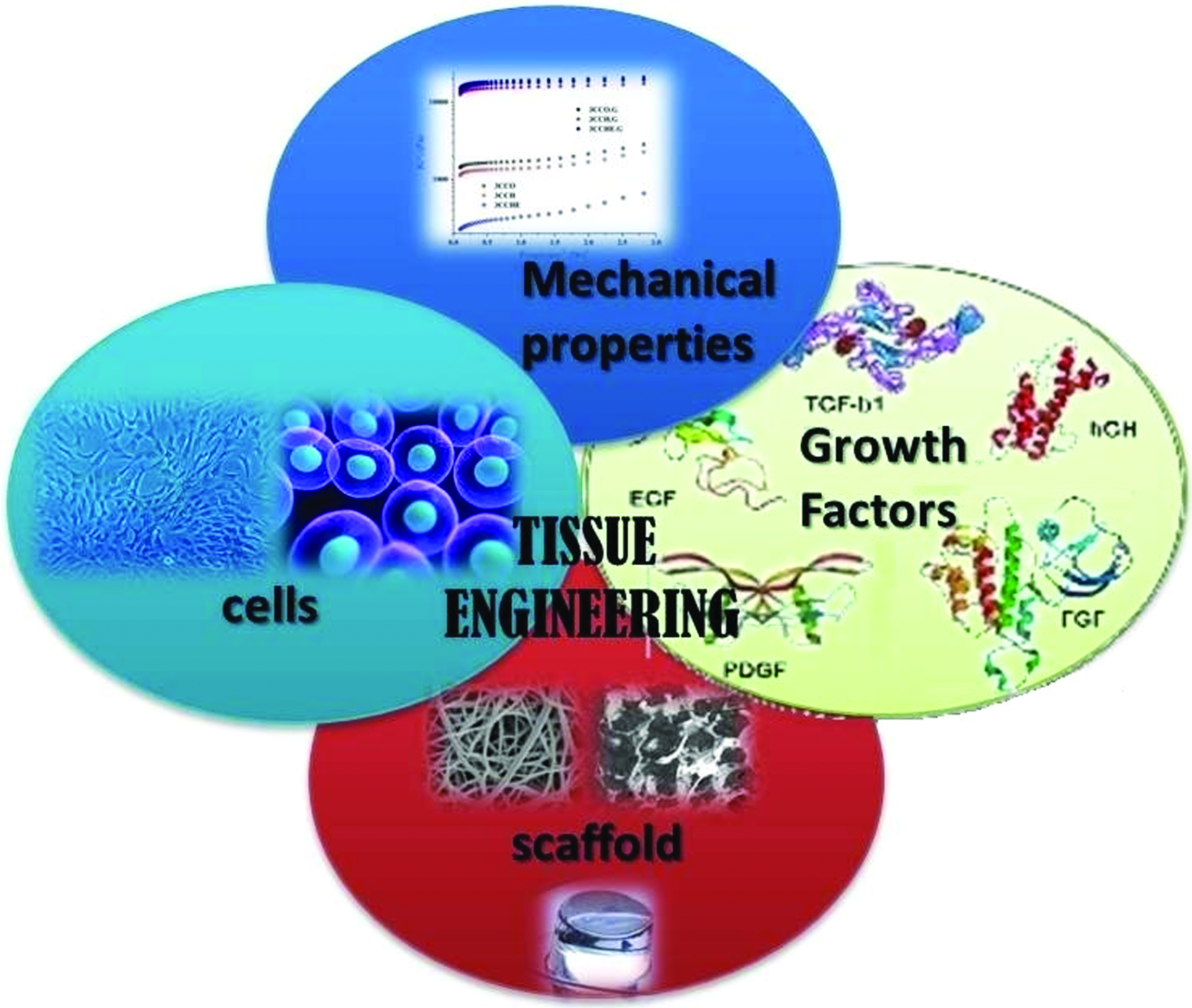
The elements of tissue engineering for cartilage restoration. Color images are available online.
Cells
The cells used in tissue engineering can be extracted directly from the cartilage (which yields chondrocytes) or obtained through chondrogenic differentiation of MSCs extracted from various mesenchymal tissue types (e.g., adipose, periosteum, bone marrow, or synovia) and subsequently treated with growth factors. However, chondrogenic differentiation is highly complex, so in many studies of cartilage tissue engineering, researchers have preferred to use mature chondrocytes. Moreover, when they proliferate they differentiate.
Chondrocytes are an attractive choice for seed cells to be used in scaffolds for cartilage tissue engineering because they produce both ECM and the subsequent type II collagen that formed it. These cells can be collected in healthy regions of articular cartilage where there is no mechanical load, and then cultured and expanded in vitro. However, this procedure has a major drawback: once chondrocytes have passed to a two-dimensional culture, they can lose their chondrogenic phenotype and differentiate into other cell types. 64 Efficient production of hyaline cartilage demands that chondrocytes maintain their phenotype and that they do not produce any type I collagen. Another important aspect is that as patients age, the aggrecans produced by their chondrocytes become smaller and less uniform aggrecans. Thus, older chondrocytes exhibit lower biosynthetic and mitotic activities, and weaker responses to growth factors. 65
Cartilage can be integrated by inducing migration of free chondrocytes from the matrix to the tissue. For example, Pabbruwe et al. devised an implantable system based on a collagen scaffold with chondrocytes to distribute the cells between two surfaces of cartilage. 66 They isolated chondrocytes from bovine nasal septum and seeded on both surfaces of a collagen-based scaffold. They used a model of two discs of cartilaginous tissue, into which was interspersed the collagen/chondrocyte scaffold system to achieve in vitro integration. The researchers kept the scaffold in culture for 40 days, after which point the resulting tissue was analyzed histologically and biomechanically. They concluded that chondrocyte supply could be controlled, and that cartilage restored could be regenerated using a chondrocyte/collagen scaffold system. There have been several studies on the supply and distribution of cells for repairing a lesion in a joint area. Likewise, the characteristics of progenitor cells, and the mechanisms behind their chondrogenic differentiation, have also been investigated. For instance, Solchaga et al. have attempted to repair cartilage tissue through differentiation of MSCs at the damaged site. 67 They studied human MSCs, methods for isolation and expansion of these cells, and their qualitative and quantitative differentiation into chondrogenic cells.
Growth factors
Several growth factors are generally associated with maintenance of the chondrocyte phenotype and with chondrogenic differentiation of stem cells in vitro, including transforming growth factor β1 or β2 (TGF-β1 or TGF-β2), insulin-like growth factor (IGF-1), growth/differentiation factor-5 (GDF-5), and bone morphogenic proteins (BMPs; BMP-2, BMP-4, and BMP-7). 68
When chondrocytes are expanded in culture, treated with growth factors, and seeded in a damaged hyaline tissue, TGF-β1 drives large-scale synthesis of DNA and GAGs due to increased cartilage expression of type II collagen. In addition, BMP-2 and BMP-4 stimulate the formation of cartilaginous tissue and GDF-5 triggers increased production of prechondrogenic precursors and the transcription factor sox-9.64,69 Interestingly, in MSCs, BMP-4 stimulates chondrogenesis, thus promoting their differentiation into mature chondrocytes. 70 The growth factor, IGF-1, has anabolic effects on chondrocytes, prompting them to synthesize ECM by favoring production of type II collagen and PGs, preventing the release of PGs and inhibiting their degradation. Interestingly, Fortier et al. evaluated the efficacy of IGF-1 for promoting ECM biosynthesis. After seeding mature chondrocytes with or without IGF-1, they observed considerably higher levels of aggrecan mRNA and type II collagen in the IGF-1-treated cells, which maintained their characteristic rounded phenotype. They also observed in the IGF-1-treated cells, a dose-dependent increase in total collagen and GAGs, as well as lack of type I or IIA procollagen, which indicated that these cells had not undergone dedifferentiation. 71 Other studies by this group suggested that fibrin composites with IGF-1 grafted in extensive articular defects reinforce the bonds between damaged cartilaginous tissue and subchondral bone, while increasing the proportion of chondrocytes, as demonstrated in a greatly improved histological score. 72 After 4 weeks, the transduced chondrocytes already showed a 100-fold increase in type II collagen; at 8 weeks, there was already a greater amount of articular cartilage covering the lesion, and the histological scores were improved compared with the controls. 73 van Beuningen et al. have investigated other growth factors and concluded that, in vitro, biosynthesis of PG chondrocytes is stimulated by molecules associated with BMP-2 and TGF-β1 and that, in vivo, these factors also influence PG metabolism in hyaline cartilage. 74
Researchers have explored the influence of type II collagen on the chondrogenic response of ECM. For example, Bosnakovski et al. studied bovine bone marrow MSCs cultured in different hydrogels based on type I collagen, type II collagen, and alginate. 75 They treated the cells with media: a serum-free medium and a TGF-β1-supplemented medium. They observed chondrogenic differentiation in the type II collagen hydrogels after 72 h of culture, and this differentiation increased with time, as verified by the presence of GAGs and type II and type I collagen in the ECM. They also found that TGF-β1 had strongly facilitated chondrogenesis. As optimal conditions for expression of the chondrogenic phenotype, the researchers used TGF-β1, dexamethasone, and type II collagen. They concluded that type II collagen only induces and maintains the expression of MSC chondrogenicity, whereas TGF-β1 improves MSC differentiation.
Scaffolds
The scaffolds used in cartilage tissue engineering are based on carbohydrates (e.g., alginate, chitosan, poly-L-lactide acid/poly(glycolic acid) [PLLA/PGA], agarose, and hyaluronic acid) and proteins (e.g., collagen and gelatin). Growth factors are used to stimulate the development of cells that will be implanted onto the scaffold and to maintain their chondrogenic phenotype. 76
The main objective with engineered tissue is to mimic the structure and function of native tissue. Accordingly, a scaffold for cartilage repair must enable growth and proliferation of chondrocytes or MSCs, allow for free movement of these cells throughout its structure, and exhibit similar mechanical properties to native hyaline cartilage. Furthermore, implantable devices must be biocompatible: thus, all scaffolds designed for clinical use, and their biodegradation products, must be innocuous to the host. Thus, when planning scaffold fabrication, researchers must consider any possible local effects on subjacent tissue, as the scaffold gradually breaks down and is replaced by host cells. Crucial factors to consider include the possible release of chemical crosslinking agents and effects of degradation by-products on local pH levels. Moreover, tissue repair scaffolds must be sufficiently porous to be preloaded with cells and enable subsequent ingrowth of new tissue. Furthermore, they must be mechanically robust enough to withstand implantation as well as the mechanical loads typically placed on the joint surface. Last, once implanted, scaffolds must be able remain in place; indeed, if they are easily dislodged, then the cells or growth factors that they deliver locally will have minimal utility.
Ensuring that cartilage scaffolds persist long enough so that, over time, they become fully replaced by neocartilage is complicated, because osteochondral devices must fulfil the requirements of two different tissue types: bone and cartilage. Bone may tolerate a device over a longer period, but if subchondral bone is not regenerated quickly enough, the overlying cartilage will not be repaired correctly. In a review of matrices for cartilage repair, Coutts et al. emphasized that scaffolds should allow for cell attachment both to aid in retention of the implanted cells and to facilitate ingrowth by native cells. 77
Mechanical properties
Mechanical properties such as compression, fluid-promoted shear stress, and hydrostatic pressure must be considered when designing a system for joint cartilage repair. Since tissue engineering aims to improve patients' quality of life, functionality of cartilage replacements is crucial—namely, to reduce the costs, or delay, of a possible joint arthroplasty or other interventions.
Mechanical stimulation is very important for the development of cartilage in infants and children,78,79 as well as for in vitro chondrogenesis and tissue regeneration in adults, through the positive control of genes and the maturation of MSCs.80,81 Efficient growth of neocartilage demands a specific combination of values for Young's modulus, lubricant coefficient and viscoelasticity.82–85 The similarity in mechanical properties between the graft material and the native tissue is cardinal for the functionality of the new cartilage at the macroscopic and microscopic levels. It is especially important in the repair of relatively large defects, because the mechanical loads must be supported effectively by the underlying tissue.
When the chondrogenic phenotype is maintained in vitro, cartilage can be regenerated by simply using an appropriate combination of scaffold and cells. However, to graft a scaffold in vivo, the implanted material must permanently bond to the local native tissue under natural joint conditions. Interestingly, subjecting the scaffold to biomechanical loads before implantation generates an adequate phenotype.
Walking causes articular cartilage to sustain myriad cyclic mechanical loads, including hydrostatic pressure. Accordingly, chondrocytes must live under constant pressure. In a study on this phenomenon, Hu and Athanasiou produce tissue-engineered constructs over agarose in vitro and then subjected some of the cells to hydrostatic pressure (10,000 Pa) at 1Hz for 4 h per day, 5 days per week, for 8 weeks. In lacunae, the cells that had withstood the pressure exhibited higher levels of collagen, a more-rounded phenotype, and lower levels of GAGs than did the control (not subjected to pressure) cells. When simulating the mechanical environment in which chondrocytes should grow naturally, they are able to secrete growth factors and differentiate them by an appropriate route. 86
Scaffolds for Cartilage Tissue Engineering: Components and Architecture
During the advent of tissue engineering, in the early 1990s, researchers explored different combinations of genes, cells, proteins, growth factors, and porous scaffolds. Biomimetic scaffolds have emerged to try resembling articular cartilage in terms of structure, function, ECM, and they are promising materials for restoration and/or repair of cartilage tissue. These biomaterials are designed to simulate the natural environment of ECM, which provides diverse chemical, physical, and biological signals for cell growth and function.87,88 Thus, creating an optimal cellular microenvironment for proper growth of 3D cartilage requires a biomimetic scaffold that has readily tunable mechanical and physical properties, adheres strongly to cells, and releases growth factors. Moreover, a scaffold for articular cartilage repair must be highly porous, with a pore size that facilitates cell adhesion, cell proliferation, and ECM production, and connections between pores to enable exchange of nutrients among cells. 89 Last, the constituent material must be biocompatible with cartilage; bioresorbable, with an adequate rate of degradation; and have mechanical properties and an architecture similar to those of cartilage, to promote the regeneration of native tissue and ensure a clinically useful size and shape.
Scaffold components
Attractive building blocks for tissue-engineering scaffolds include natural polymers, proteins, carbohydrates, and hydrogels owing to their biocompatibility and biodegradability. In addition, these compounds facilitate binding of implanted scaffolds to cartilage tissue in situ.
Hyaluronic acid
Hyaluronic acid is a fundamental component of cartilage tissue matrix. Crosslinked forms of hyaluronic acid have been studied as cartilage repair scaffolds. For example, Butnariu-Ephrat et al. implanted marrow MSCs in a hyaluronic acid-based glue in damaged goat cartilage, and then, shortly after surgery, observed that the repaired tissue appeared different than neighboring, normal articular cartilage. 90 Knudson et al. determined that hyaluronic acid oligosaccharides induce chondrocytic chondrolysis, including total loss of stainable PG-rich matrix and activation of gelatinolytic activity. 91 Other groups have used scaffolds based on hyaluronic acid (either alone or combined with calcium phosphate), loaded them with MSCs, and implanted them in rabbits for cartilage repair, achieving good results.92–94 However, the cartilage formed using these implants appeared thinner than the host's normal cartilage. Solchaga et al. compared the outcome of osteochondral defects in rabbits filled with a fibronectin-coated hyaluronan-based sponge (ACP™) with or without autologous bone marrow. The fibronectin-coated hyaluronan-based scaffold organized the natural response and facilitated the integration of the neocartilage with the tissue. 94 Marcacci et al. used a 3D hyaluronic acid matrix to culture autologous chondrocytes, and then implanted this matrix in a human knee without the use of a periosteal flap, which enabled a reduction in transplant-site morbidity by using arthroscopy as the surgical technique compared with classic autologous implant. 95
Solchaga et al. compared in another study the effect of two seeding cell methods (vacuum-aided seeding technique and passive infiltration) on the retention rate of human mesenchymal stem cells in hyaluronic acid sponges. 96 In all in vitro tests, the vacuum-aided seeding technique presented better results than the passive one. The objective of this research was to establish a simple and reproducible protocol for uniform seeding of cells in preformed porous scaffolds in large scale (14-mm diameter by 6-mm thickness).
Collagen
Type I collagen is a natural component of skeletal tissues such as bone tissue. Accordingly, collagen-based scaffolds have certain advantages over other types: for example, they allow for contact between the preloaded cells and endogenous cells located in joint tissues.97,98
Collagen-based biomaterials can be fabricated by enriching a collagen solution with biomolecules such as elastin, 99 chitosan,100–102 or GAGs.103–105 The collagen required for scaffolds can be extracted from biological tissue by using acidic,106,107 neutral saline,108,109 or proteolytic110–112 solutions. Proteolytic solutions are not highly recommended because they alter the molecular structure of this biomolecule blocking terminal telopeptides, leading to an irremediable reduction in assembled tropocollagen fibrils. 113 Interestingly, endogenous proteases can be inhibited during solubilization of type I collagen acidic solution. 114 Regardless, the extraction technique that provides the highest yield is solubilization of type I collagen acidic solution containing pepsin, which leads to minimal blocking or denaturing of telopeptides.114,115
For decades, collagen-based scaffolds have been widely used for characterization of chondrocytes and stem cells in vitro 116 as well as in vivo, in rabbits,97,117 sheep, 118 horses, 119 and dogs. 120 Collagen matrices that contain GAGs have been explored for gene therapy. 121 Encouraging results have been obtained using collagen fiber scaffolds to deliver chondrocytes 97 or BMPs 117 in rabbits. For instance, Frenkel et al. reported that, 6 months postimplantation in rabbits, a collagen fiber-based scaffold loaded with chondrocytes induced articular-type repair similar to native tissue. 97 Analogously, Sellers et al. described cell-free, BMP-2-loaded type I collagen implants that afforded excellent repair. They later assessed defects of clinically relevant size, applying appropriate means of implant retention, to confirm the clinical efficacy of repair. 117
Deponti et al. assessed autologous chondrocytes for seeding of a collagen scaffold. 122 Initially, they isolated chondrocytes from infant hyaline cartilage and then rapidly seeded these cells in collagen-based sponges immersed in medium. To optimize the seeding conditions, they evaluated the effects of adding fibrin glue on cell survival within the sponge and tested different time periods of in vitro scaffold maturation. They subsequently expanded the cells in vitro, resuspended them in fibrinogen, seeded them into collagen scaffolds, and finally, cultured them for 1, 3, or 5 weeks. They evaluated the different cultures to determine the optimal time for the rescue of chondrogenic phenotype, which they determined to be 3 weeks. Ultimately, they developed a collagen fibrin adhesive sponge that could efficiently support cell survival and synthetic activity in culture, and they demonstrated the feasibility of converting modified specimens into tissue with chondral properties in vitro.
Chitosan
Another popular constituent material for biomimetic scaffolds is chitosan, 123 which is a partially deacetylated polymeric derivative of chitin, which is commonly found in the cell walls of fungi and in the shells of crustaceans. 124 Chitosan comprises a network of β(1–4)-linked glucosamine units that also contains N-acetyl-glucosamine units. The ratio of glucosamine units to N-acetyl-glucosamine units determines the degree of deacetylation in the polymer, which varies from 30% to 95%. The molecular weight of extracted chitosan ranges from 300 to 1000 kD, depending on its source and the method used for preparation and purification. The solubility of crystalline chitosan in aqueous solutions is pH dependent: above pH 7, it is practically insoluble, but from pH 6 downward, it begins to become soluble, due to protonation of its free amino groups.125–127
The PGs and GAGs in cartilage, which are anionic, partake in electrostatic interactions with chitosan, which is cationic. This phenomenon is extremely important for retention and concentration of growth factors at the implant site, as the growth factors are related to GAG (mostly with heparin) and it is very convenient that the scaffolding based on this polymer incorporates a chitosan complex. 127 Chitosan oligosaccharides also stimulate macrophages, both in vitro and in vivo.
For orthopedics, chitosan has been widely used in combination with materials such as calcium phosphates, hyaluronic acid, poly-L-lactic acid, alginate, polymethyl methacrylate, and growth factors for bone and cartilage restoration and/or regeneration. Chitosan is also frequently used in tissue engineering to work with cells. 128 Reported examples of chitosan-based scaffolds for cell culture included gels, 129 sponges, and fibers, 130 porous materials based on a mixture of chitosan and ceramics, 131 and polymeric materials that contain gelatin or collagen I or II,132,133 added to facilitate cell seeding and enhance the mechanical properties of the implant.
Jeon et al. compared a chitosan-based scaffold with a poly(lactic-co-glycolic) acid (PLGA)-based scaffold for in vivo cartilage repair in nude mice. 134 They seeded the scaffolds with cells, and then implanted them subcutaneously in the animals. The chitosan-based scaffold maintained its volume up to 12 weeks postimplantation, and had degraded on formation of mature cartilage, at 16 weeks. In contrast, the PLGA-based scaffold material exhibited good cartilage development at 4 weeks postimplantation but was reabsorbed/absorbed by the 12th week. The authors postulated that the porosity of chitosan may have delayed the formation of new cartilage, but its longer life compared with PLGA enabled greater maturation of the ECM network.
Polylactic acid
Caterson et al. reported that marrow MSCs cultured in polylactic acid or polylactic acid/alginate scaffolds and treated with exogenous TGF-β1 undergo chondrocytic differentiation. 135 Along these lines, Frenkel et al. created, and tested in rabbits, a scaffold based on D,D-L,L-polylactic acid and collagen, which featured separate tissue-specific environments for the regeneration of cartilage and bone. 136 As with other polylactic acid scaffolds, its mechanical properties provided secure positioning at the implantation site, thus obviating additional fixation. The composite, treated with BMP-2, induced the growth of high-quality, articular-like tissue that remained up to 24 weeks postimplantation and integrated well with host tissue. This group of researchers have performed similar studies in large animals.
Elastin
Elastin is a protein that affords strength and flexibility to the connective tissue in cartilage, ligaments, skin, and so on. It comprises ∼800 amino acid residues 137 arranged in hydrophobic domains and crosslinking domains. It is normally found in ear and nasal cartilage and also found in other articular cartilages. This structure provides mechanical properties to connective tissue while enabling elastin to form reticulated, stable structures with surrounding molecules. 138 Human elastin sequences have been widely used in polymer synthesis for tissue engineering—namely for providing control over the functional properties of peptides and the mechanical properties of biomaterials. A class of elastin-derived polypeptides that have been widely used in tissue engineering is elastin-like polypeptide (ELPs), which are encoded by the tropoelastin gene and comprise repeating units of the pentapeptide VPGVG. 137 They offer excellent control over protein functionality, as their molecular weight and amino-acid sequence can be controlled with high precision at a synthetic and genetic level. Moreover, ELPs are biodegradable, biocompatible, and immunogenic, thus explaining their widespread use in tissue engineering. 139
Betre et al. added ELP to a matrix for use in cultivation of chondrocyte monolayers, which enabled longer periods of growth without the dedifferentiation inherent to other methods. They found that noncrosslinked ELP facilitated rapid accumulation and retention of matrix associated with chondrocytes. The ELP solution underwent transition and coacervation at 35°C, ultimately trapping primary chondrocytes within the matrix. Since the transition of ELP was reversible, the authors could recover the matrix and the cells (for subsequent seeding in monolayers) after 10 days of culture by simply stirring them gently at room temperature. Encouragingly, the chondrocytes retained their phenotype for up to 4 weeks, as corroborated by their accumulation of type II collagen and GAGs. 140 Later, Betre et al. also demonstrated the utility of ELP for the culture and chondrocytic differentiation of stem cells. 141
Alginate
Alginate is a linear anionic natural polymer composed of repeating units of disaccharides—specifically, blocks of the homodimers (1 → 4)-α-L-guluronic acid (GG) and (1 → 4)-β-D-mannuronic acid (MM), and heterodimer (1 → 4)-β-D-mannuronic acid—(1 → 4)-α-L-guluronic acid (MG). 142 It is naturally abundant in brown algae (25% to 45% by dry weight), from where it is extracted.1,2 The quantity and quality of extracted alginate depend on the algae species, the type and age, and the extraction method.
Given its natural abundance and biocompatibility, alginate is commonly used for biomaterials such as scaffolds for cell seeding.143,144 Purified alginates are widely used in the pharmaceutical industries as stabilizers in solution and dispersion of solid substances. In the biomedical field, they are also used for various purposes, such as drug controlled release, 145 encapsulation of cells, 146 scaffolds for tissue engineering, either in ligaments or tendons.147,148 Alginate in solution has negative charges unless there are nonmonovalent cations that promote its “natural criss-cross” due to contraction according to the famous model of the egg box. However, this is beneficial for the 3D structures prepared for the cartilage with this material because higher density of negative charge promotes better and greater cell adhesion.53,149 Li and Zhang studied the biocompatibility of chitosan/alginate scaffolds in studies on the morphology, proliferation, and function of chondrocytic HTB-94 cells. 150 They reported that the scaffolds promoted cell proliferation and improved the expression of chondrocytes, and they concluded that the chitosan/alginate hybrid could be an alternative to chitosan as a scaffold material for cartilage engineering. 151
PGA, poly(lactic acid), and PLGA
Some researchers have explored the interactions between chondrocytes and the poly-(α-hydroxy ester) polymers PGA, poly(lactic acid) (PLA), and PLGA,39,98,152–154 all of which are FDA approved. These polymers are all degraded by hydrolysis. Although PGA is more crystalline and less hydrophobic than PLA, it also degrades more quickly. 155 Sittinger et al. studied the interactions between either of these polymers and human chondrocytes at constant pH for 12 days, and concluded that PLA was less cytotoxic than PGA. 152 Chu et al. seeded perichondrial cells in PLA meshes, and then grafted the loaded scaffolds into rabbit femoral condyles. 156 They observed firm hyaline cartilage at 6 weeks postgrafting. In a similar study, Ma et al. seeded bovine chondrocytes on PGA scaffolds and observed cartilaginous morphology after 12 weeks. 157 Moreover, the mechanical properties of the engineered tissue resembled those of native tissue.
Freed et al. arrived at the conclusions that the growth of bovine chondrocytes in PLA matrix was twice as high as in the PGA in less than 2 months, however, after 6 months of study, the cell population was almost similar in both matrices. The difference in first stages is due to the fact that PLA degradation occurs much slower, and, for this reason, there was not so much space for proliferation. 158
Sherwood et al. 3D-printed a PLGA/PLA-based heterogeneous chondral scaffold as a biomimetic of articular cartilage. 159 They characterized the structure for porosity, composition, architecture, and mechanical properties, finding local variations. Thus, the upper scaffold, which comprised D-L-PLGA/L-PLA, was 90% porous and featured macroscopic channels to promote homogeneous cell seeding. Using a porosity gradient and materials to avoid scaffold delamination, they assembled a transition region. The lower area (like the bone tissue), made of L-PLGA/TCP to promote growth of bone tissue and good mechanical properties, was 55% porous. The authors seeded chondrocytes in the upper scaffold area (like cartilage). Histologic and biochemical analyses revealed that cartilage tissue had formed at 6 weeks of in vitro culture. According to the authors, the mechanical properties (tensile strength) of the bone portion of their osteochondral scaffold resembled those of cancellous human bone, thus highlighting the promise of such frameworks for clinical applications, including total arthroplasty.
Scaffold architecture
Scaffold architecture is critical for enabling strong attachment of cells to the biomaterial, favoring their proliferation and favoring good mechanical properties. For a given scaffold, the choice of architecture depends on the target implant site and the intended function in the body. 46 Porous scaffolds can be assembled via numerous techniques, including gas foaming, temperature-dependent phase separation, membrane lamination, fusion molding, solvent melting, and fiber bonding. However, research on these techniques remain too focused on process optimization, rather than on biomaterial design.
Fibrous scaffolds
One class of scaffolds that have been explored for repair of damaged cartilaginous tissue is fibrous scaffolds. Li et al. electrospun and characterized a set of fibrous polymer scaffolds, based on biodegradable poly-(hydroxy esters), which, when prepared under optimal electrospinning conditions, they considered suitable for tissue engineering applications. 160 Similarly, McCullen et al. fabricated a trilaminar fibrous scaffold from poly(ɛ-caprolactone) (PCL) by electrospinning, varying the conditions and thickness of the fibers. 161 Owing to their robust support for restoring the damaged cartilage, promotion of cartilaginous tissue growth in vitro, and superior mechanical properties (compared with those of homogeneous material), such laminar scaffolds appear highly promising.
Porous scaffolds
Porous scaffolds also have been explored in the context of tissue engineering. For instance, Wu and Ding studied the in vitro degradation of porous (87% ± 3%) 3D scaffolds based on amorphous poly (D, L-lactide-co-glycolide), which they exposed to phosphate buffer solution at 37°C. 162 The authors evaluated the extent of degradation based on four stages: quasistability, loss of force, loss of weight, and finally, fracture. They observed encouraging results for the mechanical properties of the wet material. Liao et al. assembled a biodegradable porous scaffold from chondroitin sulfate methacrylate (CSMA)/polyethylene glycol methyl ether, ɛ-caprolactone acryloyl chloride (PECA)/graphene oxide (GO) for cartilage tissue engineering and studied its degradation rate, swelling ability, conductivity, and mechanical properties, reporting good values for all parameters with a pore size range of 100–200 μm. 163 They also tested their CSMA/PECA/GO scaffold in vivo, observing suitable tissue and affirming the utility of this scaffold. Last, Jonnalagadda et al. developed porous scaffolds based on PCL/PGA, which exhibited pore sizes between 20 and 120 μm with 99% of porosity approximately for repair of articular cartilage. 164 It demonstrated similar mechanical properties to those of human articular cartilage, plus high levels of chondrocyte adhesion, proliferation, and GAG secretion.
Porous multilayer scaffolds
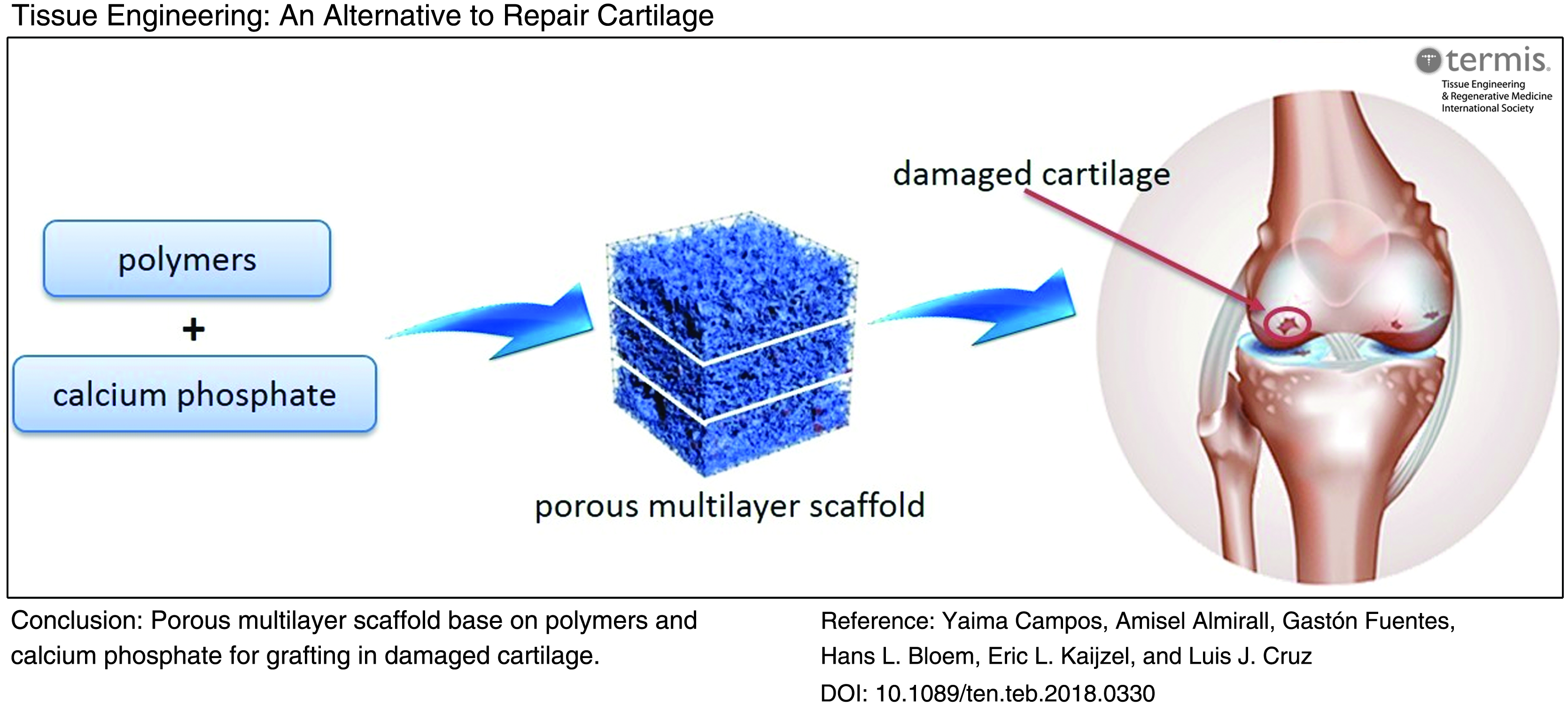
An alternative to seeding cells on a monolayer scaffold is to use a multilayer graft that promotes a higher growth of cells and guides the simultaneous regeneration of bone, cartilage, and a calcified cartilage intermediate. 165 Porous multilayer scaffolds are advantageous because they require a smaller population of chondrocytes than the traditional cell-based monolayer approach, resemble natural cartilage in complexity, and enable mimicking of the mechanical properties of the natural tissue in each layer (Figs. 3 and 4). The “calcified layer” could be filled with calcium phosphate (hydroxyapatite, β-tricalcium phosphate, octacalmium phosphate, etc.) to promote the osteointegration and osteinduction of the bone tissue, which is close to subchondral bone.

Porous multilayer scaffold for regenerating cartilage tissue. Color images are available online.

Porous multilayer scaffold based on polymers and calcium phosphate for cartilage tissue engineering. Color images are available online.
Camarero-Espinosa et al. recently designed and built a multilayer scaffold with nanometric features, in which they sought to mimic the mechanical properties, structure, and chemical signals of hyaline cartilage. 166 Their scaffolds provided control of the orientation, morphology, and phenotype of seeded chondrocytes. Furthermore, these scaffolds promoted the growth of new tissue that resembled articular cartilage and favored the formation of localized apatite nuclei, which facilitated integration of the scaffold into subchondral bone. Ng et al. devised a technique for creating agarose-based scaffolds by layering, reporting that such scaffolds demonstrated biomechanical properties suitable for chondrocyte seeding, and potentially, for tissue engineering applications. 167
Analogously, Wu et al. obtained collagen-based scaffolds via layer-by-layer casting and subsequent freeze-drying, generating a multilayer material with properties similar to hyaline cartilage. 168 The upper layer comprised collagen exclusively, whereas the lower layer was composed of collagen and hydroxyapatite. Between these two layers were intermediate layers, in which the collagen/hydroxyapatite ratio varied gradually. The authors cultured knee-joint chondrocytes from New Zealand rabbits in the upper layer, and after 2 weeks of culture, observed by immunohistochemistry and histology that the cells had maintained their phenotype. The general approach of a bilayer graft that guides the simultaneous regeneration of bone and cartilage has since been tested using various biomaterials and scaffolds,169–182 and growth factors.183–186 These studies underscore the need for a consistent biological barrier between the neocartilage and bone region. However, a bilayer design alone may not be sufficient to form structurally and functionally appropriate cartilage and bone, even when many studies support this structure.97,172,184–186 Thus, trilayer scaffolds with cartilage, interface, and bone regions have been designed for osteochondral regeneration.38,187–189
For instance, Heymer et al. tested a trilayer scaffold (produced by Kensey Nash Corporation) that included a hydrophobic interface that separated the cartilage region (type I collagen fibers plus hyaluronic acid) from the bone region (type I collagen fibers plus PLA). 190 The authors seeded stem cells in the hyaluronic acid region above the interface and cultured them for 3 weeks, ultimately observing formation of cartilage-like tissue. Moreover, although calcified cartilage formation was not observed, the three layers remained distinguishable and structurally stable after culture. Alternatively, Jiang et al. reported a scaffold composed of an agarose hydrogel and bioactive PLGA/45S5 glass composite microspheres, which supported the regiospecific coculture of chondrocytes and osteoblasts. Their design enabled in vitro assembly of three compositionally distinct yet structurally continuous regions containing cartilage, calcified cartilage, and bone-like matrices. 191
Kon et al. developed an acellular trilayer osteochondral scaffold to control the spatial distribution of hydroxyapatite and collagen to facilitate the cartilage-to-bone transition. The upper layer comprised 100% type I collagen; the middle layer, 60% type I collagen and 40% hydroxyapatite; and the lower layer, 30% type I collagen and 70% hydroxyapatite. 188 The authors joined the layers via freeze-drying and then tested them in an adult equine osteochondral-defect model. At a 6-month implantation, they observed distinct nonmineralized and mineralized regions, and the new tissue was very well integrated with the subjacent tissue, including cartilage and bone. 187
Levingstone et al. investigated the structural and microarchitectural properties of a porous multilayer scaffold and the biological behavior of the material in vitro, determining the biocompatibility, attachment, and proliferation of cells on the scaffold, and the ability of cells to infiltrate through the porous and distribute evenly throughout the construct, demonstrating the potenciality of this kind of biomaterial for osteochondral repair. 32
Porous multilayer scaffolds: mechanical properties
During the scaffold design process, the focus is oriented to the replication of native healthy tissue complexity and mechanical properties. This complexity could be achieved by introducing in the scaffolds gradients of morphology, composition, and function. Regular use of a joint subject to loads makes cartilage to generate mechanical, physicochemical, and electrical signals that allow chondrocytes to exert their synthetic and degradative activity and, consequently, to remodel cartilaginous tissue. However, when a joint is not used regularly, the matrix changes, causing a loss in structural integrity and mechanical functionality. Furthermore, aging causes alterations in matrix composition and in chondrocytic activity that can significantly compromise the function of cells and tissue, leading to gradual degeneration of cartilage. 61
Collagen fibers192–195 and scaffolds 196 have been characterized for mechanical function. Parameters that depend on specimen dimension are often described, including round load or maximum load.192,195,196 These factors are important, as clinical applications may demand larger scaffolds than those evaluated in laboratory tests. There are also studies on intrinsic properties of materials, including tangent module192,194–196 (which does not depend on the sample dimension) provides important information depending on the scaffold application. Other mechanical properties that must be assessed include mechanical-load tolerance, either in vitro or in vivo197,198 and viscoelasticity, both of which influence the function of cartilage, ligament, and other soft tissues.
The mechanical properties of nontraditional scaffolds have also been reported.63,68,199 For instance, Schaefer et al. studied a tissue engineering system based on chondrocytes that they had cultured in a bioreactor and subsequently seeded in a polyglycolic scaffold combined with an osteoconductive support. 138 They grafted this system into a large osteochondral defect in the femoropatellar groove in adult rabbits. Six months postimplant, the material supported physiological loads, had developed subchondral tissue, and exhibited a Young's modulus like that of normal hyaline cartilage. 200
Conclusion
Researchers have designed diverse scaffolds for tissue engineering applications with the long-term goal of clinical cartilage repair. However, despite their best efforts to mimic natural cartilage tissue, they have not yet reached its level of structural and functional complexity. Among the greatest achievements to date has been the development of porous scaffolds that enable internal and external cell adhesion and proliferation and that can withstand the mechanical loads applied to the damaged areas. Nevertheless, despite this and numerous other milestones, state-of-the-art materials continue to suffer from drawbacks—most importantly, once implanted, they fail over time, as demonstrated in animal studies. Thus, scientists are tasked with creating a new generation of scaffold biomaterials that are better suited for long-term cartilage regeneration in vivo. So far, multilayer cell/scaffold complexes based on natural polymers have had greater similarity in terms of composition and properties to cartilage and have achieved an appropriate restoration of the native tissue.
Footnotes
Acknowledgments
The authors acknowledge the financial support from the European Union through the Erasmus PLUS project code NL01-KA 107-008639 for Y. Campos (doctoral fellowship) and G. Fuentes and A. Almirall (staff mobility). Also, L.J. Cruz thanks the financial support of the VIDI personal grant (project number 723.012.110), project grants from the EU Program H2020-MSCA-2015-RISE (644373–PRISAR) and MSCA-ITN-2015-ETN (675742–ISPIC), H2020-MSCA-ITN-2014 TargetCaRe (642414), H2020-MSCA-2016-RISE (734684–CHARMED), and H2020-MSCA-RISE-2017 CANCER (777682).
Disclosure Statement
No competing financial interests exist.