




Abstract
Three-dimensional (3D) hydrogel systems integrating different types of stem cells and scaffolding biomaterials have an important application in tissue engineering. The biomimetic hydrogels that pattern cell suspensions within 3D configurations of biomaterial networks allow for the transport of bioactive factors and mimic the stem cell niche in vivo, thereby supporting the proliferation and differentiation of stem cells. The composition of a 3D hydrogel system determines the physical and chemical characteristics that regulate stem cell function through a biological mechanism. Here, we discuss the natural and synthetic hydrogel compositions that have been employed in 3D scaffolding, focusing on their characteristics, fabrication, biocompatibility, and regulatory effects on stem cell proliferation and differentiation. We also discuss the regulatory mechanisms of cell-matrix interaction and cell-cell interaction in stem cell activities in various types of 3D hydrogel systems. Understanding hydrogel compositions and their cellular mechanisms can yield insights into how scaffolding biomaterials and stem cells interact and can lead to the development of novel hydrogel systems of stem cells in tissue engineering and stem cell-based regenerative medicine.
Impact statement
Three-dimensional hydrogel system of stem cell mimicking the stemcell niche holds significant promise in tissue engineering and regenerative medicine. Exactly how hydrogel composition regulates stem cell fate is not well understood. This review focuses on the composition of hydrogel, and how the hydrogel composition and its properties regulate the stem cell adhesion, growth, and differentiation. We propose that cell-matrix interaction and cell-cell interaction are important regulatory mechanisms in stem cell activities. Our review provides key insights into how the hydrogel composition regulates the stem cell fate, untangling the engineering of three-dimensional hydrogel systems for stem cells.
Introduction
Stem cells derived from the inner cell mass of developing blastocysts, adult stem cells, and direct reprogramming of somatic cells show unique characteristics, including self-renewal and lineage differentiation.1–4 Tissue engineering that integrates both stem cells and biomaterials provides a versatile and promising approach for stem cell therapies for the treatment of diabetes, 5 Alzheimer's disease, 6 Parkinson's disease, 7 liver fibrosis, 8 cartilage repair, 9 sickle cell anemia, 10 and multiple sclerosis. 11 However, it remains a challenge in the field of tissue engineering to control stem cell fate for proliferation and differentiation into a specific cell type using biomaterial.
Hydrogels have been increasingly explored for use as three-dimensional (3D) culture scaffolds to promote stem cell viability and regulate stem cell fate. With high water content and crosslinking polymers, hydrogels can serve as physical scaffolds and generate chemical signals to construct stem cell niches, in which the 3D culture architecture mimics a cellular microenvironment in vivo. In comparison to a conventional two-dimensional culture, which cannot maintain the physiological features of cells,12,13 3D hydrogel systems have been shown to recapitulate the stem cell niches and thereby maintain stem cell functions, including cellular adhesion, 14 migration, 15 proliferation, 16 and differentiation, 17 which ultimately improves the cell transplantation outcome. 18 In the process of gelation, hydrogels can be physically or chemically fabricated for cell encapsulation and seeding. 19
According to their origins, the compositions of hydrogels can be fundamentally categorized as natural or synthetic. Natural compositions of hydrogels derived from the extracellular matrix (ECM), such as collagen, fibrin, hyaluronic acid (HA), and alginate, possess superior intrinsic properties of biological recognition. 20 The synthetic compositions of hydrogels without any element of original ECM, in contrast, are artificially synthesized with chemical materials, including poly(ethylene glycol) (PEG) and poly(vinyl alcohol) (PVA), which display adjustable mechanical properties and architectures. 21 The diverse compositions of hydrogels play roles in regulating stem cell functions, including adhesion, proliferation, migration, and differentiation, through different biological mechanisms. To date, considerable effort has been undertaken to probe the underlying mechanisms of hydrogel compositions in regulating stem cell fates through reciprocal cell-matrix and cell-cell interactions.
In this review, we first discuss the hydrogel compositions, including natural and synthetic ones, which have been broadly applied in the engineering of 3D hydrogel systems for stem cell culture. We then compare the characteristics, fabrication, and biocompatibility of hydrogels made of different types of compositions (Table 1), highlighting their support for stem cell adhesion and growth and their regulatory effects on the lineage commitment of stem cells (Fig. 1). We also discuss the mechanisms of 3D hydrogel systems through cell-matrix interaction and cell-cell interaction in regulating stem cell fates (Figs. 2 and 3).
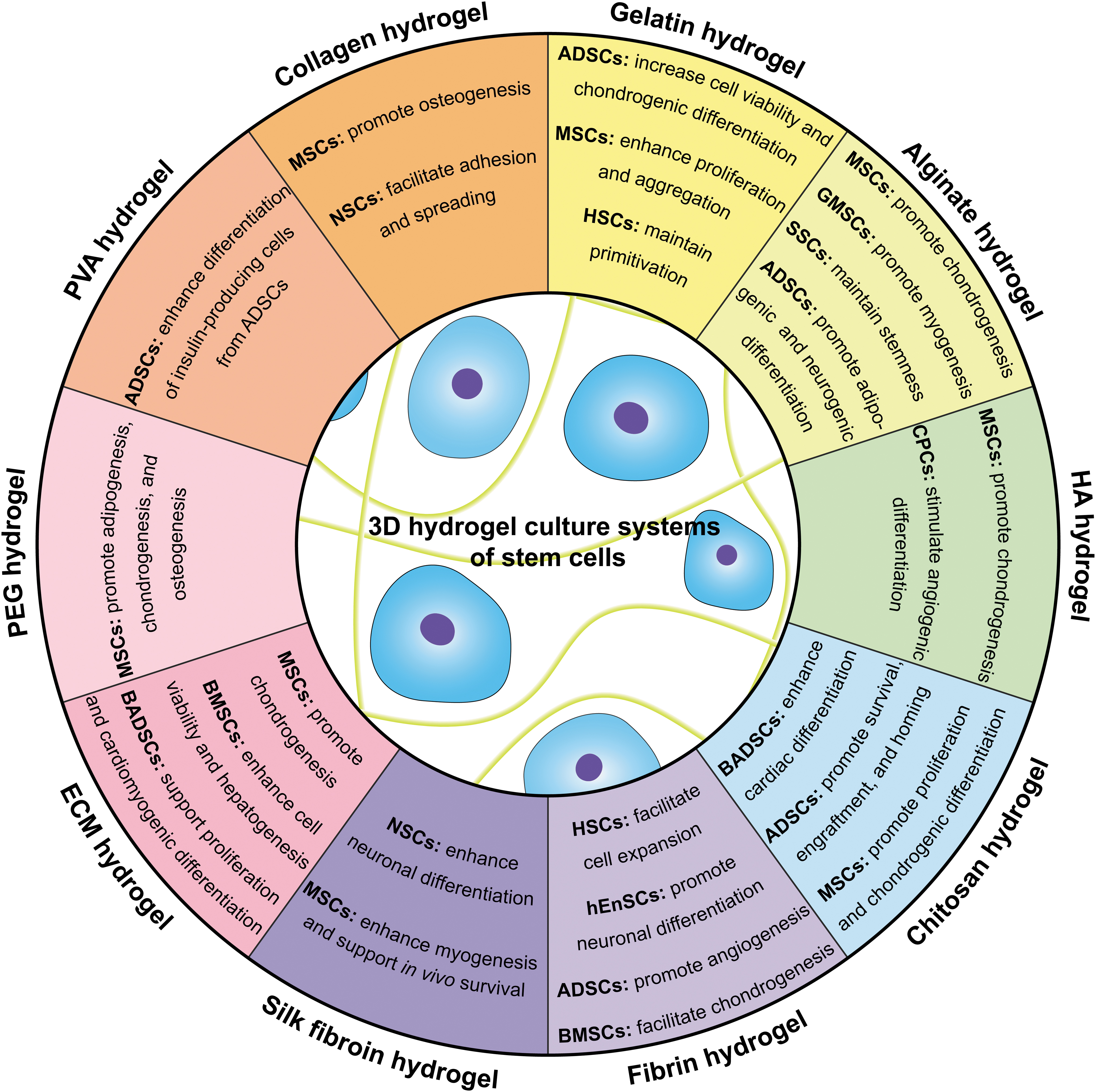
Regulation of stem cell fate by the composition of hydrogel. Three-dimensional hydrogel systems fabricated through gelation of natural compositions, including collagen, gelatin, alginate, HA, chitosan, fibrin, and silk fibroin, have been revealed to improve functions of stem cells and promote tissue regeneration. Because of the hydrogel composition- and cell type-specific regulation of stem cell fate, appropriate combination of hydrogel composition and cell type may lead to significant improvement of stem cell activities and tissue regeneration outcomes. Although less investigated on the regulation of stem cell fate, the synthetic compositions, including PEG and PVA, can also promote the specific differentiation of stem cells in hydrogel culture systems. 3D, three dimensional; ADSCs, adipose-derived stem cells; BADSCs, brown adipose-derived stem cells; BMSCs, bone marrow-derived mesenchymal stem cells; CPCs, cardiac progenitor cells; ESCs, embryonic stem cells; GMSCs, gingival mesenchymal stem cells; HA, hyaluronic acid; hEnSCs, human endometrial stem cells; HSCs, hematopoietic stem cells; MSCs, mesenchymal stem cells; NPCs, neural precursor cells; NSCs, neural stem cells; PEG, poly(ethylene glycol); PVA, poly(vinyl alcohol); SSCs, spermatogonial stem cells.

Cell-matrix interaction of hydrogel plays a critical role in regulating stem cell fate.
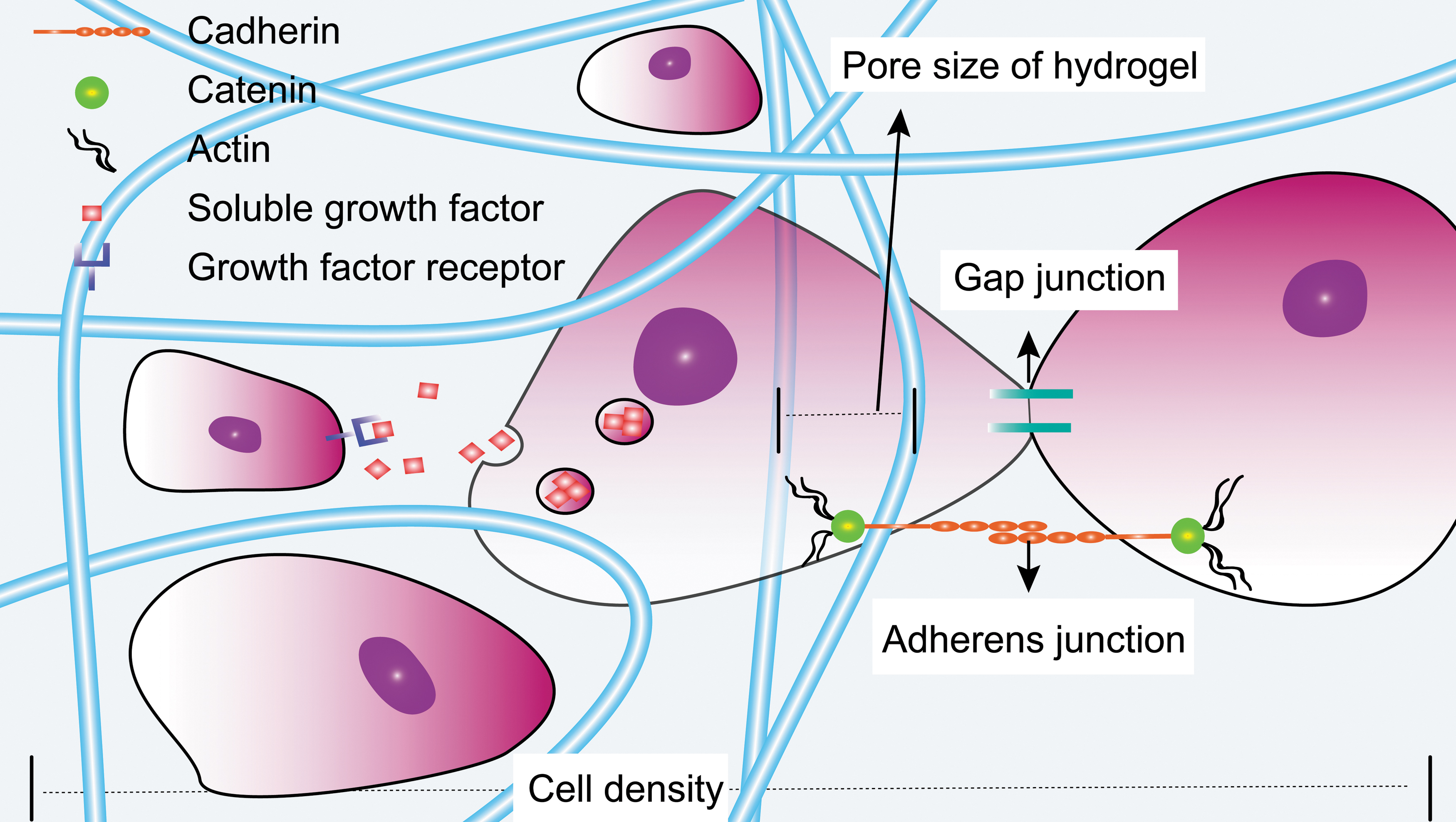
Cell-cell interaction in a hydrogel system influences stem cell activities. Cell-cell interaction includes cell communication mediated by interaction of secretory signaling molecule and cell receptor, and direct cell contact through gap junction or adherens junction. Cadherins are a major type of cell adhesion proteins that mediate the formation of adherens junctions through the extracellular domains, while the intracellular domains bound to catenin. The cell density and the pore size of hydrogel also determine the efficiency of cell-cell interaction, which in turn regulates the proliferation and differentiation of stem cells.
Overview of Natural and Synthetic Hydrogels
ECM, extracellular matrix; HA, hyaluronic acid; PEG, poly(ethylene glycol); PVA, poly(vinyl alcohol).
Natural Composition-Based Hydrogels Possess Inherent and Superior Biocompatibility
Collagen hydrogel
Collagen, which has a triple helical structure and two nonhelical regions at either end of the helix, is the dominant component of ECM. It covers at least 29 types, of which type I has been broadly used for the fabrication of collagen hydrogel for a 3D culture.22–24 In mammals, collagen constitutes about 30% of proteins in the body. 25 It is generally extracted from collagen-rich tissues, such as rat tail and calf skin, through several essential procedures, including acetic acid dilution, pepsin digestion, filtration, salt precipitation, and dialysis.24,26,27 Only in the context of specific temperature and pH can collagen hydrogels be crosslinked through ionic interaction to construct a relatively stable microenvironment serving as stem cell culture scaffolds.28,29 Cell-compatible procedures enable collagen hydrogels to encapsulate stem cells and incorporate other additional biomaterials, such as HA 30 and chitosan, 31 during the gelation process.
On the one hand, the inherent characteristics of collagen hydrogels contribute to the regulation of stem cell activities. Collagen hydrogels exhibit strong binding capacity and excellent biocompatibility because of the presence of various types of integrin domain receptors, thereby supporting neural stem cell (NSC) adhesion and spreading (Fig. 1). 32 As a result of their potential chondral-inductive capacity, stem cell-laden collagen hydrogels are extensively applied in cartilage repair research.26,33 Interestingly, collagen hydrogels can also promote the differentiation of mesenchymal stem cells (MSCs) into osteoblasts (Fig. 1). 29 On the other hand, stem cells can remodel the microenvironment by degrading the ambient collagen hydrogels, a process that is mediated by the secreted matrix metalloproteinase (MMP). 23 During this process, the degradation by-product endostatin is generated to modulate the stem cell phenotypes. 34 Although collagen stiffness varies widely depending on polymerization temperature, concentration, and origin of extracted tissue, the relatively poor mechanical properties of collagen limit the application of collagen hydrogels in tissue engineering. 35 Besides the cumbersome purification procedures, the relatively high cost of materials and inherent immunogenic responses are other hurdles for collagen hydrogel's application in regenerative medicine. 25
Gelatin hydrogel
Gelatin, which is a collagen derivative fabricated through acid or alkaline hydrolysis, has been extensively investigated in biomedical applications because of its low cost, ease of functionalization, and outstanding biocompatibility. 36 The collagen denaturation process endows gelatin hydrogel with lower immunogenicity as well as some physiochemical characteristics shared by collagen hydrogel. 37 Similar to collagen hydrogel, thermal, enzymatic, and photo-initiated crosslinking allow in situ gelation of gelatin hydrogel under mild gelation conditions. Several chemical groups in gelatin hydrogels, including carboxyl and amine groups, can induce covalently crosslinking gelation and exhibit a similar affinity to growth factors, which partly contribute to the potential for high growth factor loading. 38 Gelatin hydrogel also contains intrinsic RGD motifs for cell adhesion and MMP-recognized sites for mediating the enzymatic degradation of the gel.39,40 The naturally degradable feature of gelatin grants the hydrogel system an environmental dynamic that improves the application of stem cells. In this regard, Cheng et al. formulated a gelatin-based hydrogel system for sustained release of stem cells during cell delivery, which consequently promoted angiogenesis in vivo. 41 It has been reported that gelatin hydrogels increased chondrogenesis of adipose-derived stem cells (ADSCs) and cell viability in vivo (Fig. 1).41,42 When formulated into microspheres, gelatin hydrogels with a proper size could enhance MSC proliferation and aggregation (Fig. 1). 43 Furthermore, gelatin hydrogels could be chemically modified to fabricate hydrogel with various structures that suit a wide range of stem cell culture. For example, gelatin methacrylate (GelMA)-based hydrogel fabricated by reacting the amine groups of gelatin on methacrylate groups was found to regulate the functions of primitive hematopoietic stem cells (HSCs) in the presence of stem cell factors (Fig. 1). 44
To date, GelMA mechanical properties can be tuned by adjusting the gel concentration and methacrylation degree. 45 However, a lack of macroporosity in GelMA hydrogel may reduce cell viability and migration. Nanotopographical patterning by wet electrospinning enabled a fabrication of GelMA/alginate hydrogel nanofibers that significantly enhanced MSC motility, adhesion, and proliferation.38,46 Altogether, although poor mechanical properties are one of the main shortcomings; a growing body of evidence has demonstrated that gelatin hydrogel is an attractive biomaterial scaffold because of its outstanding biocompatibility with stem cell functions. 47
Alginate hydrogel
Alginate, originally derived from brown seaweed, is an anionic and natural heteropolysaccharide consisting of α-L-guluronate and β-D-mannuronate.48,49 The alginate gelation occurs under mild reaction conditions with an addition of divalent cations such as calcium or barium.50,51 In contrast to collagen hydrogels, alginate hydrogels have no intrinsic cell binding domains. 52 Hence, alginate hydrogels are usually conjugated with Arg-Gly-Asp (RGD) peptide to enhance cell-matrix interaction, improving stem cell adhesion and growth. In addition, the modified alginate hydrogels show biocompatibility improvements for adipogenic differentiation, neural differentiation, cartilage repair, reproductive medicine, and muscle tissue engineering (Fig. 1).53–57 For instance, Chang et al. designed a complex culture system combining alginate hydrogels with RGD-chimeric proteins. The chimeric-RGD-alginate in the presence of TGF-β3 enhanced the chondrogenesis of MSCs. 58 Another limitation of alginate hydrogel is its poor degradability. In mammals, they are unable to be naturally degraded through an enzymatic reaction. 59 With the aim of improving the degradation of alginate hydrogel, a biodegradable alginate macromer was synthesized by partial oxidation, in which the degree of oxidation was able to control the overall degradation time of alginate hydrogel. 60
Hyaluronic acid hydrogel
HA, which can be synthesized by membrane-bound synthases, exists in the ECM of tissue. 61 HA, which can be extracted from bovine eyes, 62 umbilical cords, 63 and rooster comb, 64 serves as an ideal biomaterial and has therefore been applied in wound healing and nerve, cartilage, and bone tissue regeneration.65–67 Moreover, it is a nonsulfate and linear polysaccharide with disaccharide repeating units of β-1, 4-D-glucuronic acid–β-1,3-N-acetyl-D-glucosamine. 68 Because of the rich array of special functional groups, such as glucuronic acid carboxylic acid, primary and secondary hydroxyl groups, and N-acetyl groups, it can be functionalized into thiol-modified HA, haloacetate-modified HA, and dihydrazide-modified HA, depending on the research aim. 69 In addition, HA can be easily fabricated into hydrogel by forming hydrogen bonds, owing to its abundant hydrophilic groups, including hydroxyl, carboxyl, and acetamido groups. 62 It is also feasible to fabricate a photo-cross-linkable methacrylated HA (MeHA) hydrogel by incorporating methacrylate groups into the HA backbone through the hydroxyl groups.70,71
Since HA is a crucial component of synovia that plays an important role in maintaining chondrocyte function, 72 it has been explored to direct and enhance the chondrogenesis of MSCs (Fig. 1). 73 The biomarkers CD44 and CD168, expressed on MSCs, could interact with extracellular HA to control cell behaviors.74,75 For instance, Bian et al. demonstrated that the cell-matrix interaction between HA and CD44 significantly enhanced chondrogenesis and neocartilage formation. 76 Nonetheless, as is the case with collagen hydrogels, the relatively low mechanical property of HA is one of its drawbacks. 77 Another disadvantage is its rapid degradation, which stems from the hydrolysis of ester linkages and hyaluronidase digestion. 78 To overcome this obstacle, Jha et al. successfully controlled the matrix degradation rate by incorporating MMP-13 cleavable peptides in the HA hydrogel, making it more suitable for the engraftment of cardiac progenitor cells (CPCs) through vascular integration (Fig. 1). 79
Chitosan hydrogel
Chitosan, which is a deacetylated derivative of chitin that can be isolated from shellfish such as crab and shrimp, has served as a type of biodegradable biomaterial for about 30 years. 80 Structurally, chitosan is a polymer composed of N-acetyl-D-glucosamine (acetylated unit) and β-(1–4)-linked D-glucosamine (deacetylated unit). Amino and hydroxyl groups, which are the reactive functional groups in chitosan, contribute to the form's flexibility and ease of functionalization. Functionally, chitosan can be formulated into various types of scaffolding biomaterials such as conduits, films, and hydrogels. Chitosan hydrogel has proved to be an attractive biomaterial in tissue engineering and regenerative medicine because of its injectability, enzymatical degradability, and excellent biocompatibility. 81 It was found to not only enhance engraftment, survival, and homing of ADSCs by mediating the chemokine recruitment and ROS scavenging 82 but also to promote cardiac differentiation of brown adipose-derived stem cells (BADSCs) by increasing collagen synthesis (Fig. 1). 81 In addition, chitosan hydrogel has been extensively applied in cartilage repair research because of its structural and biocompatible similarities to the glycosaminoglycans of native cartilage ECM. Porous chitosan hydrogel occupied with a thermoresponsive polymer promoted MSC proliferation and chondrogenesis as a consequence of deposition of glycosaminoglycan and collagen contents (Fig. 1). 83
Similar to other natural hydrogels, the relatively low mechanical properties of chitosan hydrogels hamper their applications. Chemical cross-linking is a conventional approach to improve the mechanical properties of hydrogels and increase their biocompatibility, but it frequently involves toxic chemical cross-linkers. A fabrication of chitosan-based composite hydrogel without the incorporation of toxic cross-linkers has been found to support stem cell proliferation and differentiation by improving their mechanical properties. 41 Alternatively, incorporation of a nanofiber scaffold into the chitosan hydrogel may also improve the scaffold mechanical performance. Nanofiber, which is a biomimetic scaffold used to support cell adhesion, migration, and differentiation, can be fabricated with natural or synthetic polymers through electrospinning.84–86 Although the characteristics of electrospun nanofibrous matrices heavily rely on the setup of solution parameters and processing parameters such as polymer concentration and flow rate, the merits of nanofibrous scaffolds include a nanoscale fiber diameter, programmable pore size, tunable fiber distribution, and relatively high range of stiffness. The construction of cellular and nanofibrous structures based on the well-distributed nanofibers improved the mechanical performance of chitosan hydrogel and benefited the MSC adhesion and stretching. 87 Moreover, the addition of cellulose nanofibers (CNFs) could also change the rheological properties and self-healing ability of chitosan hydrogel. This self-healing hydrogel with a low CNF amount facilitated the diffusion of nutrients and oxygen, thereby enhancing oxygen metabolism and neural differentiation of NSCs. 88
Fibrin hydrogel
Fibrin typically derived from either pooled plasma or single-donor plasma is a polymer of fibrinogen. 89 The gelation of fibrin from fibrinogen monomers mimics the natural blood-clotting procedure, which is triggered by the addition of Factor XIII and thrombin solution at a given temperature of 37°C.90,91 The mechanical properties and biochemical properties of fibrin hydrogels depend on the concentration of the precursor solution and the type of trigger.92–94 Fibrin hydrogels not only have inherent flexibility and uniform distribution but also contain native cell-recognizing and binding sites that can support cell adhesion and viability.95–97 Therefore, fibrin hydrogels have been found to possess great biocompatibility with stem cells, including ADSCs, 98 bone marrow stromal cells, 99 human endometrial stem cells, 90 and cord blood HSCs (Fig. 1). 100 Furthermore, this kind of injectable hydrogel has been applied in wound healing, muscle tissue engineering, and organoid formation.101–103 Recently, Broguiere et al. used fibrin hydrogels and components from Matrigel to design a well-defined 3D matrix that was appropriate for a long-term culture of organoid containing both progenitor and differentiated cells. 104
Interestingly, Neuss et al. discovered that MSCs could degrade fibrin by secreting fibrinolytic enzymes, including tissue plasminogen activator, urokinase plasminogen activator (uPA), uPA receptor, and plasminogen activator inhibitor. 105 However, the rapid degradation and relatively poor mechanical properties of fibrin hydrogels have hampered their applications in tissue engineering.98,106 Therefore, it would be beneficial to develop hybrid and functionalized fibrin hydrogels to control their degradation rate and stiffness. For instance, hydrogels combining fibrin and HA might provide a better scaffold for chondrogenic differentiation of bone marrow-derived mesenchymal stem cells (BMSCs) and articular cartilage repair. 107 Moreover, incorporation of electrospun nanofibers into fibrin hydrogels could improve mechanical performance and electrical conductivity, thereby increasing the adhesion and proliferation of human endometrial stem cells. This suggests that the nanofiber-embedded fibrin hydrogel is a promising biomaterial scaffold for maintaining stem cell activities. 108
Silk fibroin hydrogel
Silk fibroin, produced from Bombyx mori or Antheraea assama silkworm silk, can be manufactured into biomimetic and in situ gelling hydrogel in a broad range of tissue engineering applications. 109 To fabricate a 3D scaffold, silk fibroin can be treated through several approaches, including sonication, enzymatic cross-linking, redox reaction, and lyophilization.110,111 The Bombyx mori-originated hydrogel mainly contains GAGAGS sequences in proteins, whereas Antheraea assama-originated hydrogel contains polyalanine motifs alternated with glycine-rich sequences. Research findings have suggested that a β-sheet structure form in hydrogel cross-linking can provide a long-term stable niche for stem cells. However, the possible generation of large β-sheet aggregates may have an adverse impact on hydrogel mechanical properties, thereby leading to poor mechanical properties. 112 Furthermore, weak gelation performance in terms of requirements for a long gelation time and a relatively high concentration of precursor solution, and a lack of cell-adhesive domains may be other constraints of silk fibroin hydrogels. To overcome these limitations, Yan et al. adopted small peptides as biological gelators to cross-link silk fibroin hydrogel, which significantly improved gelation performance and also functionalized the hydrogel with cell-adhesive RGD peptides. Specifically, the gelation time was reduced to 20 min and gelation concentration was reduced to 2.0%. 113
Despite several disadvantages, silk fibroin hydrogel remains an attractive candidate in regenerative research because of its programmable degradability, low immunogenicity, distinguished biocompatibility, and low cost.114,115 The silk fibroin hydrogel significantly supported the survival of intracerebrally transplanted MSCs and further promoted sensorimotor functional recovery in poststroke mice, improving the neuroprotective effect of MSCs (Fig. 1). 116 When loaded with TGF-β1, silk fibroin hydrogel with proper stiffness efficiently enhanced the myogenesis of MSCs (Fig. 1). 117
Silk fibroin nanofiber hydrogel significantly increases the potential of a hydrogel system in regenerative medicine owing to its relatively high range of scaffold stiffness, which modulates angiogenic, myogenic, and osteogenic lineage commitment of BMSCs, 118 and its tunable topography, which supports neurite alignment and nerve growth. 119 Even so, the high mechanical properties resulting from electrospun nanofibers are probably not suitable for neural differentiation of stem cells. To better investigate the synergistic role of hydrogel stiffness and topography in regulating stem cell fate, Bai et al. adopted annealing processes to produce nanofiber hydrogel with soft stiffness without altering topography. The results showed that this biomimetic hydrogel had the potential to enhance the NSC differentiation into neurons instead of glial cells (Fig. 1). 120 However, the electrospun fabrication technique requires well-trained researchers to generate high-quality nanofibers, thereby posing more safety and efficiency issues than the comparatively simple fabrication process required for a hydrogel scaffold.
Extracellular matrix hydrogel
ECM hydrogel can be generated through the decellularization of primary tissues of the umbilical cord, 121 pancreas, 122 lung, 123 placenta, 124 bladder, 125 and heart. 126 The decellularized-processed tissue can then be solubilized and pH neutralized at a physiological condition for spontaneous and homogeneous gelation, which is a collagen-dominated self-assembly process and is partly mediated by proteoglycans, glycosaminoglycans, and other ECM proteins. 127 In comparison to the conventional natural hydrogel, which has a single component, tissue-specific ECM hydrogel contains physiochemical features of original ECM components suitable for the determination of stem cell fate. In some examples, ECM hydrogels derived from cartilage promoted the differentiation of MSCs into chondrocytes with an upregulation of chondrogenic genes, including SOX9, COL1, COL2, COL10, and ACAN (Fig. 1). 128 In addition, heart-derived ECM hydrogels supported the proliferation and cardiomyogenesis of BADSCs, and liver-derived hydrogels enhanced the cell viability and hepatogenic differentiation of BMSCs (Fig. 1).129,130
Compared to Matrigel derived from rat chondrosarcoma, ECM hydrogel has been found to have less immunogenicity and greater safety owing to its natural ingredients and the decellularization process that ensures the removal of other cell types and donor antigens. 131 In a recent clinical trial of ECM hydrogel in the cardiac field, no significant adverse events were observed in patients with postmyocardial infarction, who had received a localized injection of porcine myocardium-derived ECM hydrogel. 132 Although the underlying relationship between structural sophistication and biofunction remains largely unknown, the development of ECM hydrogel has opened up a new avenue for engineering biocompatible scaffold for 3D culture of stem cells because of its structural support, compositional complexity, and biosignal resemblance to native tissue.
Matrigel
Although Matrigel is not strictly classified as one type of hydrogel, its natural ingredients from ECM make it comparable to other natural hydrogels regarding the biocompatibility of stem cell culture and differentiation. Matrigel that was first found in rat chondrosarcoma (EHS tumor) consists of all components of basement membrane ECM (such as Laminin, Collagen IV, Perlecan, and Nidogen/Entactin), proteases (such as 72 kDa MMP-2 and 92 kDa MMP-9), and growth factors (such as TGFβs, FGF, EGF, PDGF, and IGF).133,134 Thus, the biological functions of Matrigel are supported by the various kinds of components. For example, Laminin promotes stem cell differentiation. 133 However, owing to its intricate compositions, Matrigel contents vary from batch to batch, which potentially make experimental results unrepeatable in vitro. 135
Typically, Matrigel gelation occurs above 24–37°C environment, resulting in matrix stiffness varying from 0.12 to 0.45 kPa. 136 Because the Matrigel stiffness resembles the ECM of neural tissue,137,138 it has been employed to study the neural differentiation of stem cells. Compared to other biomaterials, Matrigel provides a more suitable scaffold for embryonic stem cells (ESCs) differentiating into neural precursor cells. 134 In addition, neural-induced MSCs in a combination of Matrigel were applied in cell therapy for spinal cord injury in a dog model. 139 To date, Matrigel has become one of the most popular biomaterials employed in 3D cell culture and in organoid formation involved in the research of tumor, angiogenesis, and embryogenesis, whereas the potential immunogenicity and pathogen transmission from the origin of Matrigel should be carefully examined before its application in clinical therapy.104,140,141
Synthetic Composition-Based Hydrogels Offer the Advantage of Immunogenicity Deficiency and Property Tunability
Poly(ethylene glycol) hydrogel
PEG, a hydrophilic and linear polymer with a wide range of molecular weights, has been applied in cell culture systems since the 1970s owing to its advantages, including low cytotoxicity, a well-engineered structure, and tunable mechanical and chemical properties.64,142,143 Indeed, it is practical to modify and functionalize the terminal hydroxyl ends of PEG with various functional groups, including maleimide, amine, thiol, azide, and vinyl sulfone.144,145 Many subtypes of PEG such as cell-adhesive PEG hydrogels, enzyme-sensitive PEG hydrogels, and growth factor-bearing or binding PEG hydrogels have been developed to meet research purposes for various types of cell culture. 142 For example, studies have shown that acrylate or methacrylate moiety of PEG could be functionalized to produce cell-responsive hydrogels. 146 However, if not conjugated with biopeptides or functional groups, the PEG hydrogels manifested low cell adhesion and biocompatibility as a result of the shortage of cell-matrix interaction. 147 Enzymatically crosslinked PEG hydrogels could be efficiently developed with a certain stiffness to mimic the basic physical properties of Matrigel. These PEG hydrogels functionalized with ECM components, such as fibronectin and laminin, have been found to significantly increase the self-renewal ability of intestinal stem cells. 148 Moreover, it has been found that adipogenic, chondrogenic, and osteogenic differentiation of human MSCs in 3D hydrogels could be induced by the PEG functional groups t-butyl, carboxylic acid, and phosphate, respectively (Fig. 1). 149
Poly(vinyl alcohol) hydrogel
PVA, which is coupled with hundreds of hydroxyl functionalities, is a simple linear and semicrystalline polymer derived from the hydrolysis of polyvinyl acetate.150,151 Specifically, the gelation of PVA can be achieved after repeating five freeze-thaw cycles, in which the aqueous PVA solution freezes under −20°C for 1–24 h and thaws at 23°C ± 1°C for up to 24 h. The physical properties of the gel are determined by the cross-linking (crystallite) density of PVA, concentration of PVA, and number of freeze-thaw cycles. 152 Apart from conventional repeating freeze-thaw cycles in gel fabrication, chemical cross-linking with aldehydes or radiation is also an effective method to produce PVA hydrogels. 153 However, PVA hydrogels fabricated by these three methods are not compatible with stem cells lacking cell survival microenvironment. To address these problems, Schmedlen et al. conjugated a cross-linkable group into PVA pendant hydroxyl chains to generate photoinitiated PVA hydrogels. Eventually, they created a well-defined niche for stem cell culture by controlling the polymer concentration, photoinitiator concentration, and exposure to UV light. 153 Moreover, Qin et al. designed a photo-sensitive hydrogel based on norbornene-functionalized PVA. The hydrogel formation could be tuned within 1 min of UV exposure, which made it easier to encapsulate cells in situ and showed high cellular biocompatibility. 154 Similarly, Enderami et al. employed the PVA hydrogels with a surface modified by platelet-rich plasma to support the ADSC differentiation into insulin-producing cells (Fig. 1). 155 However, the development and utilization of PVA hydrogel are still hindered by its nonbiocompatible gelation condition for cells, and its resistance to protein adsorption and cell adhesion, creating the need for further research on PVA modifications. 156
The Mechanisms of the 3D Hydrogel Systems in Regulating Stem Cell Fates Include Cell-Matrix Interaction and Cell-Cell Interaction
As we discussed earlier, the features of hydrogels mostly depend on their compositions, which in turn determine their functions in the regulation of stem cell self-renewal and differentiation. Each kind of hydrogel has its unique advantages and limitations (Table 1). In general, natural hydrogels possess inherent biocompatibility, although they have difficulties in purification, potential immunogenicity, and pathogen transmission. Artificially synthesized hydrogels are more tunable in scaffold physiochemical characteristics, but they often fail to create a fibrous ECM-like architecture. Hence, some researchers have adopted novel strategies combining natural and synthetic compositions of hydrogels to construct a more rational culture system. 157 In fact, the optimization of the 3D hydrogel system requires not only the simple generation of a hybrid hydrogel but also a thorough understanding of regulatory mechanisms of cell-matrix interaction and cell-cell interaction within the hydrogel.
So far, many attempts have been made to investigate the regulatory mechanisms of hydrogels on stem cell fate. It is possible to construct a 3D hydrogel model that is more suitable for stem cells or further application in tissue engineering and regenerative medicine if the underlying mechanisms of cell-matrix interaction and cell-cell interaction can be uncovered. On the one hand, the physical and chemical properties of culture systems should be considered since the cell-matrix interaction plays a vital role in cell behavior (Fig. 2). For example, hydrogels provide basic mechanical cue like stiffness, which may trigger the downstream cell signaling. On the other hand, the cell-cell interaction, influenced by the encapsulated cell number and pore size of the hydrogel, can also regulate stem cell fate (Fig. 3).
Cell-matrix interaction
Hydrogel stiffness regulates stem cell fate through mechanotransduction and cell signaling
Hydrogel stiffness, one of the key mechanical properties, plays an important role in regulating stem cell fate in hydrogel (Fig. 2). Hydrogels with tissue-specific stiffness may recapitulate biomechanical and physiological features of stem cell niche in vivo and support stem cell self-renewal and differentiation. For example, hydrogels that mimicked muscle stiffness were found to be better scaffolds for self-renewal of muscle stem cells and muscle regeneration, indicating a better strategy of stem cell-based therapies for muscle-wasting diseases. 158 In addition, soft and stiff hydrogels were found to increase neurogenesis and osteogenesis of stem cells, respectively, 159 suggesting that each type of cell differentiation favors tissue-specific matrix stiffness. Interestingly, even after changing the culture conditions, the stem cells still possessed the mechanical memory that maintained the elasticity information of the previous environment. 160
Furthermore, matrix stiffness regulates stem cell fate through mechanotransduction and cell signaling (Fig. 2). Cells may respond to the stiffness and transduce the mechanical cue through cell signaling pathways, including RhoA, Rac, Cdc42, GTPases and Hippo pathways.161,162 YAP/TAZ was found to be activated by Ras-related GTPase RAP2, but inhibited by SWI/SNF complex to avoid overactivation of the Hippo pathway.163,164 Interestingly, Cosgrove et al. showed that HAVDI peptide-modified MeHA hydrogels could attenuate the YAP/TAZ mechanosensing in MSCs through cell-matrix interaction. 165
Since hydrogel stiffness has been shown to govern the cell behaviors, it is pivotal to fabricate a 3D hydrogel system with a tissue-specific stiffness ranging from 0.1 to 100 kPa, which mimics the specific physiological ECM of stem cells. 159 Technically, it has become possible to control hydrogel stiffness by changing the type of hydrogel composition and the cross-linker, as well as the material concentration and its molecular weight for the specific type of stem cell. 166 For example, Huebsch et al. and Wang et al. fabricated hydrogels with stiffnesses of 60 and 1 kPa for the bone and cartilage differentiation of stem cells, respectively.167,168
Other important mechanical properties, such as nanotopography and stress relaxation, have also been found to regulate cell behavior.30,169,170 For example, Chaudhuri et al. designed a type of hydrogel with a tunable stress relaxation rate by combining different concentrations of cross-linker and calcium. They found that MSCs cultured in hydrogels with faster stress relaxation enhanced cell spreading, proliferation, and osteogenic differentiation through adhesion-ligand binding, actomyosin contractility, and mechanical clustering of adhesion ligands. 171 Further investigation of the molecular mechanisms of cell-matrix interaction can help strengthen our understanding of stem cell fate, as driven by matrix mechanical properties.
Stem cells degrade hydrogel substrates to remodel the stem cell niche
Cell-matrix interaction is a mutual and bidirectional relationship between the cell and the matrix. In addition to responding to the extracellular signals, stem cells can even modify and remodel the cell niche by secreting proteinase for better accommodation in the niche (Fig. 2). The dynamic status of the spatiotemporal niche may have a profound impact on cell behavior. Recently it has been revealed that apart from the deposition of protein secreted by stem cells within hydrogels, the cell-engaged degradation and modification of hydrogel substrates had a significant impact on cell fate decisions (Fig. 2).172,173 Under a specific culture condition, stem cells in degradable hydrogels manifested a higher potential for cell differentiation and expressed a higher level of cell markers than that in nondegradable hydrogels. 174 These findings could be explained by the fact that stem cell differentiation undergoing structural and functional alterations of the cells coupled with pericellular niche remodel best suit the needs of cell-matrix interaction. However, the dynamic degradation is frequently accompanied by alterations in matrix properties such as stiffness, swelling, and pore size, which directly or indirectly control cell fate decisions. 175 By decoupling degradability from other parameters like matrix stiffness, hydrogel degradability was found to determine multicellular invasion and migration, indicating that hydrogel degradation is an important regulator of cells. 176
In most natural hydrogel systems, stem cells can naturally secrete proteinase to degrade and remodel the surrounding material to suit their own needs. However, most synthetic hydrogels are nondegradable; thus, it would be necessary to modify the synthetic hydrogel backbone. To date, enzymatic hydrolysis 177 and photodegradation 178 are practical approaches for producing degradable hydrogels because of their unique individual advantages. Incorporation of MMP-sensitive peptides into hydrogel backbone is one of the most popular approaches, as it improves the degradation of a weakly degraded hydrogel, resulting in better cell accommodation. 179 Compared to enzymatic degradation and hydrolysis degradation, photodegradation has advantages in tuning spatiotemporally in velocity and hydrogel properties. 180
The rate of degradation should be also considered in the design of degradable hydrogels for stem cell culture. Like hydrogel stiffness, the degradation rate is tunable by altering the monomer concentration and molecular weight. 68 Faster degradation may lead to lower reservation of ECM compositions, whereas slower degradation affects the proper distribution of substrates secreted by the cells.181,182 Although the effects of different degradation rates on the cell functions have not been investigated as thoroughly as that of the matrix stiffness, it has been uncovered that the degradation rate could regulate cell adhesion, cell differentiation, bone formation, and tissue healing.183,184 In fact, the spatial and temporal persistence of MSCs in transplantation could be achieved simply by changing the degradation rate of the hydrogels. 185 Hydrogel modifications are often necessary to keep the degradation rate, especially when the fibrin hydrogels or PEG hydrogels are selected for the 3D culture system, in which compositions degrade either too fast or slowly.
Bio-inspired motifs of hydrogels support stem cell adhesion, survival, and lineage-specific differentiation
Hydrogels are not only the physical scaffolds for stem cells but also provide essential bioadhesive domains recognized by the cells. When cultured in hydrogels, stem cells can bind to matrix ligands containing the RGD domains through the integrins (Fig. 2). The cell-matrix interaction subsequently supports cell survival, spreading, and growth. 186 Therefore, the cell adhesive capability in 3D culture systems should be considered an important parameter of hydrogel development. It is noticed that the size of integrin is ∼8–12 nm, 187 suggesting that the design of RGD nanopatterns with RGD binding sites should be limited to about 10 nm. Moreover, the modifications of hydrogels with bio-inspired motifs by matrix polymerization, chemical conjugation, and functionalization can greatly improve the cell adhesion. 149 It is also helpful to quantitatively increase the RGD peptide density in hydrogels to enhance the cell adhesion, migration, spreading, proliferation, and focal adhesion formation.95,188 Apart from RGD density, nanospacing is another parameter that should be taken into consideration in regulating cell behavior. RGD incorporated in hydrogels with a smaller nanospacing has been found to result in prominent cell spreading. Larger nanospacing might decrease the hydrogel affinity for the cells, although it could induce the osteogenesis and adipogenesis of stem cells. 189
To boost bioactivity for regulating stem cell fate, many other bio-inspired motifs have been exploited to engineer hydrogels. The RADA-16II peptide, 190 IKVAV peptide, 191 N-cadherin-mimetic peptide, 192 and Foxy5 peptide 193 tethered in hydrogels have shown the potential to promote the angiogenesis, neurogenesis, chondrogenesis, and osteogenesis of stem cells, respectively. The capacity of these biomimetic motifs to direct specific lineage differentiation of stem cells is dependent on the dose and concentration. For example, N-cadherin-mimetic peptides enhanced cartilage matrix deposition and early chondrogenesis within hydrogel in a dose-dependent manner. 192 It has been also revealed that cell signaling pathways, such as Wnt signaling, mediated the effects of bio-inspired motifs on the lineage specification of stem cells. Similarly, HAVDI peptide-immobilized hydrogel enhanced chondrogenesis by suppressing the canonical Wnt signaling through upregulation of GSK-3β and reduction of β-catenin/LEF-1/TCF complex. 194 Both the vasoactive intestinal peptide (VIP) and Foxy5 peptide could promote osteogenesis differentiation through Wnt signaling pathways. VIP peptide triggered the canonical Wnt/β-catenin signaling pathway, which was associated with an upregulation of Wnt6, Wnt9b, Slpi, and Sfrp2. 195 In contrast, the Foxy5 peptide, a synthetic Wnt5a-mimetic ligand, activated a noncanonical Wnt pathway by downstream RhoA and ROCK signaling factors, thereby enhancing mechanotransduction. 193 Moreover, the bio-inspired motifs tethered hydrogel can not only facilitate the cell-matrix interaction in vitro but also improve the performance of stem cell implantation in vivo. Recently, Clark et al. inserted a hydrogel with GFOGER, a targeted motif of α2β1 integrin, which could dramatically boost the engraftment efficiency and reparative activities of implanted stem cells in bone repair. 196 Collectively, the incorporation of bio-inspired motifs into hydrogels has been shown to have a profound effect on stem cell fate, especially on cell viability and lineage commitment differentiation.
Incorporation of growth factors enriches the chemical signals of 3D hydrogel systems to enhance stem cell activities
ECM is a complicated extracellular system consisting of a large body of glycosaminoglycans bound by many kinds of growth factors, which can be incorporated into the 3D hydrogel systems.197,198 However, if added to the culture medium directly, free growth factors such as basic fibroblast growth factor could undergo rapid denaturation and degradation.199–201 Therefore, the immobilization of growth factors into the hydrogel backbone is an increasingly popular approach for mimicking the stem cell niche (Fig. 2). In addition, the spatial and temporal cues provided by growth factors had a profound impact on cell fate as well. 20 The advent of the micropattern technique has enabled the spatiotemporal distribution of growth factors in hydrogels that modulate the stem cell functions.202,203
Each type of growth factor has a specific role in regulating stem cell fate, and some share a common role, thereby they can be added together into the same culture system to produce a synergistic effect on the cells. TGF-β and BMP have been found to coregulate the lineage specification of stem cells.204,205 Levenberg et al. cultured ESCs in a biodegradable scaffold incorporated with a combination of growth factors, including retinoic acid, TGF-β, and insulin-like growth factor, which promoted the differentiation of stem cells into tissue-specific 3D structures with characteristics of neural tissue, cartilage, and liver, respectively. 206 BMP-2 can promote osteogenesis of stem cells in either soft or stiff substrate, 207 while TGF-β3 regulates stem cell chondrogenesis by inducing the expression of chondrogenic genes.208,209 Moreover, blood vessel formation is a popular topic of tissue engineering, through which many angiogenic growth factors have been explored. VEGF, a well-known signaling protein in angiogenesis, is typically exploited in blood vessel formation in the 3D hydrogel systems. 210 Likewise, N-acetyl-seryl-aspartyl-lysyl-proline (Ac-SDKP), which mediates blood vessel formation, has been used with stromal-derived factor-1 (SDF-1) in the model of chronic heart failure. 211 Moreover, heparin and TGFβ1 can promote CPC angiogenesis. 212 Taken together, the incorporation of various types of growth factors into hydrogel substrates may enhance the biological function of the hydrogel system in regulating stem cell fate.
Cell-cell interaction
Cell density and pore size of the 3D hydrogel system are important for effective cell-cell interaction
ECM components account for 10–20% of total tissue volume in the central nervous system,213,214 indicating that cell-cell interaction is also involved in regulating stem cell activities besides the cell-matrix interaction. It has been revealed that the cell signals from neighboring cells might override the matrix signals to maintain the stem cells in a quiescent state in the 3D hydrogel system. 215 Cell-cell interaction plays an important role in maintaining the stemness of the cells through secreted signaling molecules such as growth factors, and direct contacts such as the gap junction and adherens junction (Fig. 3).216,217 Cadherin, a large family of transmembrane proteins, including E-cadherin, N-cadherin, N-cadherin 2, and P-cadherin, mediates the adherens junction. Each type of cadherin protein plays a unique role in stem cell behavior. In the developmental stage of mesenchymal chondrogenesis, N-cadherin is a key regulator in cell-cell interaction, 218 whereas P-cadherin is essential to promote the generation of clonal spheres of retinal stem cells in vitro. 219 Recently, Cosgrove et al. obtained a HAVDI-conjugated hydrogel backbone containing a conserved sequence of N-cadherin. It was demonstrated that N-cadherin interaction could change the mechanical effect on stem cell differentiation through ECM-inducing mechanotransduction. 165
In addition, cell seeding density is a key parameter that determines the cell-cell interaction in hydrogel (Fig. 3). A high density of cells may lead to cell apoptosis, while a low density of cells may fail to support cell-cell interaction. Lee et al. applied a cell density resembling the cardiac tissue in vivo to engineer a hydrogel system with suitable mechanical properties, which accomplished a contractile function in the engineered cardiac tissue. 220 It has also been verified that cell seeding density could regulate the lineage commitment of stem cells. The chondro-inducing activity of stem cells was linearly related to cell density, 221 and higher cell density induced the upregulation of osteogenic genes. 222 However, cell differentiation conditions can change the cell density-dependent effect on lineage commitment. When cultured in adipogenic-osteogenic induction condition, stem cells with lower density showed more osteogenesis induction, and the higher density of the cells caused them to favor adipogenesis. 223
Pore size, which is another physical feature of hydrogels, can also influence the cell-cell interaction (Fig. 3). Indeed, efficient cell-cell interaction may not be accomplished without a proper pore size of the hydrogel. A large pore size accelerates the transport of nutrients and other chemical cues, but decreases cell migration. 224 Studies have shown that proper pore size could induce stem cell differentiation into specific lineages, such as angiogenesis (50–150 nm), 225 osteogenesis (>300 nm),226–228 and chondrogenesis (90–250 nm). 229 Technically, the pore size of hydrogels can be measured by scanning electronic microscope (SEM) and mercury porosimetry. 230 However, samples of SEM require strict lyophilization, which could deconstruct the original spatial structure and lead to the measure error. 231 Al-Abboodi et al. fabricated hydrogels with tunable porosity by injecting cellulase enzyme in situ. 230 This method enabled clinicians to tailor the hydrogel structure based on the tissue nature even after transplantation, thus making the personalized tissue engineering possible. Thus, a 3D hydrogel system could be tailored for better cell-cell interaction by tuning the cell density and the pore size of hydrogel.
Outlook and Perspective: Applications of 3D Hydrogel Systems of Stem Cells in Tissue Engineering and Regenerative Medicine
The physical and chemical features of 3D hydrogels, which are mainly determined by the hydrogel compositions, are able to regulate the stem cell functions. However, exactly which hydrogel property dominates the cellular effect and what properties synchronize to regulate the cells remain poorly understood. In the case of cell-matrix interaction, when the cells remodel the stem cell niches through degradation, the pore size and stiffness of hydrogels might also change in the same system, making it difficult to distinguish the sole effect of degradation and the composite effect of several properties on stem cell function. Recently, methods have been developed to decouple substrate stiffness from other characteristics, including protein tethering 232 and surface chemistry. 233 These methods benefit deeper analysis of the specific roles of hydrogel properties and their internal interactions, and may lead to the optimization of the various functions of a 3D hydrogel system for tissue engineering and regenerative medicine. To date, significant efforts have been made to apply 3D hydrogel systems to research fields of organoid culturing, 3D bioprinting, and stem cell delivery.
Organoid culture
One of the most exciting applications of the 3D hydrogel systems of stem cells is the generation of organoid, an organ analogue that recapitulates the structure and function of an in vivo organ and provides an opportunity to study organ-level physiological and pathophysiological functions 234 The formation of this 3D organotypic model relies on self-organization of stem cells through cell-matrix interaction, cell-cell interaction, and cell lineage specification. 235 To fulfill a list of requirements of organoid formation, a methodology has been established to promote cell-matrix interaction and cell-cell interaction. It focuses on the utilization of ECM-mimetic scaffolds such as Matrigel with spatiotemporal dynamics for regulating stem cell fate. However, the batch-to-batch variability in protein compositions of Matrigel can potentially result in low reproductivity and poor tunability of the organoid culture. In addition, concerns about the potential immunogenicity caused by the Matrigel ingredients may limit organoid transplantation. To address these drawbacks, a number of studies using natural and synthetic hydrogels as alternatives to Matrigel have provided evidence that hydrogel scaffolds could support the long-term culture of organoids such as epithelial, intestinal, and kidney organoids.104,148,236,237 Compared with Matrigel, cellulose nanofibril hydrogel performed equally for hepatic differentiation of liver-derived adult stem cells in an organoid culture because of the rapid shear-thinning and self-healing behavior. 238 Moreover, a composited hydrogel based on PEG hydrogel and gelatin hydrogel immobilized with ECM-derived proteins efficiently promoted MSC vasculogenic and osteogenic differentiation. This versatile platform has been found to be superior to the Matrigel model in engineering organ analogues, including prevascularized bone constructs and liver organoid, because of the customizability of covalent immobilization of tissue-specific proteins. 239
Alternatively, ECM hydrogel can also provide a tissue-specific niche capable of directing organoid growth. For example, Giobbe et al. recently have employed an ECM hydrogel that was fabricated from the decellularization of small intestine tissue for the endoderm-derived human organoids. This hydrogel-based organoid culture not only directed organoid growth with a more stable transcriptomic signature compared to Matrigel but also made the in vivo organoid delivery safer. 240 Although problems in organoid culturing in terms of functional immaturity and low reproductivity have not yet been fully solved, the development of ECM hydrogels will enable the broader application of hydrogel-based organoids in regenerative medicine.
Three-dimensional bioprinting
Three-dimensional bioprinting is defined as an art combining biomaterials and cells in tissue engineering with enormous potential to replicate native tissues and organs. In principle, this biofabrication technology can deposit and localize the loading bioink layer by layer through computer-aided bioprinting processes, including droplet-based bioprinting, laser-based bioprinting, extrusion-based bioprinting, and stereolithography bioprinting. The biomaterial acts as a bioink to construct the preprogrammed 3D architecture and to provide a stem cell niche regulating the cell fate in these bioprinting processes. As a result of the biocompatibility, bioprintability, and accessibility of chemical modifications, 241 the 3D hydrogel systems have been increasingly studied for bioprinting of stem cell-laden scaffolds for regeneration of various tissues and organs such as neural tissue, 242 vessel, 243 cartilage, 244 and bone. 245 Despite significant progress, more efforts are needed to address questions regarding uncontrolled cell distribution and low-resolution cell deposition. Instead of conventional layer-by-layer deposition, the utilization of a gelatin-norbornene hydrogel with appropriate photo-cross-link kinetics allowed direct cell embedding, thereby offering high cell loading density and uniform cell distribution. 246
Importantly, the technical optimization of 3D bioprinting of stem cell-laden hydrogel scaffolds also needs to address how different matrix properties influence cell activities and what compositions of hydrogel regulate stem cell fates. For example, increased hydrogel viscosity or shear stress may improve hydrogel printability for higher shape fidelity, while decreasing the cell motility and viability.247,248 It was reported that the utilization of various kinds of hydrogels had a distinct impact on cell viability and directed differentiation of stem cells in the postprinted constructs. 249 Microchanneled gelatin hydrogel fabricated by 3D extrusion bioprinting has been shown to direct cell alignment and elongation and to enhance myocardial differentiation of MSCs. 250 In addition, MeHA hydrogel could support cell survival and promote osteogenic differentiation of MSCs. 251 Composite hydrogels have more advantages in both aspects of bioprinting performance and cellular regulation. A bending bioink composed of alginate hydrogel and dentin matrix with high printability increased cell viability and enhanced odontogenic differentiation of human dental stem cells. 252 PEG-gelatin-composited hydrogel loaded with TGF-β1 could not only improve scaffold mechanical properties and printing resolution but also significantly enhance chondrogenic differentiation of postprinting MSCs. 253 Although 3D bioprinting is still in the early stages of development, it has the potential to interface with an increasingly optimized stem cell-laden hydrogel system and could contribute extensively to various fields of translational medicine.
Stem cell delivery
Stem cell delivery is a practical procedure for stem cell-based therapies, which have opened a new avenue in treating some currently untreatable diseases.254–256 Unlike scaffold-free cell delivery, where single cells or cell sheets are injected at the body site, hydrogel scaffold-based cell delivery retains cell population in a 3D structure with niche cues and relatively high mechanical compliance that can be injected or implanted as a whole system. Indeed, studies have demonstrated that injectable hydrogel given through minimally invasive administration efficiently improved cell retention and increased cell viability of stem cells in vivo.257,258 Chemical cross-linking polymeric hydrogel that can be directly injected to the desired location of the recipient before gelation, is frequently exploited as a scaffold in stem cell delivery. The sol-to-gel transition occurs in situ after delivery and is triggered by various stimuli such as temperature, light, pH, and enzyme, whereas irreversible chemical cross-links of the hydrogel may produce a comparatively static matrix structure that potentially impairs stem cell activities and restricts cell infiltration. Besides, the hydrogel will lose injectability after gelation because the sol-to-gel transition is time dependent, which makes it difficult to precisely manipulate.
As for hydrogels with a short gelation time, latent obstruction of syringes may occur. Much effort has been devoted to addressing these limitations in stem cell delivery. It has been revealed that a void-forming hydrogel engineered by the addition of porogens could mediate pore formation to facilitate the infiltration and expansion of stem cells within the hydrogel. 167 Notably, a dynamic polymeric hydrogel with reversible physical cross-linking may flow upon shear stress, but resolidify rapidly after force removal. 259 The unique feature enabled the migration and infiltration of stem cells as well as the sustained release of loaded cargoes to control the functionality of implanted stem cells. 260 Alternatively, supramolecular hydrogel holding small molecules together through physical cross-linking is also structurally dynamic, rendering outstanding self-healing features and injectability for controlling the release of hydrophobic agents. 261 It has been found that dynamically cross-linked supramolecular single-chain polymeric nanogels provided a tunable dynamic substructure for protecting stem cells from deleterious mechanical shock, suggesting that the conformational development of 3D hydrogel system for stem cell delivery is an effective strategy to regulate cell-matrix interaction. 262
A 3D hydrogel system incorporated with exogenous bioactive agents such as small molecules, drugs, and proteins may serve as an interface to regulate stem cell fate during cell delivery. Studies have provided proof of concept for the feasibility of cell niche-mimetic hydrogel for stem cell delivery. The incorporation of magnesium (Mg) particles into hydrogel solutions rendered hydrogel scaffolds with optimized porosity, which consequently enhanced stem cell viability and vascular infiltration in vivo. Moreover, the generation and release of Mg2+ after the Mg particle degradation also benefited osteogenic differentiation. 263 In another Mg2+ and dexamethasone-loaded hydrogel system, Mg2+ triggered the positive feedback circuit of dexamethasone activation further promoting the in situ osteogenesis. 264 Recently, the graft of dopamine motifs provided the hydrogel with sufficient adhesiveness to corneal surface and improved cell spreading and viability of encapsulated ASC, serving as the first sutureless stem cell therapeutic delivery for corneal regeneration. 265
A number of questions remain to be addressed regarding the tolerability and safety of the 3D hydrogel system in stem cell delivery before therapeutic application. As a foreign implant, stem cell-laden hydrogel may potentially evoke a foreign body reaction (FBR), an immunological process that occurs when an implant is rejected and can subsequently interrupt the structural and functional healing. The hydrogel-induced host response mainly involves the primary protein absorption, subsequent recruitment of neutrophils, monocytic infiltration, macrophage activation, and cascade secretion of inflammatory and anti-inflammatory cytokines. 266 Increasing evidence has verified that the physiochemical properties of an implanted hydrogel, such as the component, shape, size, stiffness, and chemistry, could trigger and influence the FBR. Synthetic hydrogel induced a chronic infiltration of neutrophils dependent on scaffold stiffness and size, while the natural matrix elicited a type-2-like immune reaction because of high expression of CD206 on macrophages and an upregulation of Th2-associated genes on CD45+ immune cells (such as Arg1, Gata3, Chil3, Il4, Il13, and Cd163). 267
Hydrogel functionalized with positive-charged peptides has been found to cause a high infiltration of multiple immune cell populations (such as monocytes, macrophages, and polymorphonuclear myeloid-derived suppressor cells), substantial release of cytokines (such as IL-6), high-degree collagen deposition, and vessel formation, whereas hydrogel with negative-charged peptides only elicited a minor host response. 268 To mitigate the FBR, macrophages that can polarize into two functional phenotypes (proinflammatory and anti-inflammatory subtypes) are potential targets to modulate the host response by tuning hydrogel characteristics, such as mechanical adjustment and surface chemistry modification.269,270 For example, the incorporation of triazole created a hydrogel surface to resist the FBR by reducing macrophage recognition and fibrosis. 271 Moreover, Wang et al. engineered a photoinitiated HA-based hydrogel with tunable macrophage adhesive sites. This composite hydrogel could temporally activate αvβ3-integrin expressions on macrophages and consequently promote anti-inflammatory macrophage polarization to achieve the mitigation of the FBR. 272 The development of immunoregulatory hydrogels can improve the safety and tolerability of stem cell-laden hydrogel systems in cell delivery.
To date, hydrogel scaffold-based delivery of stem cells has already been applied in preclinical therapeutic studies to treat a variety of disorders such as neurological, 273 cardiovascular, 274 and urinary diseases. 275 In addition, it has been found that GelMA hydrogel-based delivery of induced pluripotent stem cell (iPSCs)-derived NSCs could promote axonal regeneration after spinal cord injuries. Notably, iPSCs and iPSC-differentiated cells are important stem cell sources, although the regulatory mechanisms in hydrogel scaffold-based delivery remain to be explored in future work. 276
Furthermore, hydrogel scaffold-based delivery of stem cells has already been applied in clinical studies and has showed promising outcomes. For example, after stem cell implantation with hydrogels, patients with osteoarthritis showed articular cartilage regeneration and recovery in walking capabilities after a 7-year follow-up. 277 These studies demonstrated the feasibility of applying hydrogel scaffold-based cell delivery to support stem cell self-renewal and differentiation in vivo, and that the development of delivery system to regulate stem cell fates could be balanced with the need to control the immunogenicity of hydrogel for safety in therapy. Current work continues to investigate the cell phenotypes and well-known molecular markers upon cell delivery. It is expected that genome-wide analysis of the mechanism of hydrogel in regulating stem cell fate will lead to discoveries of novel molecular signatures for more efficient evaluation of the therapeutic effects of hydrogel scaffold-based cell delivery. The continuous effort to uncover the relationship between hydrogel composition and underlying mechanism of cellular function will also lead to more efficient and safer manipulation of regulatory mechanism to take advantage of matrix property and regenerative capacity of stem cells.
Footnotes
Acknowledgments
We are grateful to the members of the Huang lab, especially Meiyang Li, for helpful comments and discussion.
Disclosure Statement
No competing financial interests exist.
Funding Information
This work was supported by the National Natural Science Foundation of China grant 81671396, Natural Science Foundation of Guangdong Province grant 2017A030313780, and funding from the “Yangfan Project” of Guangdong Province to C. Huang.