




Abstract
Tissue engineering has a great application prospect as an effective treatment for tissue and organ injury, functional reduction, or loss. Bioactive tissues are reconstructed and damaged organs are repaired by the three elements, including cells, scaffold materials, and growth factors. Graphene-based composites can be used as reinforcing auxiliary materials for tissue scaffold preparation because of their large specific surface area, and good mechanical support. Tissue engineering scaffolds with graphene-based composites have been widely studied. Part of research have focused on the application of graphene-based composites in single tissue engineering. The basic principles of graphene materials used in tissue engineering are summarized in some research. Some studies emphasized the key problems and solutions urgently needed to be solved in the development of tissue engineering and discussed their application prospect. Some related studies mainly focused on the conductivity of graphene and discussed the application of electroactive scaffolds in tissue engineering.
In this review, the composite materials for preparing tissue engineering scaffolds are briefly described, which emphasizes the preparation methods, biological properties, and practical applications of graphene-based composite scaffolds. The synthetic techniques, with stressing solvent casting, electrospinning, and three-dimensional printing, are introduced in detail. The mechanical, cell-oriented, and biocompatible properties of graphene-based composite scaffolds in tissue engineering are analyzed and summarized. Their applications in bone tissue engineering, nerve tissue engineering, cardiovascular tissue engineering, and other tissue engineering are summarized systematically. In addition, this work also looks forward to the difficulties and challenges in the future research, providing some references for the follow-up research of graphene-based composites in tissue engineering scaffolds.
Impact statement
Regeneration and repair of tissue and organ injury has become a new research hotspot in recent years. Tissue engineering scaffolds prepared with graphene-based materials have good biocompatibility, excellent mechanical properties, and strong cell orientation, which can fully induce the proliferation and differentiation of seed cells. This review briefly describes the basic materials for the preparation of tissue engineering scaffolds, and focuses on the preparation, performance, and application of graphene-based materials in tissue engineering, providing sufficient understanding of graphene applied in regenerative medicine.
Introduction
Trauma, disease, immunity, and aging can cause the injury, functional decline, and loss of tissues and organs, which is the key factor of threatening human health.1–4 Tissue engineering mainly attaches seed cells with genetic information to scaffold materials and then combines growth factors to construct three-dimensional (3D) complex of cell–scaffold materials, which can be implanted into the body to improve tissue and organ damage and restore its own structure and function.5,6
The ideal scaffold material should have three characteristics7–9 : (1) Excellent mechanical properties, resisting the pressure of surrounding tissues, and maintaining the original shape and integrity of the tissue. (2) Porous structure, promoting cell-to-cell signal transduction, facilitating the exchange of nutrients, oxygen, and metabolic wastes. (3) Good biocompatibility, ensuring that the scaffold materials have no obvious cytotoxicity, inflammation and immune rejection, and promoting the proliferation and differentiation of seed cells.
At present, the scaffold materials for tissue engineering include natural biomaterials, synthetic biomaterials, and graphene-based materials (Fig. 1). Among them, natural biomaterials have the basic physiological characteristics, such as good biocompatibility, low toxicity and immune rejection, excellent hydrophilicity, and cell affinity, which can effectively promote seed cell growth, differentiation, and expression of related factors.10–13 The synthetic biomaterials have uniform texture, strong plasticity, and lower immune rejection than natural biomaterials.14–16

Schematic diagram of material types of tissue engineering scaffolds. Color images are available online.
However, natural biomaterial scaffold has the disadvantages of poor flexibility, thermal stability, and mechanical support ability. Synthetic biomaterial scaffold has the disadvantages of poor seed cell affinity and adhesion, and poor ability to induce tissue cell proliferation and differentiation. The ideal tissue engineering scaffold cannot be constructed only by natural biomaterials or synthetic biomaterials.10–16
Graphene is a single-layer two-dimensional (2D) honeycomb nanomaterial formed by carbon atom with sp 2 hybrid orbit, which has large specific surface area, excellent mechanical and electrical properties, and good biocompatibility, but it is easy to self-assemble in solution. 17 Their derivatives, graphene oxide (GO), and reduced graphene oxide (rGO), have functional groups such as hydroxyl and epoxy groups on their surfaces, with excellent hydrophilicity, and can improve the dispersion of graphene in solution to reduce its toxicities.18–20
When graphene-based materials are used for regeneration treatment of tissue engineering, they can be prepared by bonding with natural/synthetic biomaterials to obtain ideal scaffolds with more stable mechanical properties and better electrical conductivity, which can induce better adhesion, proliferation, and differentiation of seed cells, and promote the regeneration and recovery of damaged tissues and organs.21–23 At present, due to their excellent biological characteristics, graphene-based composite scaffolds have been widely applied in various tissue engineering24–27 (Fig. 2).

Principle diagram and application of graphene-based scaffolds in tissue engineering. Color images are available online.
Aiming at ideal tissue engineering scaffolds, this review briefly describes the three most commonly used preparation methods and principles of graphene-based scaffolds, and expounds the excellent mechanical properties, cell orientation, and biocompatibility of graphene-based scaffolds. To some extent, the important questions about the stability, toxicity, directed differentiation of seed cells, and the immune inflammatory reaction in the application process were answered. In addition, based on the previous discussion, this review also summarizes the specific applications of graphene-based composite scaffolds in bone, nerve, cardiovascular, and other tissue engineering, and emphasizes their biological safety. The specific contents are described in the following chapters.
Graphene-Based Composite Scaffolds
Tissue engineering scaffolds based on graphene-based composite materials mainly include 2D scaffolds and 3D scaffolds. In 2D scaffolds, seed cells widely proliferate in the form of plane, and cannot vertically develop multilayer tissue structures to construct complete tissues and organs. Generally, they are used for drug testing and toxicity research in biomedicine.28,29 Three-dimensional scaffolds can effectively simulate the microenvironment of tissue and organ growth, which can provide a good biological platform for the growth of seed cells and avoid the limitation of nutrient uptake required by tissue cells due to the high surface quality.30,31
GO has been used to modify chitosan (CHT) scaffolds and polyurethane (PU)/polycaprolactone (PCL) scaffolds.32–34 It mainly relies on electrostatic interaction, covalent attachment, and nonspecific force to composite with natural biological materials and synthetic biological materials. The composite scaffold has good morphology and excellent biocompatibility, which can effectively improve the defects of natural biomaterials and synthetic biomaterials, promote the rapid regeneration and repair of damaged tissues, and expand the application of hybrid scaffolds prepared by multiple materials in tissue engineering (Table 1).
Application of graphene-based composite scaffolds in different tissue engineering
3D, three dimensional; CHT, chitosan; GO, graphene oxide; MSC, mesenchymal stem cells; PCL, polycaprolactone; PLA, polylactic acid; PU, polyurethane; SF, silk fibroin; RGD peptide, arginine, glycine, aspartic acid; rGO, reduced graphene oxide.
Preparation Methods of Graphene-Based Composite Scaffolds
The preparation methods of graphene-based composite scaffolds include solvent casting, fiber connection, freeze drying, gas foaming, electrospinning, and 3D printing technologies. This work will review three popular technologies as following (Fig. 3).
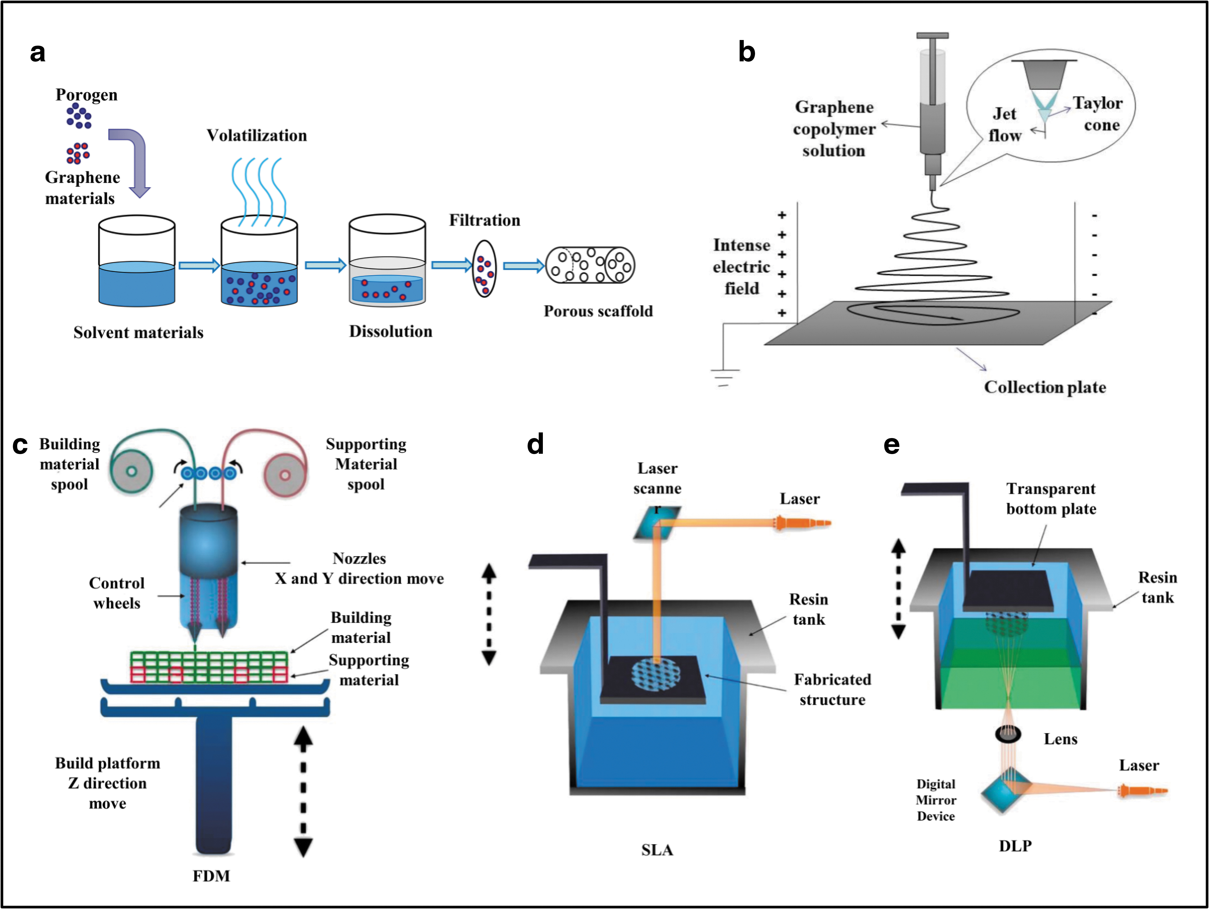
Preparation principle of graphene-based composite scaffolds.
Solvent casting technology
Solvent casting technology is one in which the porous cell scaffold with high porosity and controllable crystallinity can be obtained by evaporating and soaking the mixture of the raw materials of scaffolds (Fig. 3a) and suitable pore-forming agent with good solubility and volatility. The pore size, specific surface area, and other characteristics of graphene-based composite scaffold can be controlled by adjusting the content, morphology, and particle size of pore-forming agent, which affects the adhesion, proliferation, and differentiation of seed cells. 61
Some researchers prepared composite films doped with graphene based by improved solvent casting technology to induce tissue and organ regeneration. They found that the films with biological properties can effectively overcome the shortcomings of poor pore connectivity without reducing porosity.62,63 Mahdavi et al. 35 used this technology to prepare the gelatin-hydroxyapatite/GO biomimetic mineralized scaffold. The composite scaffold with porous morphology has stable mechanical properties and good connectivity, which provides rich nutrients for the adhesion and growth of adipose-derived stem cells in the scaffold and promotes the discharge of metabolites to induce damaged bone tissue to complete regeneration and repair.
Electrostatic spinning technology
Polymer solution or polymer fluid can overcome its surface tension in high-voltage electrostatic field to form a charged fiber jet (Taylor cone), which is pulled at high speed and evaporated by solvent, and finally solidified on the receiving plate to form nanofibers (Fig. 3b).64,65 Some researchers66–68 improved the production efficiency of electrospinning technology by optimizing electrospinning nozzle, changing electrostatic field force, and combining alternating current spinning and bubble electrospinning technology, which promotes the application of electrospinning technology in preparation of various tissue engineering scaffolds, such as cardiovascular and nerve.
Graphene composite electrospun scaffolds can improve the hydrophilicity and biological activity of scaffolds to promote cell adhesion and enhance the osteogenic performance of bone tissue engineering. 36 Meanwhile, the technical optimization of preparing graphene hybrid scaffolds by electrospinning also can promote the preparation of conductive scaffolds, which can provide relevant stimulation signals for the regeneration of nerve and myocardial tissues. Stone et al. 69 prepared rGO/polyesteramide conductive scaffolds, which effectively enhanced the growth and proliferation ability of myocardial cells.
Moreover, the surface-functionalized electrospun microfibrous scaffolds can be prepared depending on the modification of GO, these scaffolds can promote the selective adhesion of cancer cells, facilitate the capture of tumor fibroblasts, and achieve photothermal therapy according to the high photothermal conversion capacity of GO. 70
3D printing technology
Based on the digital simulation files of the target scaffolds, the computer is controlled to scan the data information; the biomaterials are printed layer by layer by light curing, paper lamination, and other technologies; and finally the scaffolds with different shapes and properties are manufactured.71,72 The 3D printing technology can be classified into fused deposition modelling (FDM); stereolithography (SLA); digital light processing (DLP) and so on. (Fig. 3c–e). 73 Compared with other preparation methods, the scaffolds prepared by this method have controllable size and shape, high porosity, and interconnectivity. Therefore, 3D printing technology can provide suitable scaffolds according to specific tissue engineering.74,75
Wang et al.42,43 provided a basis for the effective application of 3D printed graphene-based scaffold in bone tissue engineering. They found that 3D printed poly(ɛ-caprolactone)/graphene scaffolds have low immunogenicity and promote cell growth and bone deposition after being stimulated by microcurrent (10 μA), and effectively repair the skull defect of experimental rats. In addition, 3D printing technology can also be applied in cartilage tissue engineering. The GO scaffold prepared by 3D printing can promote the extension and maturity of new cartilage matrix in the scaffold and complete the construction of cartilage matrix without exogenous chondrogenic factors.76,77
Performance of Graphene-Based Composite Scaffolds
Graphene-based scaffolds have excellent mechanical properties and biocompatibility. On the premise of ensuring low immune and inflammatory response, they can induce cell directional differentiation by multiple mechanisms, so as to replace or repair damaged tissues and organs.
Mechanical properties
Scaffolds with high mechanical properties based on graphene-based materials have obvious advantages in tissue engineering applications. For one thing, they can improve Young's modulus and enhance the compressive and tensile strength of the scaffold itself.78,79 Jing et al. 52 confirmed that the increases of stress–strain capacity and tensile strength of the scaffolds are proportional to the content of GO (Fig. 4a, b). They found that the rupture resistance of PU/GO scaffolds with only 0.5 wt% (weight percent) GO meets the needs of human small vessel transplantation scaffolds. The preparation method, the addition of cellular matrix and the rationality of the scaffold porosity can also affect the mechanical properties of scaffolds.80,81
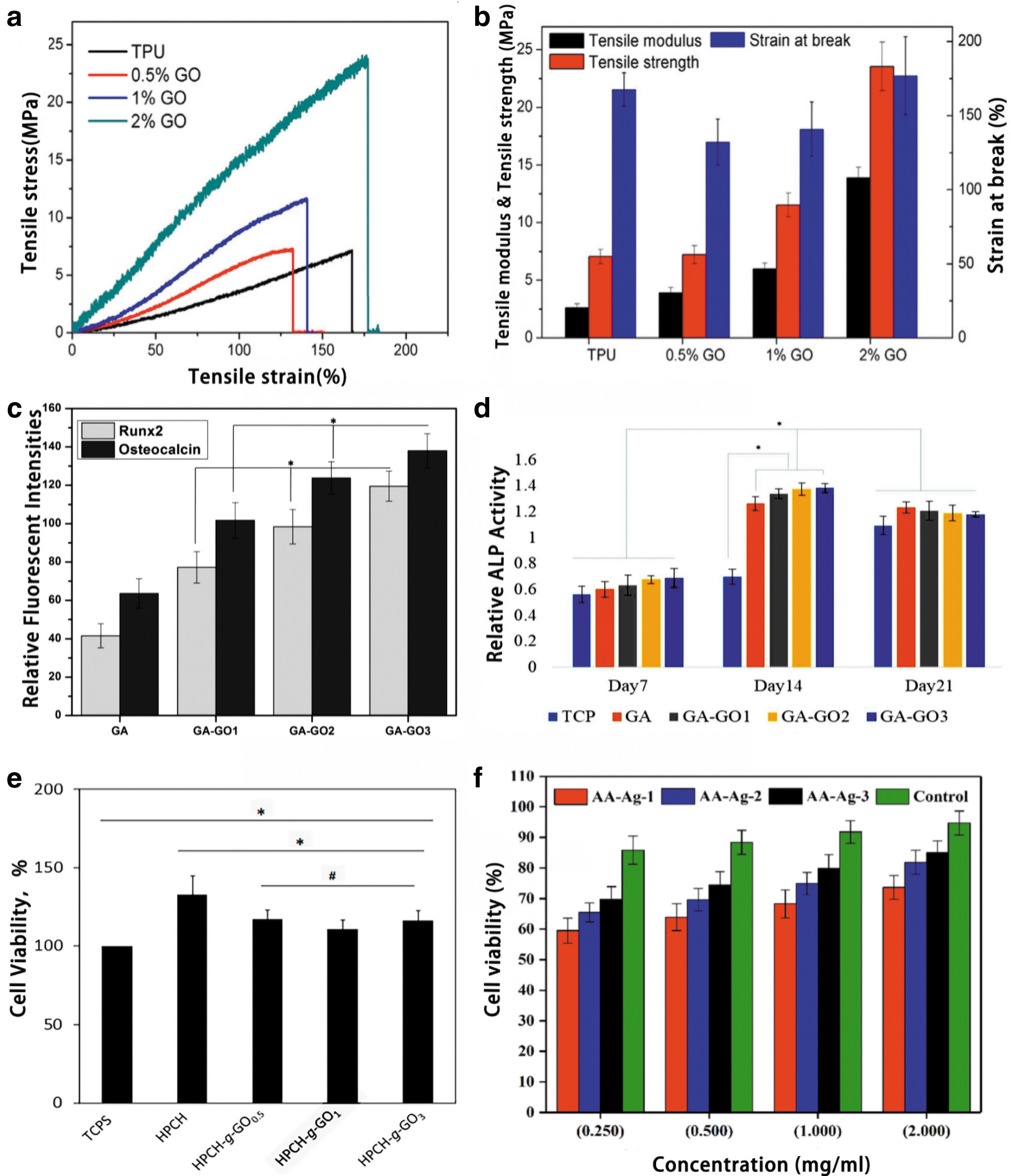
Study on the performance of graphene-based composite scaffolds.
For another, graphene-based scaffolds can enhance signal transmission in mechanotransduction pathway and induce directional differentiation of cells. 82 In tissue engineering represented by bone and cartilage, the seed cells in the scaffold convert the mechanical stimulation into electrochemical signals, and induce the migration of cell adhesion proteins, thereby regulating cell differentiation.83,84 The surface properties of graphene-based materials can also be used as a special mechanical stimulation to regulate the directional differentiation of cells. 85
Graphene-based materials can enhance the resistance of scaffolds to biochemical degradation and mechanical wear. Trucco et al. 86 prepared double-layer hydrogel scaffolds with high mechanical properties based on GO, which can simulate the structure of articular cartilage and effectively reduce the friction on the joint surface to enhance the wear resistance of the scaffolds. Soares et al. 87 confirmed that graphene coating material has high affinity for dentin, which can not only enhance the ability of resisting mechanical wear, but also protect dentin from chemical erosion.
Meantime, graphene-based scaffolds also have excellent antibacterial activity. While resisting stress wear of surrounding tissues, they can prevent biofilm formation by preventing microbial adhesion and killing microorganisms and promote the repair of infectious bone defects by enhancing the activity of alkaline phosphatase and the proliferation of stem cells. 88
Cell orientation
Mesenchymal stem cells (MSC) are a kind of seed cells with strong proliferation ability and multidirectional differentiation potential. Due to the relatively limited differentiation potential, they are more likely to differentiate into osteoblasts, chondrocytes, and nerve cells, which is of great significance for the repair of damaged tissues (Fig. 5). 89

Schematic diagram of proliferation and differentiation of MSC. MSC, mesenchymal stem cells. Color images are available online.
Tissue engineering scaffolds prepared by graphene-based composites can induce seed cells to differentiate into specific lineages through a variety of mechanisms. In addition to the use of cellular mechanotransduction pathways, seed cells can also be differentiated into specific cell types by regulating the cell microenvironment. 90 Extracellular matrix (ECM) is one of the components of microenvironment. The existence of cytokines, bioactive trace elements, and hormones can regulate cell targeted differentiation. 91 Purohit et al. 38 found that the levels of transcription factor Runx2 expressed by bone mesenchymal stem cells ( BMSCs) during osteogenesis, osteocalcin (ocn) expressed during matrix mineralization, and activity of alkaline phosphatase increased significantly with the increase of GO content in composite scaffolds (Fig. 4c, d).
In addition, graphene-based scaffolds can also restore the morphological and functional stability of damaged tissues and organs by promoting the migration of cell adhesion proteins and enhancing the conduction velocity of electrical signals. 78 Arnold et al. 41 found that the adhesion protein on the surface of GO scaffold can combine with integrin to regulate the osteogenic differentiation of stem cells by mediating intracellular and extracellular information transmission. Stone et al. 69 pointed out that the high electron transfer speed of graphene material increased the generation of action potential and enhanced the signal transmission during nerve and cardiovascular tissue. In addition, graphene materials have excellent angiogenesis characteristics, which can promote vascular differentiation of MSCs. 54
Biocompatibility
Graphene is prone to self-aggregation, and its cell damage mechanism mainly includes the following aspects: (1) Destroying the integrity of the cell membrane by cutting the cell membrane.92,93 (2) Mediating lysosomal/mitochondrial/endoplasmic reticulum-dependent apoptosis and necrosis to damage the metabolism and function of cells. 94 (3) Destroying the normal function of cells by causing DNA breakage and chromosome aberration. 95 (4) Oxidative stress leads to cytotoxic reactions such as lipid peroxidation, protein degeneration, and DNA oxidative damage.94,95 Meanwhile, the concentration, size, shape, and surface functionalization of graphene are also key factors that mediate cytotoxicity.96–98 In vitro cytotoxicity experiments, the doping of graphene-based materials within a certain content range did not cause damage to cells.
Sivashankari et al. 99 found by 3-(4,5-dimethyl-2-thiazolyl)-2,5-diphenyl-2H tetrazolium bromide (MTT) experiments that the viability of hybrid scaffolds doped with GO is lower compared with hydroxypropyl chitosan-grafted (HPCH), but compared with tissue culture polystyrene dish (TCPS), the cell viability is significantly improved (Fig. 4e). Khan et al. 100 pointed out that the increase of GO content can improve the cell density and vitality, the cell morphology is mature and the cell membrane is intact, and the expression of reactive oxygen species does not increase significantly (Fig. 4f).
Considering the clinical use of graphene-based materials in the future, their availability in vivo has become a research hotspot. In terms of biological distribution and degradation, graphene-based materials are very easy to have secondary distribution in the body and are mostly concentrated in the liver, spleen, and lung tissues enriched by mononuclear phagocytes.101,102 However, with the extension of time, graphene-based materials with small molecules can be metabolically degraded by the kidney and feces.101,102 Besides, through surface functionalization modification of graphene materials or depending on the degradation ability of peroxidases in organisms, the retention of materials in tissues can be reduced, and the occurrence probability of acute tissue injury (liver, lung, and kidney injury) and chronic pulmonary fibrosis can be further reduced.102,103
In terms of immune inflammatory response, graphene-based materials can induce the expression of inflammatory factors in vivo and reduce the clearance ability of immune cells to materials. 104 Surface modification is still a key factor to reduce immune inflammatory response, improving cell metabolism and proliferation activity. 105
Hung et al. 106 showed that doping nano gold in GO scaffold can reduce the secretion of inflammatory cytokines by reducing the polarization of M1 macrophages and inducing high expression of M2 macrophages, showing good immune compatibility and anti-inflammatory effect. Some studies 107 showed that curing CHT functionalized GO on the surface of magnesium alloy scaffolds can not only improve the corrosion resistance of the scaffolds, but also orderly regulate the immune inflammatory process induced by biological materials, and realize the proliferation of vascular endothelial cells.
Application of Graphene-Based Composite Scaffold in Tissue Engineering
Because graphene-based composite scaffolds have excellent performance, they have been widely investigated in many tissue engineering. The representative composition materials and research of graphene-based scaffolds in tissue engineering are shown in Table 1.
Bone tissue engineering
Graphene-based scaffolds not only have excellent mechanical properties required for bone tissue engineering, but also have good biological safety and ability to induce osteoblast differentiation, which can promote seed cells to complete proliferation and differentiation, and then replace or repair bone defects caused by trauma, tumor, infection, congenital malformation, and other factors. 19
Studies have shown that GO hybrid scaffolds such as GO/polylactic acid, GO/hydroxyapatite/silk fibroin, and GO/PCL/CHT/collagen can effectively promote the adhesion, proliferation, and bone deposition of bone tissue cells, and restore their normal tissue function.34,39,44 Biodegradable scaffolds are also attracting the attention of some researchers. Zhou et al. 40 prepared a kind of tissue scaffold for repairing a large area of bone injury by soft template method, and pointed out that the degradation of the scaffold can promote the rapid proliferation and spontaneous osteogenic differentiation of BMSCs to complete the repair.
In addition, graphene-based tissue engineering scaffolds can also induce stem cells to complete differentiation in the absence of growth-stimulating factors. Hermenean et al. 37 found that the expression of cell transcription factors Runx2, osteopontin (opn), and ocn in the hybrid scaffold containing GO was upregulated when studying the induction, differentiation, and repair ability of mouse skull defects. After scaffold implantation, new granulation tissue and fibrous blood vessels accumulated in the bone defect, and new bone tissue grew gradually from the periphery to the center. This confirmed that the application of graphene-based scaffolds in the early and late stages of osteogenesis could promote the proliferation and differentiation of cells and complete the repair of the bone defects (Fig. 6).

Application of graphene-based scaffold in bone tissue engineering.
Nerve tissue engineering
Graphene-based scaffold, as a bridge for proximal nerve stump regeneration axons to search for distal nerves, can simulate the composition of ECM to provide relevant stimulation signals for the regeneration of nerve tissue, and then induce differentiation and maturation of cell tissue. 108 They can promote the centralized release of neurotransmitter factors within synaptic vesicles to regulate the differentiation potential of seed cells. 109 Besides, it also supports the regeneration, development, and recovery of functional neurons by controlling the secretion of neurotrophic factors. 110
At present, aiming at cell functional defects caused by nervous system injury, graphene-based tissue engineering scaffolds can promote the regeneration and proliferation of Sertoli cells better than nerve conduits. 111 Yang et al. 45 found that GO/CHT composite scaffold can bridge the injured nerves to promote the growth and migration of nerve cells, whereas the degraded GO scaffold can further repair damaged spinal cord nerves. Heidari et al. 47 further suggested that the killing ability of the GO/PCL/gelatin nano-antibacterial conductive scaffold to Gram-positive/negative bacteria represented by Staphylococcus aureus and Escherichia coli was as high as 99%, which is beneficial to repair nerve tissue defects caused by stroke, spinal cord injury, or traumatic infection.
In addition, graphene-based scaffolds are also used to repair peripheral nerve injuries. Soleimani et al. 48 found that the concentration of graphene can promote the conductivity and cell viability of the scaffold, which can provide suitable environment and supporting growth factors for the regeneration of peripheral nerve tissue. Wang et al. 49 suggested that rGO-modified conductive nano-biomimetic scaffold can promote the proliferation and myelin formation of Schwann cells, induce myelin-specific gene expression and neurotrophic factor secretion, which is beneficial for the recovery of morphology and function of damaged peripheral nerve cells (Fig. 7).
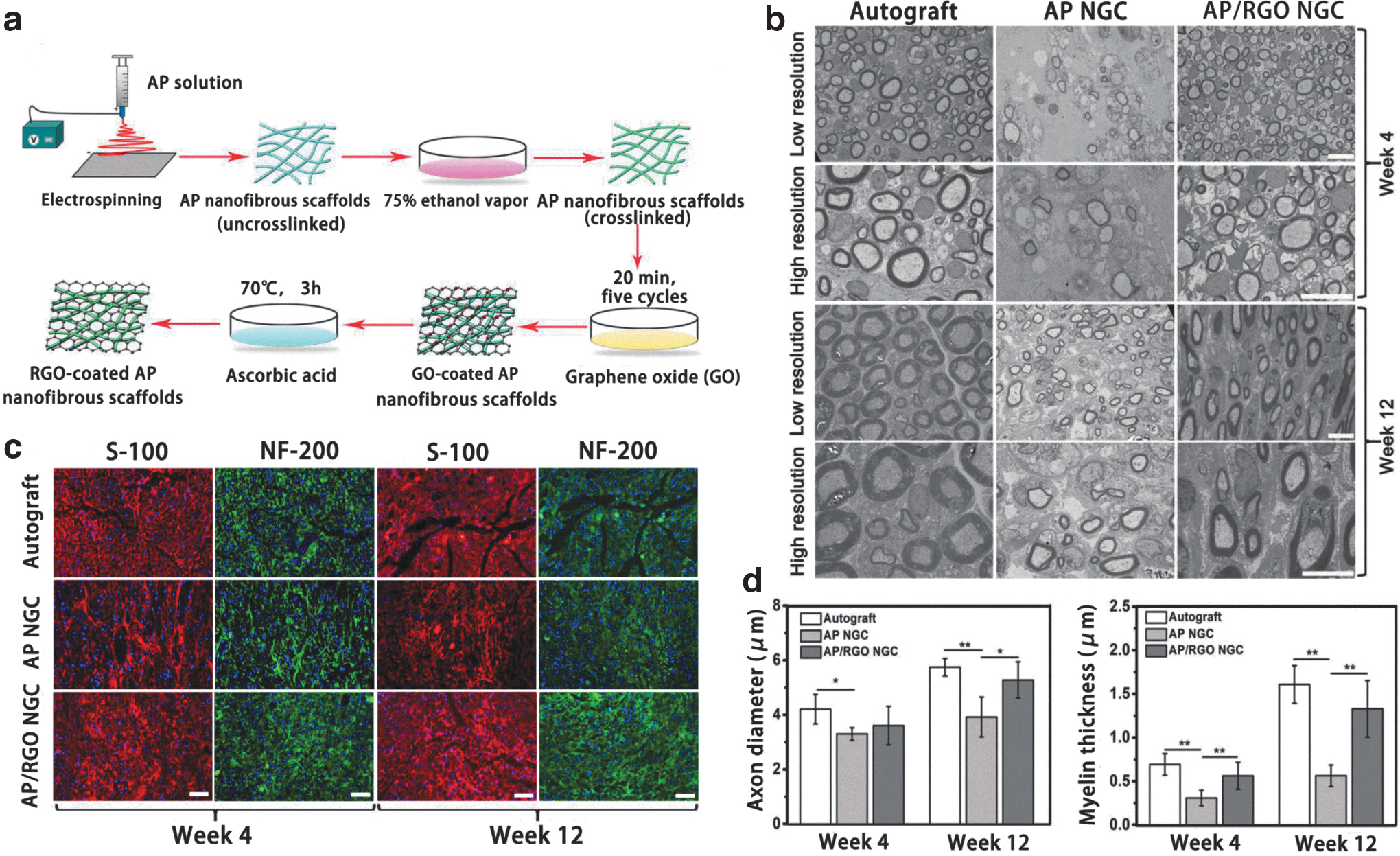
Application of graphene-based scaffold in nerve tissue engineering.
Cardiovascular tissue engineering
Depending on the electrical conductivity of graphene-based scaffolds, myogenic cells promote their proliferation and differentiation by upregulating the expression of cell–cell coupling protein and calcium-treated protein, increasing the action potential duration and calcium release peak under electrical stimulation, accelerating the autonomous pacing rate of cardiomyocytes, and ultimately repairing or replacing the damage caused by myocardial infarction. 36 The stable mechanical properties of graphene-based scaffolds also meet the stress changes caused by cardiac contraction and relaxation and can enhance the mechanical properties of myofibrils and sarcomeres. 55 Moreover, graphene and its derivatives can promote their applications in vascular tissue engineering by regulating the controlled level of reactive oxygen species in tissue cells and enhancing the expression of angiogenic factors. 112
Cardiac microenvironment has an important significance on the growth and functionalization of cardiomyocytes. Using tissue engineering scaffolds to simulate cell microenvironment has become an important way to treat cardiomyocyte injury. Jiang et al. 50 controlled the swelling rate, porosity, and electrical conductivity of the composite scaffolds by adjusting the mass ratio of CHT/GO, and upregulated the expression of cardiac troponin T (cTNT) and connexin-43 (Cx43) involved in the electrical signal transduction of cardiomyocytes to induce cell adhesion and proliferation. Hitscherich et al. 53 found that the expression of cTNT and Cx43 in mouse embryonic stem cells (MES-CM) was upregulated, and sarcomere fibers were evenly distributed in the scaffold (Fig. 8).

Application of graphene-based scaffold in cardiac tissue engineering.
Besides, based on the antibacterial properties of graphene, cardiac scaffolds with inhibitory effects on Escherichia coli, Staphylococcus aureus, and Streptococcus pyogenes were also developed to further promote the application of graphene-based scaffolds in cardiac tissue engineering. 51
Meantime, graphene materials have no obvious toxicity to vascular smooth muscle cells, and small flake GO is also conducive to the growth and proliferation of cells.113,114 Meng et al. 115 evaluated the blood compatibility of graphene films, and found that the existence of graphene can inhibit the adhesion and activation of platelets, so as to prolong the coagulation time and avoid the secondary thrombosis. Shin et al. 57 suggested GO cofunctional poly (lactide/glycolide, PLGA) nanofiber scaffolds, which have good thermal stability and hydrophilicity for promoting the adhesion and growth of vascular smooth muscle cells.
Other tissue engineering
Graphene-based scaffolds have a potential application in skin, muscle, and other tissue engineering. They can stimulate the growth of fibroblasts to promote the healing of wound skin, solving the problem of poor skin functionality caused by poor implantation, insufficient vascularization, and excessive scar repair.33,58 Graphene-based scaffolds are also used for the construction of muscle tissue engineering. Some researchers pointed out that they can induce myoblasts to differentiate into multinucleated myotubes, stimulate the adhesion and proliferation of myoblasts, and promote the spontaneous differentiation of myoblasts in the absence of muscle-stimulating factors.59,60
In addition, GO can be used for the development of photothermal therapeutic scaffolds to control the growth of tumor tissues based on its good photothermal conversion ability. 71 Zhang et al. 116 successfully prepared β-tricalcium phosphate bioceramic scaffold-modified FeO/GO nanoparticles with good thermal effect. The scaffold can induce osteosarcoma cell death rate as high as 75% and promote the proliferation and differentiation of normal bone marrow stromal cells, which is a 3D scaffold with dual functions of treatment and regeneration.
Conclusions and Prospect
In conclusion, graphene-based material is a good scaffold material. It can be combined with natural/synthetic biomaterials according to their large specific surface area. Tissue engineering scaffolds prepared by solvent casting, electrospinning, and 3D printing can provide suitable microenvironment for seed cell adhesion, proliferation, and differentiation, and are beneficial for the repair of repairing damaged tissues and organs.
The high mechanical property scaffolds prepared with graphene-doped materials can not only enhance the compressive/tensile strength and wear resistance of the scaffolds, but also convert the mechanical stimulation to electrochemical signals through the mechanotransduction pathway to induce cell differentiation. Besides, cell differentiation can also be regulated by promoting the migration of cell adhesion proteins, enhancing the conduction of electrical signals, and regulating the microenvironment of cell growth. Meantime, the biological safety of graphene materials has always been the focus of tissue engineering research.
Graphene materials can cause toxic reactions by damaging the integrity of the cell membrane, mediating apoptosis and necrosis, and oxidative stress occurring, resulting in cell metabolism and functional damage. However, within a certain concentration range, the increase of graphene material content will not cause damage to cells, and small molecular materials can undergo quick metabolic degradation through kidney and feces. Meanwhile, surface functionalization of graphene materials is still a key factor to reduce its immune inflammatory response, improve cell metabolism, and proliferation activity. Therefore, graphene materials have potential application prospects in different tissue engineering fields.
However, the toxicity of multimaterial composite scaffolds prepared based on graphene has not been fundamentally solved, and their long-term damage is not clear. Improving long-term biocompatibility is still an urgent problem to be solved. Meanwhile, the optimization of scaffold preparation technology, the improvement of performance, graphene-based material-induced cell growth, and their specific application advantages in tissue engineering are still the focus of continuous attention. In the future, it is necessary to further explore the preparation method, safety research, and the mechanism of inducing cell directional differentiation of graphene-based composite scaffolds, so as to promote the research and development of carbon-based materials represented by graphene in tissue engineering scaffolds.
Footnotes
Authors' Contributions
Conceptualization, all authors; investigation, W.X., J.D., Q.L., Y.W., and Y.L.; writing—original draft preparation, W.X.; writing—review and editing, J.D., Q.L., Y.L., and J.F.; supervision, S.Y. and Y.Y.; project administration and funding acquisition, S.Y. All authors have read and agreed to the published version of the article.
Disclosure Statement
No competing financial interests exist.
Funding InformationFunding statement:
This research was funded by the Science and Technology Innovation Project of Excellent Talents in Shanxi Province of China (201805D211001) and Central Leading Local Science and Technology Development Fund Project of China (YDZX20201400001722).