




Abstract
There are different types of skin diseases due to chronic injuries that impede the natural healing process of the skin. Tissue engineering has focused on the development of bioengineered skin or skin substitutes that cover the wound, providing the necessary care to restore the functionality of injured skin. There are two types of substitutes: acellular skin substitutes, which offer a low response to the body, and cellular skin substitutes (CSSs), which incorporate living cells and appear as a great alternative in the treatment of skin injuries due to their greater interaction and integration with the rest of the body. For the development of a CSS, it is necessary to select the most suitable biomaterials, cell components, and methodology of biofabrication for the wound to be treated. Moreover, these CSSs are immature substitutes that must undergo a maturing process in specific bioreactors, guaranteeing their functionality. The bioreactor simulates the natural state of maturation of the skin by controlling parameters such as temperature, pressure, or humidity, allowing a homogeneous maturation of the CSSs in an aseptic environment. The use of bioreactors not only contributes to the maturation of the CSSs but also offers a new way of obtaining large sections of skin substitutes or natural skin from small portions acquired from the patient, donor, or substitute. Based on the innovation of this technology and the need to develop efficient CSSs, this work offers an update on bioreactor technology in the field of skin regeneration.
Impact Statement
The manufacture of functional cellular skin substitutes (CSSs) is one of the current goals in the field of tissue engineering to improve the treatment of chronic skin injuries, thus favoring skin repair and regeneration. The main advances in the development of innovative and effective CSSs are largely focused on the selection of more adequate cellular components, biomaterials, and biofabrication techniques to be used in their biofabrication. However, the maturation of CSSs should be an essential step in obtaining a functional substitute capable of replacing the native skin. The sequential procedure from the design of the CSS to its maturation process will be reviewed. In the context of the manufacturing of novel CSSs, different technologies to biofabricate functional structures and how their maturation can be carried out by specific devices are addressed, as well as key challenges facing the design and development of CSSs.
Color images are available online.

Introduction
The skin is the largest organ of the body and comprises 15% of its entire weight. It is composed of three layers: epidermis, dermis, and hypodermis. The epidermis is the outermost layer and is responsible for the color, texture, and moisture of the skin. Its main cell type is keratinocytes, which, when mature, form four stratified layers: the stratum corneum, the granular layer, the spinous layer, and the basal layer. 1 The epidermis is separated from the underlying layer, the dermis, by the basal membrane, a highly specialized extracellular matrix (ECM) that holds the two layers together and acts as a diffusion barrier allowing the exchange of cells and fluids to maintain tissue homeostasis.
On the contrary, the dermis is divided into the papillary layer and the reticular layer, and its main function is to support the epidermis. Fibroblasts are the cells that primarily make up the dermis, and they are primarily responsible for synthesizing and remodeling the ECM. In addition, the dermis plays other roles, such as protecting deeper layers of the skin, aiding in sensation or assisting in thermoregulation, due to the presence of structures such as nerve endings, hair follicles, glands, or blood vessels. 2 Finally, the hypodermis is composed of superficial fascia and subcutaneous fat. Adipocytes are the main cell types present in the hypodermis. Their main functions are to conserve body temperature and provide mobility to the entire skin.
The skin plays an important role as an immune barrier in defending against external pathogens and as a fundamental regulator in the maintenance of homeostasis. 3 When an injury occurs, the human body orchestrates a series of cellular, humoral, and molecular events organized in four phases 4 : hemostatic, inflammatory, proliferative, and remodeling. However, chronic skin injuries, such as those caused by common or hereditary pathologies (e.g., diabetic ulcers), sacrococcygeal fistulas, or thermal trauma, such as burns, involve the alteration of the histological conformation of the skin causing the loss of its functions and causing nonhealing chronic wounds. 5
Wherefore, the restoration of skin functionality after chronic skin injuries requires specific medical care, such as (1) elimination of necrotic areas, (2) removal of excess fluid from the wound, (3) improving circulation and increasing the speed of the healing process, and (4) removing the wound infection. 6 Therefore, the treatment of these types of injuries includes cleaning and disinfecting the wound, applying antibiotics for the elimination of possible bacteria, and promoting better circulation. 7 The most frequent treatments for skin regeneration in these types of injuries are based on covering the wound with dressings, compression bandages, or skin grafts. 8 All of them have limitations of use, such as failing to provide a moist wound environment, frequent dressing changes, or the limited healthy skin areas that can be removed for the skin graft.
Therefore, it is necessary to design and develop new treatments as an alternative to these procedures. Thus, tissue engineering (TE) has focused on the development of bioengineered skin or biocompatible skin substitutes. 9 There are two types of skin substitutes, 10 acellular skin substitutes (ASSs) manufactured from biomaterials and cellular skin substitutes (CSSs), which incorporate living cells. ASSs act by protecting the wound, encouraging its regeneration, or preparing the area for subsequent grafting. 11 In contrast, the incorporation of living cells into CSSs promotes a more natural healing process through the secretion of growth factors and components of the ECM. This activity improves the relationship between the CSS and the wound, favoring the curing process and decreasing the healing time. 12
The design and development of CSSs involve the manufacture of complex structures, such as meshes, membranes, hydrogels, porous solid scaffolds, and even hybrid constructs that combine hydrogels with porous scaffolds. 13 There are a wide range of techniques from which it is possible to design the most suitable CSSs for each type of wound such as crosslinking of hydrogels, lyophilization, gas foaming, and more advanced techniques such as electrospinning or three-dimensional (3D) bioprinting.
CSSs are biofabricated from biomaterials loaded with living cells. The evolution in the field of cell therapy has allowed the incorporation of different cell types into CSSs, obtaining substitutes with similar cell and biological components to those that constitute the dermis and epidermis. All of them stimulate the growth and proliferation of fibroblasts, to have greater control of ECM hydration and improved regulation of inflammation 14 through the modulation of the immune system, increasing the recruitment and binding of macrophages and the release of cytokines. 15
However, the CSSs can present a high degree of immaturity, caused by the inactivity of the cellular components, which implies that the healing of the wound cannot occur after their grafting on the patient. 16 Immature CSSs are cell-laden scaffolds or constructs that can temporarily supply functions of the skin such as the immunological barrier or homeostasis regulator role, but they are not functional tissues, 17 as the cells have not been subjected to the same chemical and mechanical stimuli that occur in their native state in the body. Therefore, the use of immature CSSs presents numerous limitations such as low vascularization, integration failure, scarring, and immune rejection. 18
Thus, the development of mature CSSs acting like functional tissue is a major advance in the area of dermal substitutes, taking a further step toward the goal of designing and developing CSSs that mimic native skin. Thanks to the cell activation of immature CSSs, a series of events similar to the remodeling phase are triggered, such as the formation of the basal lamina and cell differentiation, thus promoting wound healing and scarring. 19 With the intention of manufacturing mature CSSs, the use of bioreactors is emerging since these devices favor the acquiring of a structure similar to the native skin by the CSSs, 20 considering them to be more effective in the treatment of skin injuries. 21
The term bioreactor appeared for the first time in the field of biotechnology, where it was defined as an optimized system that allows the manufacture of biological products on a large scale. 22 TE has taken bioreactor technology one step further, turning these devices into optimized systems capable of recreating any bioprocess, 23 such as providing ideal physiological conditions for the cultivation or maturation of natural or bioengineered tissues. 24
In recent years, bioreactor technology involved in the skin field has improved, incorporating innovative designs that can reduce the maturation time of CSSs and offer a new alternative for cases that require immediate treatment. In this way, bioreactors are postulated as an effective methodology for obtaining large segments of mature and bioengineered skin like CSSs suitable for their use as grafts for patients who have suffered chronic wounds, allowing their healing. 25
This study aims to outline the current advances in the design and development of functional CSSs. The characteristics of biomaterials, cellular components, biofabrication methodologies, and the use of bioreactors in the maturation process of CSSs are described. In addition, this work provides an overview and comparison between the main types of skin bioreactors.
Evolution in Manufacturing of CSSs by TE
Skin injuries can cause deep chronic trauma or full-thickness skin wounds that are very complex to treat due to the high risk of infection, the complexity of the treated area, or the potential for damage to the skin layers. 26 The treatment per excellence usually used in clinical practice for this type of injury is the use of autografts, 27 which involves transplanting grafts from another part of the patient's own body. The use of autografts prevents the problem of host/donor immune rejection, but this technique is highly limited due to the lack of healthy skin areas possessed by the patient after the trauma. 28 In addition, it is an invasive process, which causes a new wound to the patient, prolonging recovery time. 29 To remedy the main drawbacks of autografts, allogeneic and xenogeneic techniques namely allografts and xenografts, respectively, have been developed.30,31
Allotransplantation is focused on the use of allografts, which are obtained from healthy donors or cadaveric skin. Although allotransplant of skin is used today to treat chronic wounds, 32 the long-term durability of transplanted skin is very limited due to the immune rejection by the patient. Therefore, the main sources of innovation in this area are focused on finding a solution that blocks the autoimmune response. 33 On the contrary, it is possible to use skin xenografts from animals such as tilapia 34 or pigs. 35 This type of xenotransplantation offers an inexhaustible source of grafts for the treatment of skin wounds. 36 However, they are used less and less nowadays due to their short viability after the implant. 37 In addition, both allotransplants and xenotransplants present a risk of disease transmission. 38
In response to these limitations, TE has been focused on developing ASSs and CSSs that present a therapeutic alternative for wound regeneration. 39 Both types of substitutes must be biocompatible and offer a structure with one or more resistant layers that replace the functions of the natural skin in the wound. 40 ASSs emerged to restore the functions of damaged skin, such as preventing the loss of liquids or serving as a protective agent.41–43 Most of them are manufactured from biomaterials such as collagen, silicone, nylon, decellularized human dermal matrix,44–47 and even animal derivatives (i.e., porcine ECM, aldehyde crosslinked porcine dermis, or porcine acellular lyophilized small intestine submucosa).48,49
There are several commercialized ASSs such as Integra® (consisting of bovine collagen and shark chondroitin-6-sulfate),50,51 Alloderm™ (acellular dermal matrix derived from human cadaveric skin, collagen, elastin, proteoglycans, and vascular plexus), 52 or Matriderm® (matrix of bovine type I collagen and elastin),53,54 and each of them has a preferential use depending on the lesion or affected area. 55 Thus, Integra is used to treat pathologies such as chronic wounds and scar contractures, whereas Alloderm has been used for laryngoplasty or vaginal prolapse repair, and Matriderm is indicated for the treatment of deep dermal defects.
However, ASSs are very limited due to their low barrier function and short-term viability. In addition, some of the classes of substitutes such as Integra or Matriderm require several interventions in a row, which signifies an increased risk of wound infection.56,57 On the contrary, the use of animal derivatives and cadaveric skin entails the risk of transmitting infectious diseases. 58
To mitigate these disadvantages, the living cells were included in the development of CSSs with two-dimensional or 3D structures, promoting the natural regeneration of the skin through the secretion of growth factors and the formation of the ECM. 59 Hence, CSSs replace or restore the functions of the natural skin. It is vital in their production to use biomaterials that allow maintaining viable cells within the CSSs. 60 In this regard, new products have been developed, such as Dermagraft®, which is based on the combination of neonatal foreskin fibroblasts 61 embedded in a biodegradable matrix of polyglycolic acid (PGA) to provide a more natural process of skin regeneration and reduce the possibility of rejection. 62
The evolution of CSSs has allowed the manufacture of new products that incorporate different cell types into their structure, for example, TISSUEtech Autograft System™, which comprises autologous fibroblast and keratinocyte seeding in a hyaluronan scaffold, attempting to replicate the natural histology of human skin more accurately. This product has demonstrated positive results in the treatment of diabetic ulcers and pressure wounds in ≤1 year. 63
Biofabrication of Functional CSSs
The evolution and perfection in the manufacture of innovative CSSs are currently widely studied. The main objective is focused on the design and development of functional CSSs that imitate native skin and achieve better control of the healing process. 64 For this, it is necessary to determine the structure of the substitute most adequate for each specific wound, and select the different biomaterials and cellular components that will form the CSS. In addition, its process of manufacturing should be defined, selecting the most adequate biofabrication and maturation methodologies (Fig. 1).

Main steps in the development of a CSS. CSS, cellular skin substitute. Color images are available online.
Selection of the conformational structure
A skin wound is diagnosed as a chronic injury when it is unable to follow the normal stages of the skin repair process or gets stuck in one of them (usually inflammation). 65 Chronic wounds do not have specific dimensions, and they can have a surface area of a few square centimeters (such as ulcers) to much larger surfaces (such as burns). In addition, they can be a cut that has only affected the epidermis, or deeper cuts that have reached the hypodermis. For the treatment of these types of wounds with a CSS, it is necessary to first define the dimensions and depth of the injury 66 to determine the specific structure of the substitute to be biofabricated.
Currently, CSSs are usually manufactured from (1) hydrogels (more biocompatible and viscoelastic structures) or (2) solid scaffolds (less biocompatible rigid structures with more skin-like mechanical properties). Hydrogels are presented as highly hydrated matrices with an intricate fibrillar network, closely mimicking the structure of the native EMC. 67 They are hydrophilic structures of biopolymeric networks or protein fibrils intertwined within a hydrated network due to crosslinking. 68 Hydrogels are more suitable for the treatment of wounds that will not be subjected to constant and high stress. 69
On the contrary, porous solid scaffolds are presented as structures made up of solid fibers, such as mesh or nanofibrous scaffolds. Their porosities facilitate the migration of cells within the construct, promoting tissue growth, 70 and these fibrous structures are very similar to the physical characteristic of the ECM, resulting in a biocompatible environment where cells can grow and perform their functions. 71 Although solid porous scaffolds are not widely used, their mechanical properties may make them more suitable for the treatment of high-stress wounds. 72
Selection of biomaterials
Biomaterials used in the design and development of CSSs must be biocompatible, biodegradable, strong, durable, and ductile. 73 They are generally classified as either natural biomaterials or synthetic biomaterials (Table 1).
Advantages and Disadvantages of the Different Biomaterials Used in Cellular Skin Substitutes
CSS, cellular skin substitute; PGA, polyglycolic acid; PCL, polycaprolactone; PEG, polyethylene glycol; PLA, polylactic acid; PLGA, poly lactic-co-glycolic acid; RGD, Arginine-Glycine-Aspartate.
Natural biomaterials provide a CSS that mimics the ECM, offering molecules that interact with the cellular component. 74 An example is collagen, which is characterized by tripeptide Arginine-Glycine-Aspartate (RGD) sequences that are important in keratinocyte attachment and wound healing. 75 Also, gelatin (denatured from collagen) has been shown to have lower antigenicity than collagen, 76 making it ideal for applications where the wound requires a long healing process with a high risk of infection.
Natural biomaterials not only offer an adequate interaction with the cellular component but also offer exceptional antibacterial and anti-inflammatory properties 77 that promote and accelerate the healing process. For example, chitosan has been shown to activate hemostasis, 78 while fibrinogen has been shown to interact with vital factors in the healing process such as fibronectin, growth factors, and protease inhibitors. 79 Also, hyaluronic acid promotes the healing process due to its ability to increase fibroblast and keratinocyte proliferation. 80
On the contrary, synthetic biomaterials allow the development of CSSs with biomechanical properties similar to those of native skin that are not available with natural biomaterials. 81 Polyethylene glycol has already been used in the manufacture of CSSs to improve the structural and compositional properties of the substitute. 82 There are also other synthetic biomaterials such as poly lactic-co-glycolic acid, polylactic acid, and PGA or polycaprolactone, which provide the CSS with excellent biomechanical properties,83–85 produce an increase in cell proliferation and wound healing due to degradation of the polymer surface. 86
In summary, natural biomaterials do not offer optimal biomechanical properties, while synthetic biomaterials have a low tissue cell response. It is suggested that a single biomaterial is not able to mimic the properties of the native skin. 87 Therefore, for the development of CSSs, it is necessary to combine different types of biomaterials 88 (both natural and synthetic) to biofabricate a hybrid scaffold. Thus, natural biomaterials can be used to formulate hydrogels, which will be loaded with living cells, and synthetic biomaterials will then be incorporated to improve the mechanical properties of CSSs or to be used as a sacrificial support material.
Synthetic biomaterials used as sacrificial supports allow biofabrication of high-resolution 3D soft structures (like hydrogels), acting as a mold of the CSSs and holding the cells together. 89 After the biofabrication of the CSS, the sacrificial support remains in the structure until the maturation and even implantation stages, after which it is usually discarded. Therefore, it is important to know the characteristics of the wound and then, select the most adequate substitute structure for its treatment, the most appropriate biomaterials to biofabricate a CSS with rheological and physicochemical characteristics that best suit the wound, and the most appropriate cellular component.
Selection of the appropriate cell type
There are a wide variety of cell types involved in the wound healing process, 90 which can be included in the biofabrication of a CSS to provide growth factors and cytokines, provisional matrix protein, and the capability of responding to environmental conditions, improving wound healing and wound treatment. 91 Currently, the most used cells are differentiated adult cells such as fibroblasts, keratinocytes, and endothelial cells, and stem cells such as epidermal stem cells (ESCs) or mesenchymal stromal cells (MSCs). Each of these cells can act throughout the four phases of wound healing (Table 2). For example, fibroblasts and MSCs act in the hemostasis phase synthesizing and organizing the ECM, which leads to platelet plug formation.92,93 The formation of the platelet plug allows the release of factors such as platelet-derived growth factor (PDGF) or epidermal growth factor, which are important mediators of later stages of healing, as well as providing a temporary matrix for the infiltration of other cell types. 94 On the contrary, keratinocytes act in the inflammatory phase activating macrophages and neutrophils, while MSCs direct the activity of M2 macrophages.95,96 Macrophages and neutrophils are cells of the immune system whose main function is to destroy and phagocytose foreign bodies that enter the wound. 97 Endothelial cells are primarily involved in the process of angiogenesis. The formation of new vessels in the wound is essential for the delivery of nutrients and oxygen necessary for cellular function. 98
Cell Type Involved in the Wound Healing Process
ECM, extracellular matrix.
During angiogenesis, the activity of keratinocytes is also important as keratinocytes increase the permeability of blood vessels allowing the flow of molecules (such as oxygen) out of the vessel. 99 Finally, keratinocytes are also mainly responsible for the re-epithelialization process, 100 which is regulated by growth factors and cytokines synthesized by fibroblasts. 101 During the same re-epithelialization process, the activity of ESCs is also crucial. ESCs migrate from different locations in the epidermis (such as the hair follicle) to the wound, where they can be differentiated into keratinocytes.102,103
In recent years, CSSs have been manufactured by combining the use of fibroblasts and keratinocytes, due to the critical interaction of both cell types during the wound healing process. On the one hand, keratinocytes secrete interleukins and cytokines that promote the production of keratinocyte growth factor by dermal fibroblasts. 104 This mechanism is essential for keratinocyte proliferation during the re-epithelialization stage. In addition, keratinocytes secrete proangiogenic molecules such as PDGF that promotes fibroblast proliferation during the proliferative stage. This keratinocyte–fibroblast interaction is evidence of the joint use of both cell types in CSS structures promoting skin regeneration.
Biofabrication methodologies
The selection of the methodology to biofabricate a CSS will depend on the final structure of the substitute required. The biopolymer crosslinking methodology is used to obtain hydrogels, while other methodologies such as electrospinning or lyophilization are used to obtain solid porous scaffolds. Using these techniques, both hydrogels and porous solid scaffolds can be manufactured in sheets (monolayer) as dressings, membranes, or meshes to treat more superficial wounds, or as bilayer/trilayer structures to treat deeper wounds. 105
Hydrogels are the most used forms to manufacture CSSs. There are different ways to achieve crosslinking of these biopolymers, such as physical crosslinking, chemical crosslinking, or ionic interactions. 106 However, the crosslinking process carried out by these strategies is often cytotoxic. 107 In contrast to these crosslinking techniques, enzymatic crosslinking of hydrogels uses enzymes such as transglutaminase or peroxidase, which prevents the formation of cytotoxic residues. 108 This not only provides a noncytotoxic hydrogel but also allows them to be used as hydrogels that can be applied directly to the wound. 109 These injectable or in situ hydrogels have a great advantage since when injected before gelling, can cover all the irregularities of the wound. 110
Regarding porous solid scaffolds, there are different traditional methods used for their manufacture, such as gas foaming, lyophilization, micropattering and micromolding, electrospinning, or bioprinting.111–113
Electrospinning has proven to be a useful technique in the manufacturing process of skin substitutes, allowing robust, lightweight, and customized scaffolds to be obtained from the extrusion of biopolymeric nanofibers. 114 Another commonly used technique is lyophilization, which allows the dehydration of molds of structures, resulting in rigid biocompatible scaffolds suitable for culturing skin cells. 115 However, it is the 3D bioprinting technique that has recently taken the biofabrication of CSSs a step further, as it shows a significantly greater control over the architecture of the structures obtained, as well as offering high reproducibility thanks to the automation of the method.116,117
3D bioprinting allows the biofabrication of hydrogel-based scaffolds, solid porous scaffolds, and hybrid constructs that mix porous solid scaffolds with hydrogel structures. In this regard, some print heads of the bioprinter may be loaded with the biomaterial that forms the porous solid scaffold (usually synthetic materials), and other print heads are loaded with the hydrogel solution in which the cells are embedded (and which will fill the solid scaffold). 118 There are different bioprinting methodologies available: extrusion bioprinting, droplet/inkjet bioprinting, and laser-assisted bioprinting. Each methodology has advantages or disadvantages that make them ideal for different CSS biofabrication. 119
Finally, there are different strategies to incorporate the cellular components into the CSS, with the main challenge of maintaining their cell viability after biofabrication 120 : (1) immersing the acellular substitute in a solution that contains cells and growth medium (e.g., in hydrogels and solid porous scaffold, allowing the cells to migrate into the construct) 121 ; (2) growing the cells on a surface (such as a well) that is in contact with the substitute. In this way, the cells are expected to migrate and adhere to the surface of the construct (mainly used in membranes) 122 ; and (3) injection of a cell solution to biofabricate directly in the structure (either manually or automatically using a bioprinter). 123
CSS functionalization
After the CSS biofabrication process, a structure similar to the native skin is obtained, but it has low functionality and is therefore considered an immature structure. This is because the regulation of skin functions depends on extracellular signals that are interpreted by the cells, promoting or restricting proliferation, migration, differentiation, ECM remodeling, or tissue organization. 124 By performing the whole “in-vitro” manufacturing process, CSS cells lack all these extracellular signals such as some growth factors, intercellular interactions, or environmental stress, and/or mechanical stimuli that activate and regulate their behavior. 125
In addition, an immature CSS is characterized by having an ECM with a much lower degree of assembly than the natural ECM, not being able to withstand the tensile forces of fibroblasts and generating a different architecture of collagen fibers in the substitute. 126 Thus, immature CSSs have very different biomechanical properties from natural skin that compromise their fundamental functions such as transepidermal water retention. 127 Furthermore, although the CSS can be manufactured in two layers mimicking native skin, it lacks fundamental internal structures such as the basal lamina or stratum corneum. 128
The basal lamina holds the dermis and epidermis tightly together, preventing detachment of the layers after implantation. In addition, the basal lamina is involved in controlling the organization of keratinocytes in the epidermis, so its absence could lead to a disordered arrangement of cells within the substitute after implantation. 129 Furthermore, the absence of the stratum corneum in the substitute increases the vulnerability of the wound to bacteria and reduces the hemostatic properties of the skin. 130
Therefore, immature CSSs are unable to cover the function of the native skin, making it impossible to reactivate the natural healing mechanism in chronic wounds and increasing the chances of graft detachment. 131
To regain cellular functionality, CSSs undergo an “in vitro” maturation process in a bioreactor. This process consists of exposing the CSSs to an environment similar to that which occurs during the maturation stage of wound healing. During the maturation phase, remodeling of the ECM occurs, replacing the abundant type III collagen of the platelet plug with type I collagen. The type I collagen fibers align in the direction of forces increasing the tensile strength. In turn, fibroblasts pull through the ECM to completely close the wound, while the new epidermis stratifies to form the stratum corneum. 132
Bioreactor technology is usually used for this purpose. Bioreactors allow the replication of biological processes by applying different stimuli within an aseptic and controlled environment. In this way, it is possible to subject the cells of a CSS to the extracellular signals of which they have been deprived during the biofabrication, promoting cell maturation and recovery of functionality. 133
During the maturation process in the bioreactor, several changes in the immature CSSs occur that result in mature CSSs (Fig. 2): (1) stratification of the epithelium layer and fibroblast proliferation; (2) fibroblasts are activated, increasing the synthesis of type I and IV collagen; (3) some proteins (i.e., type IV collagen) interact with proteoglycans (i.e., perlecan) and glycoproteins (i.e., laminin or entactin) to form the basal lamina; and (4) packing and arrangement of collagen fibers parallel to the surface, increasing resistance to stress. These changes greatly increase the ability of the CSS to reactivate the wound regeneration process, in addition to the chances of success of the substitute implant.134,135

Evolution of the maturation of a CSS:
Design and Functions of Bioreactors
A bioreactor designed for TE must manage the in vivo tissue architecture, the cell–ECM and cell–cell interactions, and finally, cell viability. The bioprocesses replicated in a TE bioreactor can be very different, varying from the evolution of disease to the behaviors of a drug.136,137 More specifically, a TE graft could be matured ex vivo by a bioreactor if it could mimic the native tissue microenvironment. 138 To ensure the viability or maturation of a TE construct, a bioreactor has (Fig. 3) (1) a chamber, where the optimal conditions for the tissue are maintained; (2) support, which allows the stability of the tissue or organ; (3) a control unit (it could be a computer or a microcontroller); and (4) input and output systems that maintain the renewal of the culture medium. 139
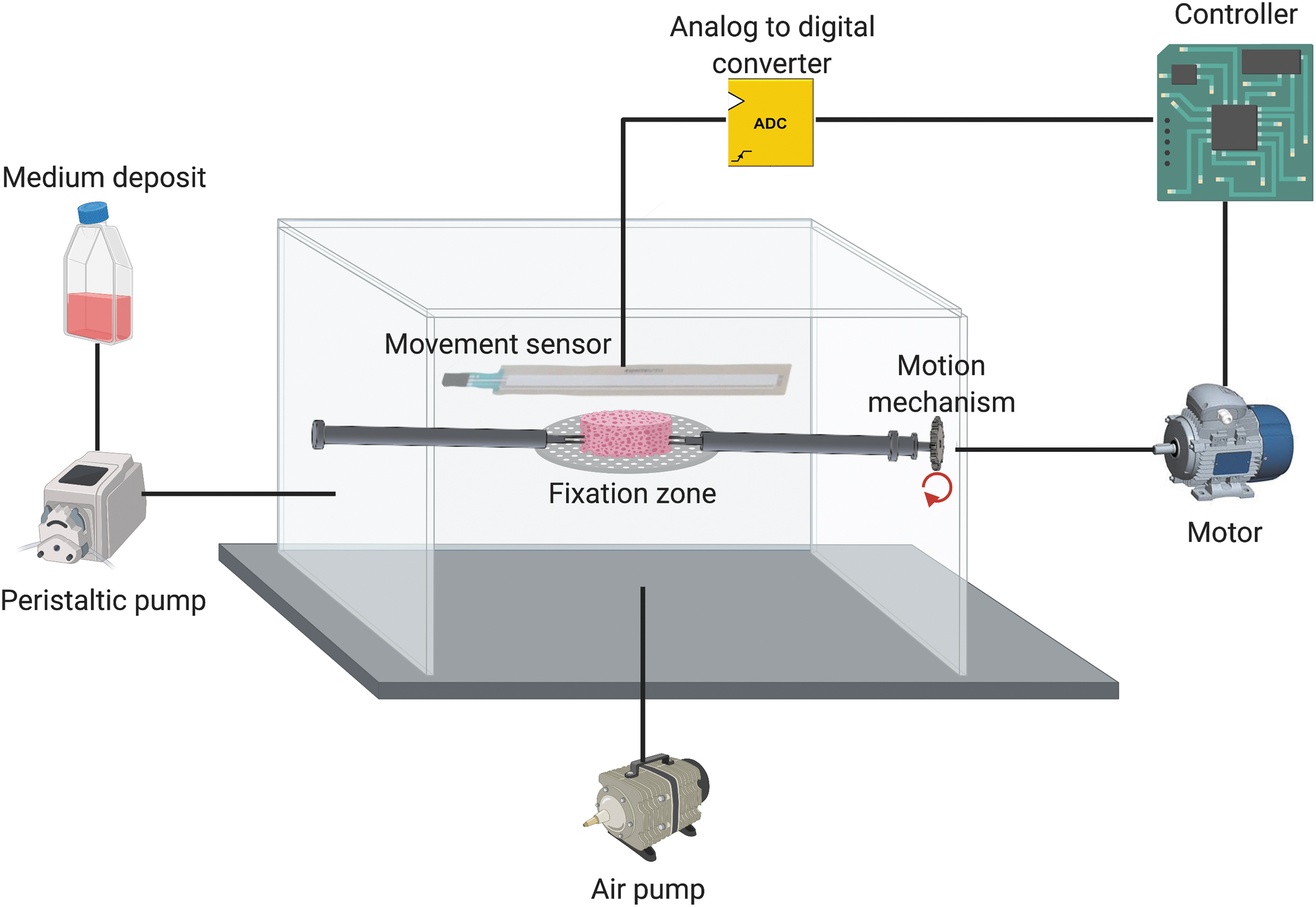
Prototype of the circuit of a bioreactor with media inputs by peristaltic/air pump and application of a mechanical stimulus controlled by a movement sensor. Color images are available online.
However, depending on the design objective, other components can be included. For example, if the design requires a reading of various stimuli, it is normal to have different types of sensors (such as movement, force, load, or temperature sensors). These sensors are connected with analog/digital converters to transform the signal into binary values (0/1). In this way, a digital signal is sent to processors that translate the signal thanks to a previous analysis or analytical theorems. Further, if the design objective is to apply a stimulus inside the bioreactor, motors are usually used together with movement mechanisms (such as gears, shafts, cams...), to convert electrical energy into the desired stimulus.
In addition, bioreactors can incorporate surveillance instruments, that is, cameras or microscopes, 140 which, together with image processors, can evaluate the state of the tissue/organ and provide relevant data on their development process.
A TE bioreactor should consider in vivo tissue/organ structure, the cellular organization within the scaffold, and cell viability. 141 Based on the variability in the design of these devices, together with their high rate of adaptability to new technologies, their functions can become almost unlimited. Thus, a bioreactor can play the role of (1) maintaining cells uniformly distributed throughout the culture volume; (2) maintaining an aseptic environment; (3) minimizing the cost of manufacture; (4) serving as a monitoring device, maintaining a homogeneous temperature, and regulating input and output of nutrients; (5) reducing tissue maturation time, for example, with mechanical or electrical stimulus, 142 to obtain greater yield and production than conventional methods; and (6) acting as organ storage or transport equipment. 143
The functionality of a bioreactor in the TE area is based on the maintenance of a biomechanical and biochemical environment that controls the transfer of nutrients and oxygen to the cells and the excretion of the metabolic products from the cells. 144 A TE bioreactor must be a sterile device that presents tightness with a controlled gas/liquid exchange. In addition, the bioreactor can carry out an external mass transfer (transfer of nutrients, oxygen, and regulatory molecules from the culture medium to the tissue surface) and an internal mass transfer (transfer of nutrients, oxygen, and regulatory molecules to the cells inside the natural or bioengineered tissue or organ) 145 through the input system, and also metabolites and remains of carbon dioxide (CO2) should be removed through the output systems. 146
These transfer rates depend on different conditions: the external mass transfer depends on the hydrostatic conditions of the bioreactor (supply rate, flow, etc.) and the internal mass transfer depends on a combination of the diffusion parameter between the cells and the convection mechanisms (scaffold structure and porosity, cell size and diffusion rate through the biomaterial).147,148 Moreover, this whole process must be carried out under favorable environmental conditions (temperature, humidity, pH...), which are usually controlled by the bioreactor itself through the reading provided by specific sensors. 149
Types of bioreactors
Bioreactors are generally classified into two main groups 150 : suspension bioreactors and immobilization bioreactors. Suspension bioreactors replicate bioprocesses where cells are suspended in a medium, while immobilization bioreactors replicate bioprocesses where cells are attached or immobilized.
One of the major aspects that differentiates these types of bioreactors is their application to animal/plant cell cultures. In a suspension bioreactor, two mechanisms are produced that can cause damage to the cells 151 : the hydrodynamic shear force, a product of agitation, and air bubbles caused by gas dispersion. For this reason, the main bioreactors used for the development of TE constructs are immobilization bioreactors. In these bioreactors, cell sensitivity to shear force decreases as they are immobilized inside a carrier, in addition to offering a microenvironment that may be more favorable than the one used in suspension tanks. 152
Moreover, immobilization bioreactors used for the development of TE constructs can be classified and differentiated according to the stimulus applied, 153 such as (1) rotating wall bioreactors, which apply a rotation to the whole chamber 154 ; (2) rotating flask bioreactors, which apply a rotation inside the bioreactor (usually by the use of an agitator) 155 ; (3) perfusion bioreactors, which apply a continuous and renewed supply of fluid 156 ; or (4) compression bioreactors, which apply a specific pressure. 157
To reproduce and control the stimuli applied, bioreactors can be equipped with different types of sensors and regulation mechanisms. In this way, a bioreactor can control parameters such as the pressure and flow of the culture medium, the mechanical stimulus used, or the temperature, volts, and displacements of the chamber itself.158,159 Depending on the types of sensors and regulation mechanisms used, bioreactors can also be automatic (operating without assistance) or semiautomatic (requiring some manual assistance).
Skin bioreactors
The skin bioreactors designed so far aim at cultivating bilayers CSSs to replicate the structure of the epidermis and the dermis, simulating the native environment of the skin. For this purpose, three stimuli that are present during wound maturation are usually replicated inside the chamber of a skin bioreactor 160 : (1) the constant supply of nutrients, oxygen, and growth factors that reach the dermis through the blood vessels; (2) the contact of the epidermis with the outside air, which is responsible for stratification; and (3) simulation of environmental conditions similar to those of the human body, to favor cell culture and viability (36–37°C and pH 7.4).
Skin bioreactors are immobilization bioreactors that allow the fixation of CSSs, using systems composed of tweezers and clamps, although they are also screwed or use special glues. 25 To replicate both the supply of nutrients, oxygen, and growth factors, as well as the contact of the epidermis with the air flow, the bioreactor cultivates CSSs at the air/liquid interface (ALI). 161 To this end, skin bioreactors perform the external mass transfer by flooding the water-tight chamber, covering the CSSs with culture medium but leaving the surface airborne. Thus, the culture medium penetrates through the pores of the CSSs, aiming to reach all the cells of the substitute. 162 Through this procedure, the maturation and differentiation of cells embedded in the CSSs are favored.
For example, the epithelial cells that can be found in some of these substitutes differentiate into other cell types such as basal cells or stratified cells of the stratum corneum. 163 Unlike the external mass transfer, the internal mass transfer will depend on the type of skin bioreactor used, since, for each type, different biomechanical conditions can be applied to CSSs to ensure their cell viability.
New designs of skin bioreactors have several functions: (1) cultivate and mature CSSs in a simulated native environment in the devices; (2) accelerate the maturation process through the application of a biomechanical stimulus; and (3) obtain large surfaces from mature small dermal grafts or even from small CSSs 164 (Table 3).
Analysis of the Different Types of Bioreactors in the Literature
The term skin types in the first column of Table 1 represents the type of skin used to demonstrate the viability and performance of each of the bioreactors. Since for the case of perfusion bioreactors different cell types are used in the conformation of the CSSs, it has been necessary to supply different types of media for the maintenance of the different cell types.
DMEM, Dulbecco's Modified Eagle's Medium; EGM, Endothelial Growth Medium; FBS, fetal bovine serum; FTS, serum thymic factor; HBSS, Hank's balanced salt solution; hDF, human dermal fibroblasts; hDMEC, human dermal microvascular endothelial cells; hEK, human embryonic kidney; hFKC, human fetal kidney cells; huVec, human umbilical vein endothelial cells; KGM, Keratinocyte Growth Medium; P/S, penicillin/streptomycin; SUPL, supplement.
Thus, most current skin bioreactors go beyond reproducing the native conditions in the skin maturation process, managing not only to mature the substitute with the ALI technique but also, through the application of tension forces based on the same principle as extender implants, 165 managing to increase the dimension of the natural skin or CSSs and decreasing the maturation time. Therefore, the design and development of a skin bioreactor involve the use of different mechanisms through which it is possible to obtain a mature CSS. According to the mechanical stimulus applied, there are two types of skin bioreactors differentiated: perfusion bioreactors (Fig. 4A) and extension bioreactors (Fig. 4B).

Schematic composition of the two types of skin bioreactors:
Perfusion bioreactors
Perfusion skin bioreactors are immobilization bioreactors focused on replicating the natural state of maturation of the skin. Most of them are used for the culture of different cell types within the same scaffold, among which endothelial cells, thanks to the supply of a specific growth medium, allow the obtaining of vascularized CSSs. An adequate vascularization in a CSS is an important prerequisite for the treatment of chronic wounds because this offers a natural system that prevents wound infection and decreases the likelihood of CSSs detachment.166,167 The manufacture of vascularized CSSs is a milestone in TE that is currently under development in animal models but has not yet reported enough results to begin their study in a clinical phase.
Perfusion bioreactors are equipped with different fixation mechanisms (clamps, screws...) that allow the immobilization of the CSS. To perform the external mass transfer and supply the liquid part of the ALI, these bioreactors usually use peristaltic pumps that, through inlet ducts, allow filling of the bioreactor chamber. 168 This type of pump is a programmable component that controls the flow rate of the culture medium that circulates through the bioreactor chamber. The pump creates a laminar flow that impacts the equivalent of the dermis, helping the medium to pass through the scaffold and reach the cells. The flow rate will depend on the shear forces exerted on the cells (typically between 0 and 5 mL/min), while the culture medium used will depend on the type of cells incorporated into the CSS. Thus, for culturing fibroblasts and keratinocytes, media such as Dulbecco's Modified Eagle's Medium (DMEM) or Keratinocyte Growth Medium (KGM) are usually used, while, if endothelial cells are incorporated, Endothelial Growth Medium (EGM) is also used. The removal of debris and renewal of the culture medium depend largely on the bioreactor system. Thus, while some perfusion skin bioreactors have a system that transports the used medium to a waste container and renews the medium from that stored in a reservoir, other bioreactors require this procedure to be performed manually. 169
For air supply of the ALI, perfusion bioreactors usually have an inlet at the top of the chamber. Being an aseptic environment, this inlet uses a gas filter that separates bacteria and particles that would compromise cell viability inside the bioreactor. 169 In addition, the inlet air must possess a CO2 concentration of 5% because exogenous CO2 can change the pH of the culture medium. This CO2 concentration maintains a pH of 7.4, which is the most appropriate level for optimal growth of CSS cells. 170
To meet these requirements, two ways are usually used 171 : (1) design a bioreactor with a size that allows it to be introduced inside an incubator, so that the air inlet is directly connected to the incubator environment (regulating the temperature between 36°C and 37°C); or (2) connect the air inlet to a CO2 pump that allows regulating its concentration in the air. With this method, an additional heating source would be needed to control the temperature in the culture. 171
On the contrary, there are different types of biomaterials used for the development of the CSSs that favor the internal mass transfer. For example, Groeber et al. 172 used a bioartificial scaffold called BioVasc173,174 synthesized from the decellularization of porcine tissue that maintains the vascular structure and presents a high capacity to generate a reliable endothelium within the tissue matrix after the reseeding with human endothelial cells. In contrast, Helmedag et al. 175 used a fibrin-based hydrogel that offers a structure with high rheological capacities thanks to the filaments that make up the fibrin network, which allow greater deformations and hardening against external stimuli. In addition, thanks to the biochemical properties of fibrin, it is used as an attractive vehicle for the supply of growth factors. 79
In this type of skin bioreactors, the initial dimensions of CSSs delimit their final measurements since no stretching of the substitute is performed; so, the required dimensions of the CSSs must be stipulated before the maturation process. Typically, the time for skin maturation in a perfusion bioreactor is 14 days, although this can be extended if the CSS requires it. Importantly, by maintaining ideal conditions for the viability of the skin's cells, perfusion bioreactors can be used as storage bioreactors for sections of natural skin that can be used later.
Extension bioreactors
An extension bioreactor is a device whose objective, in addition to mature the skin, is to increase the surface area of the CSSs by stretching small portions of skin to the desired dimensions. Thus, autologous skin transplants can be performed from small sections taken from the patient and in the case of CSSs, obtain mature bioengineered skin of greater dimensions. Furthermore, it has been shown that the application of tension on a CSS is advantageous to the mechanosensitive response of the cells, stimulating processes such as cell proliferation or the deposition of new ECM, which reduces the maturation time. 176
Extension bioreactors are also immobilization bioreactors, so they usually use fixation systems such as clamps that hold the substitute and allow the stretching. 177 To culture, a CSS at the ALI inside an extension bioreactor, a procedure like that of perfusion bioreactors is followed: a peristaltic pump usually delivers the liquid phase, while the gas phase is usually applied, filtered, either inside an incubator or through a CO2 pump.
However, inside an extension bioreactor, another process takes place simultaneously with the culture of the substitute: the extension of the bioengineered skin. To carry out the extension, these bioreactors usually use electric motors (convert electrical energy into motion). These motors are connected to the attachment systems of the substitute employing parts such as cams or gears, forming a mechanism that transfers the movement generated by the motor to the CSS. For example, a circular motion produced by the motor can be converted into a linear motion in the fixation system, thus allowing the extension of the CSS. 178
This whole process in this type of bioreactors is usually regulated by a handler or controller, preventing the application of excessive extension forces that could damage the CSS or further stretching once the desired dimensions have been reached. For this purpose, the controller uses readings provided by sensors that monitor parameters such as the dimensions of the substitute (length sensors) or the tension applied (force sensors).
The stretching process can be carried out in different ways, distinguished especially by the time taken to obtain mature skin of desired dimensions. For example, Jeong et al. 178 designed an extension bioreactor for obtaining a graft of specific dimensions within 14 days of starting the study, allowing a 5–10% daily stretching of human skin in two directions. In contrast, Huh et al. 179 used an extension bioreactor with continuous stretching in four directions for 2 h to obtain a graft of specific dimensions (Table 4).
Differences Between Perfusion Bioreactors and Extension Bioreactors
As in perfusion bioreactors, different types of biomaterials are used to promote internal mass transfer in an extension bioreactor. These biomaterials must not only favor the arrival of medium to the interior of the CSSs but must also resist the stress that will be applied. For example, Yoo et al. recommend that these scaffolds be able to stretch ∼5% of their static dimensions.
Conclusions
The development of a CSS is a sequential process, which depends mainly on the type of wound to be treated. The depth and dimensions of the wound allow the choice of the most suitable structure, the selection of biomaterials and cellular components that will form the most appropriate CSS for the type of wound. In this way, cellular components can be used to reactivate the repair of damaged skin layers, while ensuring the viability and durability of the substitute through the correct selection of natural or synthetic biomaterials.
Regarding the manufacturing methodologies, the most used structures are hydrogels and solid porous CSSs. 3D bioprinting is one of the most important methodologies thanks to its high precision and reproducibility, as well as allowing the biofabrication of CSSs in the form of hydrogels, solid porous structures, and even hybrid CSSs that combine both structures to form the substitutes. Finally, after the biofabrication of the substitute, the use of skin bioreactors to recover the cellular functionality of CSSs has been demonstrated, ending the manufacturing process, and obtaining a functional substitute suitable for testing and use in the treatment of chronic skin wounds.
Immature CSSs that have not undergone a maturation process present important limitations such as implant detachment, patient rejection, poor healing, or the impossibility of reactivating the skin's natural healing mechanism. However, thanks to the maturation of CSSs in perfusion and extension bioreactors, it has been demonstrated that it is possible to recover the functionality of the substitutes, obtaining promising results in the treatment of chronic skin wounds, as well as reactivating the vasculature in the lesion, improving the appearance of scars, and greatly increasing the success rate and reproducibility of the substitute implant.
The design of skin bioreactors is an ongoing process with a wide range of improvements, including the optimization of the media supply system. An option could be the incorporation of ducts, such as microchannels connected directly to the CSSs, which allow the adjusting of the quantities of culture medium to be used in a more precise way and also increase the efficiency of the supply.
On the contrary, the main mechanism to fix the skin samples is performed utilizing clamps, glues, or screws, which presents limitations, such as causing necrosis in the fixation areas and reducing the equivalent organ surface obtained. Therefore, it is necessary to improve these issues by looking for an alternative that allows maintaining the integrity of the entire CSS. Another aspect that presents room for improvement in the future is the design of the motion mechanisms that allow the extension of the CSS, hence determining how and which actuators would be required. While some of the designs incorporate up to four engines, other systems reduce to two, which results in a decrease in the cost of the devices.
One future development may be to try to mimic the properties of in vivo skin more, through the reconstruction of skin appendages (e.g., glands or hair follicle), introducing pigmentation and vasculogenesis to the grafts. Taking all this into account, the manufacture of CSSs for their application in humans remains a current challenge in TE, which leaves the door open to the development of future designs that will allow the maturation and obtaining of functional vascularized tissues. These CSSs would offer greater resistance to infection and help to further reduce the chances of implant detachment.
Similarly, obtaining three-layer skin CSSs that allow the maturation of these 3D CSSs is another of the current objectives in the field of skin bioreactors. The incorporation of the hypodermic layer into the structure of the CSSs offers, in addition to obtaining a substitute that more closely resembles natural skin, a new source of precursor cells located between the mature adipocytes, representing an advance in the process of skin regeneration.
Footnotes
Disclosure Statement
No competing financial interests exist.
Funding Information
This work was supported by the Consejería de Economía, Conocimiento, Empresas y Universidad de la Junta de Andalucía (B.CTS.230.UGR18, P18-FR-2470, and SOMM17/6109/UGR, FEDER Funds), by the Consejería de Transformación Económica, Industria, Conocimiento y Universidades de la Junta de Andalucía (PYC20 RE 015UGR), by the Fundación Mutua Madrileña (project FMM-AP17196-2019), and by the Ministerio de Ciencia, Innovación y Universidades, Instituto de Salud Carlos III (FEDER funds, DTS19/00143, DTS21/00098, and DTS17/00087).