




Abstract
The role of angiogenesis in health and disease have gained considerable momentum in recent years. Visualizing angiogenic patterns and associated events of surrounding vascular beds in response to therapeutic and laboratory-grade biomolecules has become a commonplace in regenerative medicine and the biosciences. To achieve high-quality imaging for elucidating the molecular mechanisms of angiogenesis, the two-photon excitation fluorescence (2PEF) microscopy, or multiphoton fluorescence microscopy is increasingly utilized in scientific investigations. The 2PEF microscope confers several distinct imaging advantages over other fluorescence excitation microscopy techniques—for the observation of in-depth, three-dimensional vascularity in a variety of tissue formats, including fixed tissue specimens and in vivo vasculature in live specimens. Understanding morphological and subcellular changes that occur in cells and tissues during angiogenesis will provide insights to behavioral responses in diseased states, advance the engineering of physiologically relevant tissue models, and provide biochemical clues for the design of therapeutic strategies. We review the applicability and limitations of the 2PEF microscope on the biophysical and molecular-level signatures of angiogenesis in various tissue models. Imaging techniques and strategies for best practices in 2PEF microscopy will be reviewed.
Impact Statement
Deep live tissue imaging provides unique opportunities to study angiogenesis and associated events in real-time. In contrast to cross-sectional data provided by conventional methods, two-photon microscopy enables high-resolution tissue imaging, data acquisition over time, real-time visualization of angiogenic events, and reduces the number of animal models used in scientific research. This review provides insights on different two-photon microscopy methods and its application in live and deep tissue imaging of angiogenesis on in vitro and in vivo tissues. We believe that the current trends in imaging can transform the investigation of angiogenesis, cancer research, and biofabrication of vascularized tissues.
Introduction and Background
Angiogenesis, or the formation of new blood vessels for supporting tissue development, growth, and repair, is regulated by a complex network of processes involving endothelial cells, pericytes, and the surrounding cells of the vascular microenvironment. 1 Vascularity is necessary for nutrient and gaseous exchange and in supporting the normal proliferation and differentiation of apposite cells. Owing to the physiological and biochemical complexities that occur during vascular network formation, there is a growing need to unravel intricate events to better understand the pathologies that underlie defective vascularization.
To appreciate the contribution and impact angiogenesis play in health and pathogenesis, it is imperative to consider optical techniques that offer unique advantages over conventional imaging capabilities obtained from single-photon fluorescence and confocal microscopy. Among the gamut of high-resolution imaging techniques, two-photon excitation fluorescence (2PEF) microscopy has gained popularity in its use over conventional microscopy due to their superior properties.2–5 Investigations into the bioengineering of vascular networks are aimed at studying growth factors and mechanical signaling pathways that induce or inhibit vascularization. As vascularity is largely responsible for the growth and development of the tissue it supports, imaging methods are used to provide visualizations and data for events that lead to well-organized vascular networks. An organized vascular network comprises of arterioles, venules, and endothelial cells in functional homeostasis over the tissue volume. Vascular maturation is determined by vessel size and diameter, density and branching, as well as extent of distribution. 6
Multicellular interactions taking place in tissues during angiogenesis have been particularly difficult to elucidate. 7 Various epifluorescence imaging modalities have been used for the visualization of vascularity and biologically critical events. However, in-depth imaging by epifluorescence and confocal microscopy has been impeded by physical limitations in tissue penetration depths. 8 Absorption and scattering phenomena occur in thick tissue specimens due to perturbations in tissue density and cell distribution. Real-time imaging of angiogenesis and their dynamically intricate processes thus becomes particularly challenging to observe in most optical microscopes. Two-photon and multiphoton microscopy endeavor to address the visual and data discrepancies in other linear fluorescence microscopy.
2PEF Microscope: Properties and Advantages
First theorized by Göppert-Mayer 9 and experimentally demonstrated by Kaiser and Garrett, 10 2PEF is widely used to image fluorescent-tagged or endogenously labeled tissues and structures. Two-photon excitation phenomenon occurs when two longwave photons arrive near-simultaneously within several femtoseconds (∼10–15 s apart) to excite a fluorophore in a single quantized event. 2PEF photons are at intense near-infrared (NIR) light for combined excitation, equaling energies required in single-photon microscopy. 11 Compared to single-photon microscopy, 2PEF utilizes photons of half the energy and therefore twice the wavelength to generate high instantaneous photon density. 12 Upon photon absorption, the excited electron returns to ground state, releasing energy in the process and resulting in the emission of light. Because of its low-energy nature, infrared photons penetrate tissues much more deeply than photon sources used in standard fluorescent microscopes. Low-energy photons are less damaging on tissue components, thereby allowing living specimens to be imaged.
A 2PEF microscope setup is composed of ultrashort pulse laser source; excitation optics for image relaying, scanning, and photon excitation; detection optics for photon counting; and electronics workstation for software data processing and analysis (Fig. 1). 13 Short laser pulses are administered by Q-switching for nanosecond pulses, but more commonly by mode-locking for femtosecond pulses. Basic image processing is normally analyzed with open-source software, such as MATLAB, LabVIEW, or ImageJ, and the dedicated and user-friendly software in commercially available multiphoton microscopes such as Leica Application Suite, LAS X (Leica™), NIS-Elements (Nikon™), Zen (Zeiss™), and Olympus Steam (Olympus™). The 2PEF microscope allows for the integration of complementary components such as second- or third-harmonic generation modalities for a fully customized multiphoton microscopy setup.15,16

Schematic of the high-speed two-photon microscope imaging system: Infrared excitation pulsed beam (optical path depicted in red) is scanned through a polygonal mirror (fast, horizontal) and a galvanometric mirror (slow, vertical) to generate raster scanning on a sample. The emitted fluorescence (optical path depicted in green) is collected by the objective lens and detected by PMT after spectral filtration by a dichroic mirror and a barrier filter. (Kojic et al. 14 © 2008, Optics Express. Courtesy of OSA Publishing.) Color images are available online.
The most common 2PEF laser source is the Ti:Sapphire laser. Certain ultrashort pulse solid state lasers such as Nd:YLF-based, Yb:KGW-based, and Er-doped fiber Cr- and Kr-ion lasers have also been adopted for other specialized applications. 17 Reported for their more efficient pump-to-laser light conversion and lower heat energy production, Yb-lasers translate into greater laser output power, beam quality, and less radiation emission. 18 Standard detectors, namely photomultiplier tube, charged-coupled device, and avalanche photodiode, receive and detect incident light in the electromagnetic spectrum through their scanning mirrors. 19
The nonlinear optical properties of 2PEF made for localized excitation virtually eliminates out-of-focus light to produce less phototoxicity and photobleaching damage, thus allowing for more confined excitation. Localized excitation produces a concentration of maximum beam intensity. The energy interplay process in 2PEF microscopy is conceptually described by a Jablonski energy diagram (Fig. 2). It provides an illustration of photon excitation on the same fluorescence dye in two-photon and single-photon microscopy. Unlike confocal microscopy, two-photon imaging eliminates the need for a pinhole. Its focal spot is therefore the only position that light is generated. This inherent feature permits tissue samples to be optically sectioned for image collection. While some photobleaching effect still occurs, it is limited to the localized vicinity of the focal plane. 21 In contrast, whole specimen bleaching occurs with single-photon excitation even if a single plane is only imaged. 22
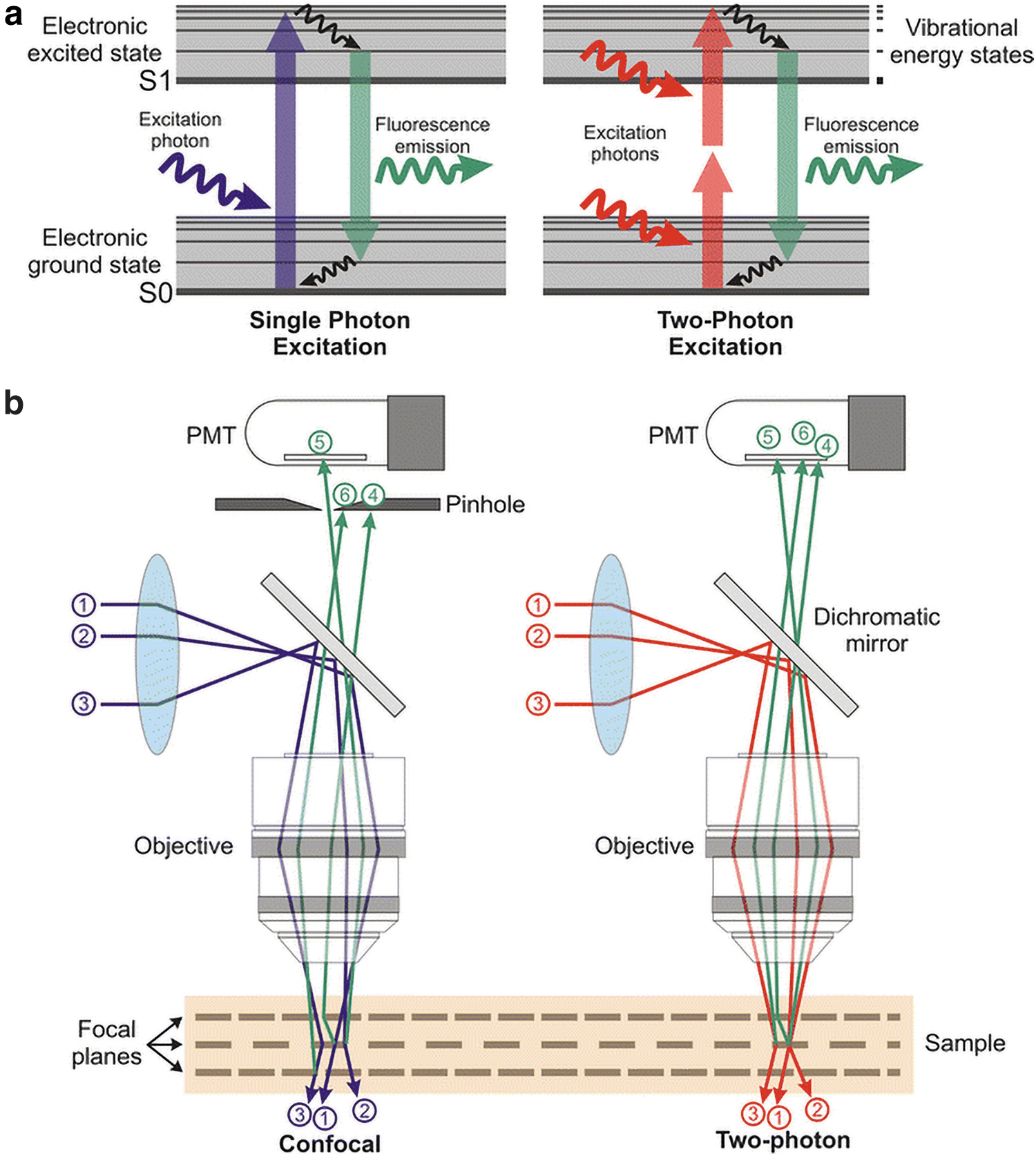
Principles of 2PEF microscopy.
Other distinct advantages include deep red or NIR excitation wavelengths for more in-depth tissue penetration; nonlinear excitation of photons for greater imaging clarity at a single focal volume; and scattered photons collected as useful signals. 20 Deep and efficient exploration in tissues are made possible through the inclusion of scattered photon signals, with the 2PEF allowing imaging depths of epithelial tissues ∼150 μm beneath the surface and up to 1 mm deep in the neocortex with a regenerative amplifier.23,24 Vasculature imaging up to 1600 μm deep in granulation tissues of mounted mice skin and cortex has been reported.25,26 Apart from fixed tissues, 2PEF is excellent for presenting cellular details of live tissue vasculature in vivo.
Second harmonic generation
Investigations on fluorescence changes in Ca2+-indicators in 2PEF excitation report that cumulative photodamage occurs in the window range of 75 fs to 3.2 ps. 27 The use of second harmonic generation (SHG) and third harmonic generation (THG) modalities for live tissue imaging negates any appreciable phototoxicity or photobleaching effect. 28 The optical power of SHG is displayed in its high sensitivity imaging of collagen fibrils. By direct measurement of image intensity as a function of laser polarization, SHG can extract structural information on the assembly of collagen fibrils. 29 Endogenous SHG-active molecules such as fibrillar collagen emit strong SHG signals intrinsically, therefore no such labeling with exogenous probes is necessary for image construction.30–33 Another advantage of SHG imaging in angiogenesis studies is the signal polarization properties for visualizing the absolute orientation, as well as the molecular organization of collagen in tissues with high degree of detail. 34 While 2PEF emission increases linearly with the number of molecules, SHG emission power scales with the number of molecules squared. 35 In addition, 2PEF radiation does not depend on molecular distribution because of its incoherent nature; in regions where dipole distributions are completely symmetric, SHG signal cancels out while 2PEF signal stays intact. Similarly, THG shares similar coherence properties as SHG but its use is restricted to tissues surface regions with large differences in refractive index. 36 THG imaging is therefore particularly useful for elucidating intracellular lipid deposition on capillaries. 37
Image delineations involving more than one tissue element have been used to investigate the interdependent growth of vessel-surrounding cell types. SHG-imaging can capture and monitor collagen fibers with keratinocytes, fibroblasts, and melanin density in engineered skin equivalents5,38 and drug-treated native skin tissues.39,40 The 2PEF/SHG system also affords the distinct advantage of visualizing angiogenic processes in tissue scaffolds that are inherently difficult to document.41–43 Microvascular networks comprising <10 μm microvessels and biological structures can simultaneously be visualized in bone repair and regenerative processes. With greater resolution power obtainable with SHG imaging, the combined use of 2PEF and SHG/THG will continue to push the boundaries of clinical and translational cancer research forward. 15
Characterization of Vascularization and Angiogenesis
Fluorophores and chromophores for imaging of vascular and related events
Understanding the physical properties and characteristics of fluorophore probes will allow for the appropriate choice of fluorescent dye crucial for successful 2PEF microscopy. 44 The ideal fluorophore exhibits the following properties: strong NIR absorption at the 750–950 nm range; high photostability; nonphototoxic, and chemical stability in cells. Fluorophores with cross-sections larger than 100 Göppert-Mayer units (GM ≡10–50 cm4 s) are preferable. Several suitable 2PEF NIR fluorophores have been prescribed, including rhodamine, fluorescein, and Bis-MSB, 45 and innovative fluorophores such as cyanine, 45 ultrabright squaraine, and squaraine-rotaxane dyes. 46 Endogenously labeled 2PEF elastin autofluorescence (500 to 550 nm) can be applied to distinguish veins and arteries in vivo.47,48 The IRDye38 and Cy5.5 fluorochrome system was successfully used in imaging skin tissue up to 150 μm deep. 49 Other dyes, including Texas Red, ICG, and YFP, have longer excitation wavelengths suitable for use in a three-photon microscopy, to achieve vasculature imaging depths of 1200 to 1330 μm. 50 In particular, YFP and Texas Red dyes are used for visualizing the dynamics of neurovascular network and tracking of microvasculature and pyramidal neurons with the multiphoton microscope. Stacked microvasculature images collected can be merged, amplified, and vectorized for data analysis (Fig. 3). Research studies of neurovascular sprouting and communication in developmental biology and disease are areas of growing interest.52,53

Stacked images collected by two-photon excitation microscope with ytterbium-doped laser using YFP for
NIR II fluorophores (1000 to 1700 nm) used in vasculature imaging afford considerable advantages in terms of reduced photon scattering, minimal interference from autofluorescent molecules, and greater penetration depths. 54 Organic NIR-II fluorophores include small-molecule dyes (SMDs) (for vasculature and lymphatic vasculature and hind limb vasculature), SMD complexes (for hind limb vasculature), and SMD-based organic nanoparticles (for whole body blood vessels, blood vessels in tumor). Inorganic NIR-II fluorophores include single-walled carbon nanotubes (blood perfusion, brain vasculature, venous vessels, and cerebrovascular imaging). 54
One study designed a strategy for developing photostable dyes with high-fluorescence quantum yield. 55 It involves two bioconjugate probes used in conjunction with 2PEF to target vascular endothelial growth factor receptor on cells. Behavioral events in angiogenesis occur during tumor progression and vascularity is needed to support the growth and metastatic capacity of tumors. 56 By elucidating vascular growth, we can better understand morphological characteristics, perform tumor staging, and document the progress of therapeutic interventions. Microvascular permeability may also be assessed and quantified by dextran-fluorescein conjugates for obtaining leakage data and the clearance constant.57,58
Chromophores such as hemoglobin and cytochromes reside inside living cells and possess undetectable fluorescence, requiring a different approach to imaging. Imaging nonfluorescent species requires a two-color excited-state absorption-based approach. 59 Chromophores with large absorptive cross section are two photon-absorbing materials that can be coupled to an excited state absorption for detection 60 and by wavelength for chromophores with distinct absorption spectra. 61 Label-free optical imaging of blood vessels and oxygenation in vivo provide important information on tumor angiogenesis and progression, hypoxia, 62 and are useful in oxygen transport studies in normal and diseased tissues. 63
Measurements in cell biology by 2PEF microscopy
Tissue- and cellular-level visualization
Endothelial cell and pericyte interactions have been studied as an in vitro model in angiogenesis. Some common applications of 2PEF include observing the effect of notch signaling between endothelial cells and pericytes on endothelial sprouting, 64 measuring vascular tortuosity, intercapillary distance, and static photopatterning imaging of sprouting vessels in deep tissues. 65 Particularly relevant in wound healing and tumor studies is the visualization of tortuous microvessels and their relationship to wound impairment and the three-dimensional (3D) reconstruction of invading capillaries (Fig. 4). Two-photon excitation imaging allows comparison studies with normal capillaries in qualitative indices such as spatial location, total length, mass displacement, and other properties. 66

2PEF microscopy for elucidating the ultrastructural processes involved in angiogenesis. Vascular plexus (0 to ∼1100 μm and integrin-expressing cells (1100 to 1600 μm)
Two-photon microscopy has been applied extensively in evaluating in vivo and reconstructed skin tissues,5,38 and in understanding skin biology, disease and other dermatological studies.67–70 In vitro, 2PEF images and analyzes developments in the dynamics of real-time endothelial cell differentiation.71,72 Specifically, 2PEF can be used to examine the effects of different culture conditions, regulatory growth factors, biomaterials, other molecular regulators, and bioengineering methods on primitive network formation via vasculogenesis—and vascular network maturation and stabilization arising through the influence of other cell types. 73 It can assess microvascular structures in skin tissue constructs designed to recapitulate human papillary and reticular dermis, with vascular network at depth profiles of 250 to 425 μm. 74
Increasingly, 2PEF microscopy has been applied for in vivo visualization and analysis of blood vessels together with melanin for investigating aging and regeneration. 69 In wound healing, invasive neoangiogenesis can be visualized through the observation and characterization of incumbent cells in capillary infiltration, as well as behavioral changes of inflammatory cells, fibroblasts, and fibrin clots. Endothelial cells regulate many Arg-Gly-Asp (RGD) expressing integrins, including avb1, avb3, and a5b1, that can be imaged with water-soluble fluorenyl probes to produce whole-mounted tissue micrographs. 75
Biochemical and molecular-level measurements
The capacity of 2PEF to measure molecular events of sensitive tissues that impact real-time human physiology is well established. Biochemical oxygen partial pressures in retinal capillaries provide an indication of oxygen delivery performance for maintaining the metabolic activity of the retina. 2PEF microscope can be customized to image retinal microvasculature and measure oxygen partial pressure. 76
With the demonstrated capability of the 2PEF microscope to measure optical redox ratio, its application for the study of associated metabolic changes alongside vascularity have led to visual delineations previously not possible with intensity-based fluorescent measurements. 77 Fluorescence lifetime imaging microscopy (FLIM) measures the fluorescence lifetime of a molecule in its excited state and informs on the molecular environments of fluorophores and changes in conformation of molecules in proximity. 78
Common sensing methodologies of FLIM and phosphorescence lifetime microscopy (PLIM) are used to investigate luminescence lifetimes. Fluorescence lifetime imaging (FLI) may be combined with Forster resonance energy transfer (FRET) to visualize proximity of donor-acceptor pairs. 79 FRET sensors provide information on protein-protein interactions and their molecular conformation in nanoscale (1–10 nm) measurements. Various FLIM-based sensors for exogenous fluorescent molecules measure microenvironmental parameters, including temperature, pH, viscosity, polarity, and mechanical forces. While FLIM-FRET biosensors provide information on ligand binding, FLIM detects fluorophore changes in molecular structure. PLIM is used in imaging oxygen and hypoxia with micro- to milliseconds (1–1000 μs) lifetimes and for sensing the presence of molecular oxygen.
FLI modalities may be integrated into other imaging microscopes: confocal, intravital, light sheet, and multiphoton for specialized imaging approaches. When merged with 2PEF, the modalities of FLIM, PLIM, and FLIM-FRET present can be widely applied to various biomedical engineering applications, cell/tissue oxygen and hypoxia, 80 cancer diagnosis, and drug treatment. 81 Commonly used biosensor probes include vascular cell-penetrating dendrimeric probes (e.g., nitroimidazole, Oxyphor-2P) (2-photon PLIM); optical metabolic imaging of endogenous fluorescence produced by NAD(P)H and FAD, and redox status by redox-sensitive small molecules and fluorescent protein-based redox biosensors (e.g., NADH:NAD+ ratio) (2-photon FLIM); FLIM-FRET-based biosensors for protein interactions and receptor-ligand binding activities (2-photon FLIM-FRET); nanosensor and fluorescent protein-based for quantitative measurements of pH, Ca2+, and other ions (e.g., ECFP and Oregon Green BAPTA-1) (2-photon FLIM).79,82
The 2PEF/SHG-FLIM setup as an imaging platform has gained the interest of at least one industrial company in dermatological and cosmetic applications. 83 Shirshin et al. validated the advantage of this extended setup with the intricate assessment of blood capillaries. 84 Detailed images of dermal papilla organization, that is, the collagen-to-elastin ratio composition in the papillary dermis was achieved with SHG, while FLIM tomography presented adjacent cross sections of capillary loops of the tissue architecture to render data on interior capillaries, red blood cells, blood plasma, and neighboring supporting tissue framework. 85
The applications of 2PE fluorescence microscopy on angiogenesis described in this review are summarized in Table 1.
Practical Applications of Two-Photon Excitation Microscopy in Angiogenesis
Clinical and bioprinting applications
An emerging and promising optoacoustic imaging technique, Macro-FLIM, is a valuable imaging tool in biological and clinical specimens. Macroscopic or mesoscopic imaging has been exploited for its importance in optoacoustic cancer imaging in clinical settings and for sensing hemodynamic changes in different brain regions. 86 Macro-FLIM fundamentally uses acoustic waves to project images through contrast mechanisms derived from tissue chromophores and exogenous contrast agents. 87 It is widely used to image patient specimens and constructs with high resolution and allows imaging of depths to a few millimeters with resolution orders of magnitude ranging from a few microns to hundreds of microns. 86 Macroscopic modality as a complementary component to 2PEF may be extended to include optical imaging of deep tissues and engineered tissue constructs. With potential applications in vascular, 88 bioprinted, 89 or biofabricated scaffold-laden tissues, 2PEF/Macro-FLIM shows promise in clinical and biomedical elucidation of molecular events beyond conventional optical depths.
The coherent Raman scattering (CRS) technique may be used in tandem with 2PEF microscopy. 90 CRS microscopy is described in two variants: coherent anti-Stokes Raman scattering (CARS) and stimulated Raman scattering (SRS). CRS microscopy utilizes two pulsed laser beams to probe human tissues with enhanced resolution, speed, and chemical sensitivity.91,92 Both CARS and SRS involve a pump and Stokes beam to elicit active vibrational states for in-phase oscillation of molecular bonds; however, while CARS generate coherent radiation at the anti-Stokes frequency difference (ωp − ωs) matched to a Raman mode of interest, SRS is an inelastic scattering process that provides quantitative information on chemical constituents.93–95 The inherent advantage of label-free methods ensures that living specimens remain relatively unaffected. The 3D imaging of 2PEF/CRS allows for the quantification of capillary cortical blood volume and the direct measurement of cerebral hematocrit volume, a complementary parameter. 93 Utilizing vibrational spectroscopy with synchronized 2PEF and SRS picosecond dual-excitation scheme, the technique also provides label-free contrast optical maps of microvascular structures, capillary morphology, and surrounding tissues.
The option to use NIR femtosecond laser pulses to elicit ultraviolet photons for 3D photopolymerization reactions have produced innovative outcomes in biomaterials and tissue engineering research. The technique known as two-photon polymerization (2PP) or lithography is controlled by optical fabrication parameters and resin properties; 96 2PP has been used for the fabrication of photosensitive materials of submicrometer (<100 nm) resolution. 97 Two-photon laser lithography has guided the assembly of blood vessel substitutes, vascular grafts, and scaffold embedding for endothelial cell adhesion. 98 Biological innovations on the various biocompatibilities of photopolymers with engineered analogs are currently ongoing 97 and are expected to contribute to the progress of 3D bioprinting and microfluidic vessel-on-chip platforms research.99,100
Current Limitations and Scope for Improvements
Despite the merits of 2PEF technology, sample defects and aberrations that arise while imaging deep sections of tissues have long been responsible for the lack of quality of high-resolution imaging. Aberrations degrade fluorescent images, and corrections are made by adaptive spatial resolutions during the imaging process. Continuous design improvement in the configuration is key to improving visualization. To achieve this, modifying the 2PEF to incorporate functional integrations with other specialized microscopy is therefore necessary. In designing methods to increase the acquisition speed and penetration depths of 2PEF, practical considerations also need to be addressed to detect the resulting fluorescence signals.
Attempts at modifying the conventional properties of 2PEF allow for improved visualization in vascular networks. Longer wavelengths of excitation in the 2PEF microscope translate to lower imaging resolutions compared to confocal laser-scanning microscopes. Technical improvisation to the 2PEF microscope has been reported to increase spatial resolution at ∼1.7 times that of a typical 2PEF for murine brain phantom. 101 The technique involves autocorrelation scanning and in situ estimation of the PSF. Information on the PSF is required for effective image deblurring. Annular illumination is used to obtain fluorescent images from biological samples and the PSF estimates in the lateral plane for deconvolution.
Background fluorescence signals obtained with 2PEF may be reduced by maximizing the frequency performance of the 2PEF optical system. The concept was first conceived based on the principle of structured illumination microscopy 102 and involves the modulation of two overlapping spots to produce structured illumination103,104; however, this proved to be difficult for 2PEF as it required 2D detectors.
Other super-resolution imaging techniques such as the stimulated emission depletion (STED) microscopy105,106 saturated excitation (SAX) microscopy107,108 and the spatial overlap modulation nonlinear optical microscopy (SPOMNOM)109,110 may be used in tandem with 2PEF to improve image contrast in deep tissues. SPOMNOM achieves background-free imaging at depths cannot be achieved by the 2PEF microscope alone. STED microscopy can elicit improvements in spatial resolution of about 60 nm although the imaging depth is limited to 100 μm. 111 With the use of core-ring illumination in SAX microscopy, 75 spatial resolution improvements in the z-direction were significant at depths of 100 μm in a tissue model compared with 2PEF microscopy. The side lobe artefacts present in engineered PSFs were reduced, and the result is a further enhancement of the spatial resolution. 3D spatial resolution was reported to be enhanced by 1.4 to 1.8 times, and out-of-focus signals reduced to a factor of 102 within the tissue samples.
Conclusion and Future Developments
Concluding remarks
As the fields of biomedical engineering and regenerative medicine continue to push the boundaries of research, there is an unprecedented demand for noninvasive and deep tissue imaging. 2PEF microscopy provides the opportunities to address the needs for noninvasive and deep tissue imaging with lower phototoxicity and photodamage. Different applications call for different 2PEF microscopy setup and configuration with added modalities to achieve optimal visualization of molecular events previously not possible with standalone microscopes. Although detailed two-photon microscopy optical design and physical configuration setup are beyond the scope of this review, we acknowledge as with all types of multiphoton laser scanning microscopes, it comes in an infinite combination of assemblies for a myriad of applications. Other higher-order nonlinear optical microscopy techniques, such as the three-photon,112–114 four-photon114,115 ultrahigh resolution microscopy enables deeper tissue imaging in greater ultrastructural detail. Technical modifications and improvements made over some common optical limitations encountered in the conventional 2PEF microscopy can produce better imaging outcomes. For example, when combined with FLIM, PLIM, and FRET modalities, the resolving power of the 2PEF/SHG could further contribute to the progress in noninvasive imaging of changes in molecular structures, ligand binding, and studies in oxygen quenchers and hypoxia levels associated with angiogenic events, respectively. With the level of detail in molecular species localization demonstrable with the FLIM setup, its use is expected to gain traction with greater awareness of its power in elucidating angiogenesis and its related molecular events.
For human samples, Macro-FLIM has been utilized in clinical settings as an optoacoustic cancer diagnostic imaging tool. The technique has also found its promise in deep tissue imaging bioengineering research where imaging beyond standard optical depths is often warranted. The option to combine 2PEF with CRS and 2PP modalities have also contributed to providing invaluable datasets with vibrational spectroscopy and support vascularization studies in 3D bioprinting and microfluidics.
Future outlook
Depending on application demands, designing a 2PEF microscope with the necessary laser and optical components in place—even with minimal configuration setup—can be a costly endeavor. Barring cost considerations, technological enhancements to the 2PEF microscope in time is expected to allow imaging of vascularity for the most demanding biomedical applications. Improvements in vascular and tissue imaging beyond currently achievable depths are expected to be made by stepwise incremental optimization of parameters, instead of quantum leaps in imaging technology. The nature of the tissue and the type of angiogenic events are major considerations on how improvements in imaging quality of live tissues can be achieved. The appropriate selection and use of fluorescence probes, advances in adaptive optics, and laser source development for producing ultrashort NIR laser pulses will elevate visualization of cellular and molecular events in angiogenesis to higher ground.
Bioengineering strategies are underway to remodel vascular network formation and distribution for long-term functionality and for supporting the integrity of whole tissues. 116 As such, the future of 2PEF microscopy is expected to combine various matrices for more elaborate structural and functional studies in, for example, tissue perfusion and tumorigenesis. These include imaging renderings as well as usable analytics that charts and measures indices: in situ signaling effect induced by growth factors, integrins and biomolecules that influence vascularization, real-time patterning, time course of vascular network development, blood flow rate, and the effects of hypoxia-induced angiogenesis and their relationship with normal cells. The regulatory interplay of hypoxia and tumorigenesis in the absence of, or reduced functionality of, blood vessels is a field of research that will rely on the increasing use of 2PEF to elucidate. We anticipate the use of the 2PEF microscope to further contribute to significant progress in the fields of angiogenesis and tissue engineering research.
Footnotes
Acknowledgments
We thank Dr. Pakorn Tony Kanchanawong (Mechanobiology Institute & Department of Biomedical Engineering, National University of Singapore) for his mentorship on advanced quantitative microscopy techniques. We also acknowledge the funding support for Marcus Lee, Muniraj Giridharan and Sathya Kannan through NUS Research Scholarship.
Disclosure Statement
No competing financial interests exist.
Funding Information
We acknowledge funding by National University of Singapore (Faculty of Dentistry); Ministry of Education, Singapore (R-221-000-118-720 and R-221-000-118-133); Agency for Science, Technology, and Research (A*STAR, Singapore) Advanced Manufacturing and Engineering (AME) Research Grant (R-221-000-122-305, Project No.: A1883c0013) and National Additive Manufacturing Innovation Cluster (NAMIC) Research Grant (R-221-000-120-592, Project ID: 2017068).