




Abstract
In vitro
ovarian follicle culture is a reproduction technique used to obtain fertilizable oocytes, for overcoming fertility issues due to premature ovarian failure. This requires the establishment of an in vitro culture model that is capable of better simulating the in vivo ovarian growth environment. Two-dimensional (2D) culture systems have been successfully set up in rodent models. However, they are not suitable for larger animal models as the follicles of larger animals cultured in 2D culture systems often lose their shape due to dysfunction in the gap junctions. Three-dimensional (3D) culture systems are more suitable for maintaining follicle architecture, and therefore are proposed for the successful in vitro culturing of follicles in various animal models. The role of different methods, scaffolds, and suspension cultures in supporting follicle development has been studied to provide direction for improving in vitro follicle culture technologies. The three major strategies for in vitro 3D follicle cultures are discussed in this article. First, the in vitro culture systems, such as microfluidics, hanging drop, hydrogels, and 3D-printing, are reviewed. We have focused on the 3D hydrogel system as it uses different materials for supporting follicular growth and oocyte maturation in several animal models and in humans. We have also discussed the criteria used for biomaterial evaluations such as solid concentration, elasticity, and rigidity. In addition, future research directions for advancing in vitro 3D follicle culture system are discussed.
Impact statement
A new frontier in assisted reproductive technology is in vitro tissue or follicle culture, particularly for fertility preservation. The in vitro three-dimensional (3D) culture technique enhances follicular development and provides mature oocytes, overcoming the limitations of traditional in vitro two-dimensional cultures. Polymer biomaterials have good compatibility and retain the physiological structure of follicles in the 3D culture system. Utilizing hybrid in vitro culture materials by merging matrix, hydrogel, and unique patterned materials may facilitate follicular growth in the future.
Introduction
Folliculogenesis, or follicle development, is governed by several interacting process and cell types. Follicular development is assisted by interactions between the oocyte, theca cells, granulosa cells, and cumulus cells (Fig. 1).1–3 In vitro culture of extracted ovarian tissue or isolated immature follicles is an alternative strategy in assisted reproductive technology.4–6 This could be beneficial for patients with cancer who are at risk of losing their fertility due to ovarian damage caused by chemotherapy or radiotherapy.7–9 As the technique requires the implantation of cryopreserved ovarian cortical strips, in vitro fertilization of mature oocytes and embryo transfer reduce the risk of reintroducing cancer cells.10–16
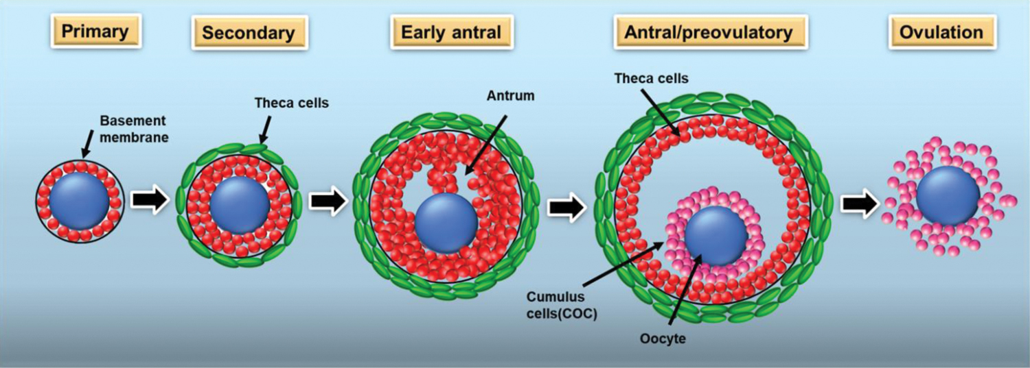
A schematic illustration of the ovarian follicle at different stages of development.(2,3) Color images are available online.
Ovarian follicle isolation and in vitro culture have been investigated as a potential alternative strategy for resolving the limitations of traditional ovarian tissue transplantation.17,18 In vitro culture has been attempted and studied in various animal models, such as pig, cow, mouse, and dog, and in humans.19–22 In vitro two-dimensional culture (2D) of mouse ovarian follicles and the successful ovulation of the mature oocyte were also reported.23,24 In vitro 2D culture, on the other hand, causes the follicular structure to collapse due to the distribution of granulosa cells on the dish surface. 25 In vitro three-dimensional (3D) ovarian follicular culture was developed to overcome the problems associated with 2D ovarian follicle culture.26–29
The advantages of the 3D system include the preservation of the spherical form of ovarian follicles and their improved vitality and development.30,31 The properties and functions of a variety of biomaterials, including alginate (ALG), collagen (Col), Matrigel, fibrin (FIB), and composite hydrogels, were investigated to develop a strategy for the optimal 3D culture of ovarian follicles.32–36
At present, natural and synthetic polymers are currently being employed for in vitro follicle culture.9,37 Choosing suitable and effective polymers with appropriate compatibility and mechanical properties is essential for maintaining follicle structure and morphology. In addition, the culture systems can impact follicle development. Therefore, in this study, we comprehensively reviewed the numerous in vitro 3D culture techniques used for follicle development and oocyte maturation, including microfluidics, hanging drop, and 3D printing. We also focused on polymer biomaterials used for building hydrogels for 3D culture systems that provide a suitable microenvironment for follicular growth, oocyte maturation, and hormone secretion. In addition, the criteria for choosing biomaterials, including material concentration and stiffness, and future research directions for advancing in vitro 3D follicle culture system are discussed.
In Vitro 3D Follicle Culture
2D and 3D culture systems for follicle growth
The 2D and 3D in vitro culture of preantral follicles have been extensively studied (Fig. 2).38–40 However, the adhesion of granulosa cells to the tissue causes the 2D cultured follicles to flatten in the long term. This increasingly disrupts the gap junctions in the follicle complex.41,42 The significance of cell spatial configurations has encouraged research into 3D culturing techniques. 43 The gene expression profiles of 3D cultured cells differ from those of traditional 2D cultured cells and are more similar to that of in vivo cells. 44 Despite the potential benefits of 3D culture, the implementation of such a system remains debatable. The key concerns are the types of biomaterials to be used and their properties, such as toxicity, permeability, and the ability to be molded and manipulated properly during follicular loading and retrieval. 45

A comparison of follicular growth in a two-dimensional system and three-dimensional system. This schematic is reprinted from Desai et al 56 with permission from Springer Nature. Color images are available online.
Furthermore, the survival of the follicle and oocyte maturation determine their biological utility. The animal species and follicular culture period should be considered while evaluating the applicability of 3D culture techniques.46,47
Maintenance of follicle morphology
Matrix- and hydrogel-based 3D culture techniques are being extensively studied for maintaining follicular architecture and acquiring a better understanding of follicular growth requirements.27,28,46,48 The ability of 3D systems to preserve the spherical architecture to sustain crucial cell–cell and cell–matrix interactions within the surrounding stromal tissue allows the isolated follicles to mature properly.49,50 In addition, it prevents oocyte mortality. Trophic substances generated by granulosa cells persist around the oocyte, promote oogenesis, and potentially stimulate the formation of new local gap junctions. 51 Oocyte development and cytoplasmic meiotic competence require gap interactions between the oocyte and the granulosa cells. 52
The granulosa cell growth and certain metabolic processes are mediated by oocyte-derived secretions.53,54 The oocyte neither transport particular amino acids nor carry out activities, including cholesterol or glycolysis production, without the assistance of granulosa cells in delivering crucial components. The dissociation of the gap junction and intercellular interaction during in vitro culture causes premature ovulation and subsequent degeneration of the liberated oocytes.55,56
In vitro 3D culture systems
Microfluidic culture
Microfluidics and 3D cell culture technologies have increased the potential of in vitro modeling.57–59 Microfabrication, which enables precise cell manipulation at the microscale or nanoscale, has been a significant technical achievement. 60 Microfluidics has applications in the field of reproductive biology, including that in oocyte fertilization, embryo culture, and sperm sorting.61–67 Microfluidic devices utilized for sperm analysis and other in vitro fertilized-on-a-chip innovations, in contrast, require a standard interface. Researchers used a nonplanar polydimethylsiloxane microfluidic device to generate ovarian microtissue by encapsulating early preantral follicles in core-shell microcapsules (Col and/or ALG) obtained from deer mice (Peromyscus). 57
A microfluidic chip has also been created to culture a single human preantral follicle. 68 The pressure-assisted network for droplet accumulation is a multichannel fluidic system and a hanging drop cell culture component to produce homogenous 3D microtissue (Fig. 3A). 69 A microfluidic system with controlled flow rate has been used to encapsulate the ovarian follicle in a 3D two-layer (core and shell) structure to support the production of the early oocytes, both structurally and hormonally (Fig. 3B). 70 The oocytes were successfully encapsulated using this microfluidic technology, which maintains the size and cellular compartmentalization of the oocytes.

Matrix-free technique
Matrix-free technique is an alternative method for 3D culture to overcome the difficulties in removing the follicles from the gel, and to avoid the subsequent loss of healthy follicles.71–73 The key matrix-free 3D cell culture techniques include the forced floating method and the hanging drop method.74–76 Roller bottle systems, rotating-wall vessels, and orbiting test tubes maintain 3D follicle architecture.73,77,78 However, these systems are not very effective and handling large number of follicles would be a delicate and labor-intensive process. Follicles could be retrieved and propagated in vitro until the late preantral/early antral stage utilizing human ovarian cortex strip culture in serum-free media. 79
In a recent study, ovarian tissue pieces were cultured for 3 weeks in the anti-Mullerian hormone (AMH) system. In the AMH-modulation group, follicle diameters, steroid hormone levels, and paracrine factor production increased significantly compared to the control group. The secondary follicles retrieved from the cultured ovarian tissue formed clusters and progressed to the antral stage after 6 weeks of group culture. Oocytes produced from AMH-modulated cultured follicular clusters reached the MII stage, with a normal-sized first polar body and meiotic spindle formation. 80
3D printing
3D printing is the method of introducing material in layers to create 3D solid structures from a digital program using a printer (Fig. 3C).81–83 3D printing enables to produce complex shapes using less material and high reproducibility than that using traditional manufacturing methods. 84 Photolithography, stereolithography, magnetic 3D bioprinting, and direct cell extrusion have been some of the techniques applied for 3D bioprinting of cells.85–87 3D printing systems for bioengineering of reproductive tissues is a promising strategy for in vitro follicle culturing, ovary tissue transplantation, and menopausal hormonal therapy. 88 However, inkjet 3D printing is limited due to nozzle blockage, which limits a steady flow of the ink, and the decrease in cell viability. Three-dimensionally printed microporous hydrogel supports murine follicle survival and function in a bioprosthetic ovary. 89
A bioprinting was optimized for producing ALG microbeads comprising a cumulus-oocyte complex (COC) and its validity was tested in sheep. 90 In addition to preserving cumulus integrity, COC encapsulation possessed high efficiency and reliability. A 3D printed ovary was created by utilizing gelatin-methacryloyl (GelMA) bioink, and the exogenous follicles were implanted in the GelMA scaffolds. The ovarian follicles gradually developed and consequently produced mature oocytes. 91
Hydrogels for 3D follicle culture
Hydrogels assist various functions such as regulating cell behavior and guided cell expansion, and provide a microenvironment for the transplantation of isolated follicles or ovarian tissue (Fig. 3D).92–95 The various types of polymers used to fabricate matrices for in vitro follicle culture are listed in Table 1.
Summary of Three-Dimensional Follicle Culture in Hydrogels from Various Types of Animal Species: Follicle Development and Oocyte Maturation
GV, germinal vesicle; GVBD, germinal vesicle breakdown; MII, metaphase II; NA, not available.
Hyaluronic acid-based hydrogel
Hyaluronic acid (HA) is a glycosaminoglycan present in the extracellular matrix (ECM), particularly in the soft connective tissues. 105 HA has several applications; it is used as a drug delivery carrier in gene therapy and as a scaffold for cell carriers.106–109 HA hydrogel is used either alone or in combination with ECM (ECM-HA) for follicle growth in vitro. 110 Germinal vesicle (GV) breakdown (GVBD) and estradiol secretion were considerably greater in ECM-HA than that in HA after 12 days of culture, while the number of MII oocytes retrieved remained markedly greater in the control. 110 The early effects of hyaluronic acid-based hydrogel (HABH) scaffold supplied with basic fibroblast growth factor and vascular endothelial growth factor (VEGF) on heterotopic auto-transplanted fresh ovarian tissues in rats have been studied. 111
In the ovarian tissue encapsulated with HABH, the mean number of follicles at all developmental stages and the expression level of VEGF significantly increased post-transplantation (p < 0.05). Ovarian encapsulation with HABH alone increased follicular survival, restored hormone levels, prevented or reduced ischemia-induced follicle loss, and aided angiogenesis. Hyaluronic acid-alginate (HAA) hydrogel and ovarian cells (OCs) were used to culture mouse ovarian follicles; the performance of HAA hydrogel was compared to that of the ALG and fibrin-alginate (FA) hydrogels (Fig. 4A

Chitosan-based hydrogel
Chitosan (CS) is a partially deacetylated derivative of chitin and is a natural polymer mainly composed of D-glucosamine, widely distributed throughout connective tissues. 112 CS has excellent biocompatibility, biodegradability, and antimicrobial activity.113,114 Therefore, CS-based bioactive materials are frequently employed in tissue engineering and regenerative medicine.115,116 CS-based hydrogels at 0.5%, 1%, and 1.5% were used to simulate follicular growth and oocyte maturation. Their performance was compared to that of ALG-based hydrogels (Fig. 4B). The CS hydrogels had greater survival rates, estradiol secretion, normal meiotic spindle, and chromosome alignment (p ≤ 0.05). In addition, the number of MII oocytes was higher in 1% CS (43.08%) than that in 1% ALG (26.3%); however, the oocytes from the ALG group showed higher number of GVs (34%) than that in 1% CS group (7.6%). 96
Col-based hydrogel
Col is a triple-helix protein, which is a main component of the ECM.117,118 Col is an ideal biomaterial for in vitro experimental systems because of its great biocompatibility, unique mechanical properties, and transparency.119–121 The culture of mouse follicles in Col was established in the late 1980s. The follicle development can be sustained in Col-cultured follicles cultured; however, they require additional support to continue growing. 72 When implanted under the kidney capsule, immature follicles cultured in vitro with or without serum could develop to the mature Graafian stage. 122 These findings led to further improvements in mouse model, and models based on other animals such as pig, cow, and humans.123–128
The feasibility of using Col hydrogel matrix for ovarian follicle culture was evaluated by monitoring follicle development in matrices with varying concentrations of Col hydrogel. Col at 3 and 5 mg/mL concentrations showed follicle viability above 90%. The oocyte meiotic competence in follicles cultured in 5 mg/mL Col showed the greatest GVBD (88 ± 4%) with a higher number of GVs (11 ± 3). 98 In cases of premature ovarian insufficiency, the implantation of adipose-derived stem cells cultured on Col promised to restore ovarian function for the long term, including estradiol levels, estrus cycles, follicle formation, and fertility. 129 Culturing mouse follicles with human menstrual blood-derived mesenchymal stem cells using Col assists follicle development and oocyte maturation; moreover, this system shows improved survival rate, hormone secretion, and maturation rate. 100
ALG-based hydrogel
ALG is a polysaccharide with high biocompatibility, gel-forming ability, and nontoxicity, and is amenable to processing as 3D scaffolding materials. 132 ALG hydrogel is formed by cross-linking with calcium ions. This allows easy encapsulation of follicles under physiological conditions. ALG-based hydrogels have been widely studied for in vitro follicle culture in several animal model-based studies and in various types of culture media.33,34,99,133–137 Primate tissue architecture is reportedly maintained in ALG system, and primate follicle survival and architecture are better maintained when cultured in 2% ALG, compared to that in follicles cultured in 0.5% ALG.138,139 However, only a few oocytes were obtained from the ALG system.
In addition, the meiotic spindles were disturbed due to poor biological viscosity that prevented cell adhesion and proliferation. 140 Despite these limitations, ALG is used in combination with several natural polymers such as FIB, HA, ECM, and Col.50,70,97,99 Human and bovine ECM-ALG scaffolds enable better in vitro follicle development. 141 The concentration and hardness of ALG, as well as the nutritional factors present in the culture environment, influence follicular growth. In mouse follicular growth, introducing supplements to the ALG culture system is favorable.25,142–144
Synthetic polymer-based hydrogel
Synthetic polymers are employed in biomedical applications such as drug delivery, therapeutics, and tissue engineering.145,146 It is often difficult to modify natural polymers to obtain the desired physical properties; therefore, engineering innovative synthetic polymers with well-defined mechanical and biodegradable properties, and biocompatibility is preferred. 147 Synthetic polymers are engineered through the polymerization of monomer units or modification of functional groups on polymer chains.148,149 A copolymer of polyvinyl alcohol (SuperCool X-1000), polyvinylpyrrolidone (PVP K-12), and polyglycerol polymer (SuperCool Z-1000) may be a suitable additive for vitrification solutions, for assessing in vitro follicle development.150–154 One of the most frequently utilized tested materials for ovarian follicle culture is polyethylene glycol (PEG).155–157 Follicles were encapsulated in 7% PEG-vinyl sulfone (PEG-VS) hydrogels modified with integrin-binding peptides (Arg-Gly-Asp) and a trifunctional matrix metalloproteinase-sensitive peptide. 158
PEG-VS hydrogels could support graft remodeling and revascularization postimplantation, with 60% of the follicular pool remaining. The levitation system is a new technology proposed for 3D follicle culture. Secondary follicles subjected to 3D culture with 200 μL/mL magnetic nanoparticles showed higher viability, antrum development, lower degeneration rates, and a more consistent daily growth rate than those in the 2D control culture. 159 Functionalized degradable PEG hydrogels with ECM-sequestering peptides, imitating the original ECM component for ovarian follicle growth and oocyte maturation, are promising. After 10 days, 88.2–96.5% of secondary follicles entrapped in PEG hydrogels survived, with the average diameter ranging from 297 ± 47 to 324 ± 60 μm (Fig. 4C).
The percentage of follicles that established an antrum was 53.2–65.4%. The proportion of oocytes that matured from both size groups to MII exhibited a similar range from 28.6% to 33.3%. 104 Poly(epsilon-caprolactone) (PCL) blended with gelatin has been evaluated to assess its suitability for artificial ovary applications. 89 Patterned electrospun fibrous PCL/gelatin scaffolds support follicular growth with the typical spherical morphology. 160
Fibrin
FIB is a natural polymer composed of fibrinogen and thrombin. It has minimal inflammation-inducing properties, significant elasticity, and viscosity, along with angiogenic properties. Follicles encapsulated within FIB hydrogel emit proteolytic enzymes that break down the matrix, allowing the follicles to squeeze out. Consequently, FIB is generally combined with other polymers for the in vitro culture of follicles. 161 FIB-ALG matrices impart the dynamic cell-responsive mechanical properties required for the in vitro development of ovarian follicles. 162 FIB improves the growth of macaque primary follicles, as well as induces steroidogenesis, AMH/VEGF production, and oocyte maturation. 163
Autografting of isolated preantral follicles and OCs encapsulated in FIB is an alternative method to restore fertility. 164 The FA interpenetrating network (FA-IPN) maintained the ovarian follicle's architecture; regulating the FIB degradation rate allowed for prolonged toxicity monitoring without interrupting follicle growth. 165 FIB enriched with platelet lysate (PL) as an angiogenic and growth factor carrier to evaluate transplanted preantral follicles. 166 The FIB-PL system has the potential to improve graft vascularization and follicle survival. 167
Paulini et al reported cryopreserved human preantral follicles encased in the FIB matrix (fibrinogen, thrombin, and HA) appear to be a promising matrix for creating an artificial ovary, encouraging follicle survival and development. 168 Collagenase NB6, a highly purified combination enzyme, is used to obtain a significant number of viable human follicles. 169 Use of FIB enabled obtaining 90.7% viable follicles after 3 days of in vitro culture. Simvastatin administration or incorporating human ovarian tissue into FIB clots improved human ovarian tissue transplanting. Tissue immersed in FIB clots exhibited a better number of proliferating follicles and promoted vascular architectures that expressed both human and murine markers than untreated tissue. 170 Encapsulating the ovary with nitric oxide-releasing nanoparticles embedded in FIB hydrogels prevents ischemic damage and accelerates angiogenesis. 171
Extracellular matrix
ECM is a complex and well-organized 3D macromolecular network of biomolecules occurring in all tissues. The ECM is essential for cell behavior regulation. It acts as a scaffold for cellular constituents and activates important biochemical and biomechanical signals.172–174 It provides the essential scaffolding for the cellular constituents and initiates crucial biochemical and biomechanical cues. 175 Mouse ovarian cortical strips cultured in ECM showed improved survival rate and antrum formation. 176 Follicle viability and oocyte maturation were improved by using the appropriate material and culture media in scaffolds. Matrigel-ALG (MA) ensures better follicle survival rate and oocyte maturation rate than the FA scaffold. Therefore, MA, being rich in ECM components, provides a more favorable environment for follicle development than FA. 177
A methodology has been established for decellularizing human ovarian tissues, while preserving the intact ECM. A human-derived decellularized scaffold supported the survival of isolated human follicles, with 25% recovery rate after three weeks postgrafting. 178 Ovarian follicles cultured in ECM-derived soft hydrogel (ES-hydrogel) were significantly superior than ALG control in terms of pseudo-antrum formation rate, MII oocyte rate, normal meiotic spindle rate, and hormone secretion. 179 Human ovarian follicles could survive and proliferate in vitro with the assistance of an ECM hydrogel produced from decellularized bovine ovaries (oECM). Combination of 90% oECM with 10% ALG could be a new and prospective approach to transplant isolated human follicles in a bioengineered ovary. 180
Parameters for Evaluating Biomaterial and Supplements That Support Follicular Growth
Molecular weight and solid concentration of biomaterials
The mechanical features of biomaterials, such as viscosity and molding ability, depend on the molecular weight; they influence the utility of biomaterials for follicle culture. The role of matrix concentration was evaluated by adjusting the molecular weight of the ALG and the composition of the hydrogels.138,139 Lowering the solid content concentration improves follicular growth in the mouse model. 96 Antrum development increases dramatically when the solid content of the matrix is reduced in a mice model. 26 Reduced solid content promotes antrum development, while also increasing estradiol secretion and lowering the expression level of androstenedione and progesterone. 26 The effects of type I Col concentration (1–7%) on generating a 3D culture system for murine follicles were evaluated. 98 Follicles grown in 7 mg/mL Col had a fairly large follicle diameter.
However, the follicles were smaller than the follicles grown in 3 and 5 mg/mL of Col hydrogels. The human ovary has a dynamic structure, with a stiffer Col-rich cortex. Increasing solid content provides a biomechanically nonpermissive environment and alters mechanical signaling, which contributes to improving the growth of human follicles and maturing human oocytes. 82
The shear elastic modulus and rigidity
The effects of mechanical properties of biomaterials on follicular growth are well studied. The rigidity of the biomaterial, defined by shear modulus, describes its elastic characteristics as well as its capacity to resist deformation when subjected to a force.181,182 The mechanical properties of the ovarian ECM, notably the stiffness gradient, are essential for follicle survival and folliculogenesis. The elastic modulus (G’) of ALG hydrogel was investigated in three groups: high G’ (3% ALG), intermediate G’ (1.5%), and low G’ (0.7%). When the G’ decreased, the follicle diameter, the rate of antrum production, and the formation of layers increased. Most importantly, oocytes cultured in low G’ ALG had a high capability to resume meiosis. 183
Tethered integrin-binding peptides, in combination with gonadotropin follicle-stimulating hormone and luteinizing hormone, help in better simulation of the milieu for ovarian folliculogenesis and enhance the interactions between the oocyte and its surrounding granulosa. 184 The mechanical properties of the PEG hydrogel are significantly correlated with follicular growth, with an ultimate G’ of ∼1 kPa reported in mouse models.26,157,185 When considering the effect of matrix stiffness on human follicle growth, stiffer biomaterials are preferred.168,186,187 When softer bovine ECM was combined with harder ALG to improve the mechanical properties, the rate of follicle recovery improved in exact correlation to the ALG concentration, with the recovery rate peaking (82%) when only ALG was used. 180
Co-culture of follicles: follicle–follicle communication
Culturing follicles in groups facilitates the exchange of autocrine and paracrine secretions, along with increasing follicle–follicle communication and possibly improving growing conditions. The number of follicles growing together has a significant impact on primary follicle survival and development. The greatest follicle improvement was discovered in the group with the highest number of follicles. 188 The disadvantages of co-culture systems include the potential sharing of growth-inhibiting hormones, such as AMH, among follicles. Furthermore, co-culturing can make it more difficult to trace, monitor, and harvest individual follicles during the maturation phase.
A period of culture time with murine follicles encapsulated in 0.3% ALG hydrogels was conducted to properly understand the underlying mechanisms during follicle co-culture. When compared to small groups (5 × ), follicles co-cultured in larger groups (10 × ) displayed a unique transcriptome profile and functional pathways, such as prolactin signaling and angiogenesis-related genes. 189 The insulin-like growth factor I (IGF-I)-oxytocin (OT) system (IGF-I-OT), which controls follicular steroidogenesis and supports follicular growth, is influenced by communication between porcine and bovine follicles, which is mediated by either downregulation or overexpression of the IGF-I-OT system. 190
Summary of Present Status, Limitations, and Future Research Directions
The potential to produce immature human oocytes in vitro has numerous prospective applications; however, it is imperative to preserve the oocyte quality. 191 Ovarian tissue cryopreservation is currently available for fertility preservation. However, there are concerns about the dangers related to the surgical procedure, particularly the possibility of re-implanting malignant cancer cells. 192 Here, an in vitro culture system for follicle development and maturing immature oocytes is presented as a promising strategy for restoring female fertility without the risk of reintroducing tumor cells.21,42 The procedure depends on the clinical viability of the in vitro developed primordial follicles, because preserved cortical strips can harbor follicles at this stage. Further research is required before the clinical application of these in vitro primordial follicles; however, there are reliable methodologies for creating culture procedures that promote in vitro human oocyte development. 193
Obtaining MII oocytes from primordial human follicles using in vitro culture for in vitro fertilization is a promising approach, with the basic human follicle in vitro culture systems being currently available. 191 However, low MII rates, uncertain fertilization capacity, and safety concerns limit their clinical application. For in vitro simulation of the in vivo human follicular development environment, future research studies should focus on developing a 3D system using microfluidics and 3D printing techniques combined with bioengineering technology. The follicle development and oocyte maturation rate could be improved by adopting the whole procedure culture system. Finally, future research on the ability of mature oocytes to fertilize and produce embryos is necessary.
Footnotes
Authors' Contributions
The authors confirm contribution to the article as follows: S.K.: article preparation, original draft, review, and editing. H.P.: conceptualization, supervision of article, and corresponding author.
Disclosure Statement
No competing financial interests exist.
Funding Information
This research was supported by the National Research Foundation of Korea (NRF), funded by Ministry of Science and ICT (Grant No. NRF-2021R1A2C2007189), and the Korea Health Technology R&D Project through the Korea Health Industry Development Institute (KHIDI), funded by the Ministry of Health and Welfare, Republic of Korea (Grant No. HI14C3484, HI21C1353).