




Abstract
The craniofacial region contains skin, bones, cartilage, the temporomandibular joint (TMJ), teeth, periodontal tissues, mucosa, salivary glands, muscles, nerves, and blood vessels. Applying tissue engineering therapeutically helps replace lost tissues after trauma or cancer. Despite recent advances, it remains essential to standardize and validate the most appropriate animal models to effectively translate preclinical data to clinical situations. Therefore, this review focused on applying various animal models in craniofacial tissue engineering and regeneration. This research was based on PubMed, Scopus, and Google Scholar data available until January 2023. This study included only English-language publications describing animal models' application in craniofacial tissue engineering (in vivo and review studies). Study selection was based on evaluating titles, abstracts, and full texts. The total number of initial studies was 6454. Following the screening process, 295 articles remained on the final list. Numerous in vivo studies have shown that small and large animal models can benefit clinical conditions by assessing the efficacy and safety of new therapeutic interventions, devices, and biomaterials in animals with similar diseases/defects to humans. Different species' anatomical, physiologic, and biological features must be considered in developing innovative, reproducible, and discriminative experimental models to select an appropriate animal model for a specific tissue defect. As a result, understanding the parallels between human and veterinary medicine can benefit both fields.
Impact statement
The main objective of the review article is to discuss several methods to develop craniofacial defects in animal models that can be treated with tissue engineering strategies. Applying tissue engineering therapeutically helps replace lost tissues after trauma or a disease like cancer.
Introduction
Craniofacial reconstruction is challenging due to the region's anatomical complexity and cosmetic importance. Available surgical options are currently limited, emphasizing the need for new approaches to restore the face and skull's form, function, and esthetics when missing or damaged soft or skeletal tissues are present. 1 The development of functional substitutes for damaged tissues is the goal of tissue engineering, an interdisciplinary field that combines biology and engineering principles. Regenerative medicine, as a developing field, combines tissue engineering2–4 and self-healing principles. 5 There is a growing interest in using cutting-edge technologies and biomaterials in regenerative medicine and tissue engineering in therapeutic settings. However, there is still a bottleneck in encouraging preclinical data to secure a successful clinical application.
To serve this purpose, tissue engineering heavily relies on animal models to draw reliable and meaningful conclusions. The evaluation of the efficacy and safety of a new product/method, which allows its clinical use, presupposes the use of animal models, as closely related as possible to humans (models of large animals), in the preclinical evaluation stage that follows the step in which small animals such as mice or rats are used. Therefore, animal models must be chosen wisely to ensure they are suitable for clinical scenarios and have outcome indicators that accurately reflect them.3–5
Despite the benefits of in vitro models, such as the capability to perform systematic, standardized analysis of a wide range of cellular, biophysical, and biochemical indications in a controlled setting, these models are still limited in their ability to provide a concrete understanding of disease and disorder pathophysiology due to the lack of the natural variability that in vivo animal models present. 5 Consequently, in vitro experiments cannot yet replace the need for animal models in translational research. Hence, animal studies remain an essential requirement for the method refinement and safety and effectiveness testing needed for regulatory authorization and technological breakthroughs, such as three-dimensional (3D) bioprinting and bioreactors. 6
Sheep, goats, pigs, rabbits, and dogs are frequently used for in vivo research instead of some laboratory models, like mice and rats, because they can be operated on more quickly due to better surgical access and providing large enough tissue for mechanical experimentation. The technical difficulties presented by mice and rats frequently raise concerns about the applicability of findings to humans.7–9 Choosing an appropriate animal model can be time-consuming and challenging, influenced by the investigators' assumptions and personal preferences rather than what is best for the investigation. Unsuitable animal models are selected for scientific studies irrationally, wasting resources, causing futile death, and leading to inaccurate, redundant, and improper investigations. These issues have been reduced by developing animal models that are highly specific to the study. 10 Therefore, this review aimed to describe the animal models used frequently in craniofacial regenerative medicine as reproducible and discriminative experimental models.
Methods
Animal models were explored in this study to review their role in craniofacial tissue engineering. A checklist-based strategy was used for the search, screening, and data extraction. Preferred Reporting Items for Systematic Reviews and Meta-Analyses guidelines were also followed. 11 Two reviewers (S.A.M. and H.T.) independently searched Pubmed, Scopus, and Google Scholar for relevant articles. The search terms “Animal Models,” “Craniofacial Tissue Engineering,”" “Craniofacial Regenerative Medicine,” “Laboratory Animal Science,” and “Animals” were used to find all relevant publications until January 2023. The first step was to search and retrieve studies. Duplicates were discarded; afterward, the articles' titles and abstracts were vetted. Study inclusion criteria were then applied to those remaining articles to finalize the screening process.
The reliability between the evaluators regarding the literature screening process was calculated using Cohen's Kappa coefficient. Based on the frequency of precise agreement between reviewers, the kappa value (k) was determined. In vivo and review studies describing the role of animal models in craniofacial tissue engineering and regenerative medicine were included. In vitro studies, posters, non-English language studies, abstracts, and studies with insufficient data were excluded. After that, two reviewers (S.A.M. and H.T.) extracted the required data. A third author (A.H.) was consulted in cases of disagreement among reviewers.
Results
The initial search resulted in 6454 articles. Upon removing duplicates, 3996 remaining studies were reviewed based on title and abstract. A total of 152 records were excluded after analyzing the full text of 447 remaining articles. Two hundred ninety-five studies were included to structure this review (Fig. 1). Based on Cohen's Kappa coefficient, the evaluators were almost in complete agreement (к = 0.84).
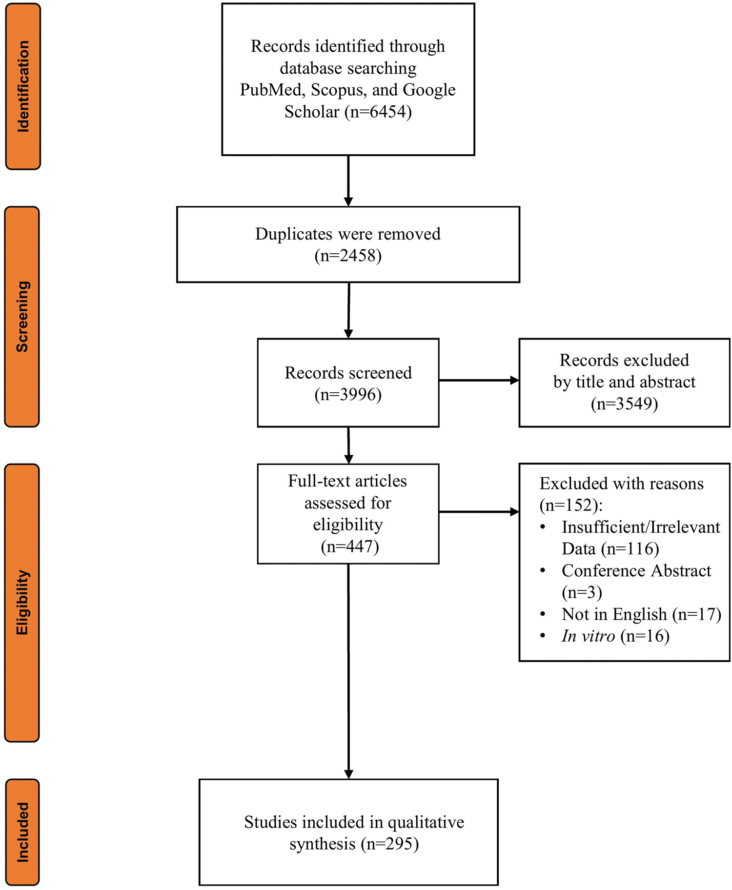
A flowchart showing the Preferred Reporting Items for Systematic Reviews and Meta-Analyses process for selecting articles for review. Color images are available online.
Craniofacial regenerative medicine
The craniofacial anatomy is highly specialized and unique to each individual. It is critical to the body's functionality and appearance, but it is vulnerable to damage caused by both genetic and environmental factors. Cancer and different forms of cranial and maxillofacial trauma frequently manifest in the head and face; one-third of all live births marred by congenital malformations involve the head and face, whether minor or significant. 2 A field of interdisciplinary study, regenerative medicine combines molecular and cellular biology, bioengineering, and material science to encourage endogenous healing and replace or regenerate cells, tissues, or organs to restore functionality. 1 Animal models are essential elements of in vivo studies for implementing biomedical research on human and veterinary patients.
These models can help with the development and pharmacokinetic investigations of medicines, toxicity studies, and preclinical exploration of tissue engineering and medical devices in humans. Regardless of the application of animal models in biomedical research, they should be developed based on principles of replacement, reduction, and refinement (3Rs). 10 As a complicated multifactor process, identifying the most appropriate model is critical in animal research. Laboratory animal models are preferred to large-scale animal models in terms of cost-effectiveness and throughput; nevertheless, large-scale animal models are required for studying the anatomy and physiology of humans, obtaining preclinical data, and ascertaining the clinical potential of vascular grafts, implants, and orthopedic bone plates.
The significant physiologic and anatomic dissimilarity between rodents and humans influences the extrapolation of findings from murine studies to human research. Large-scale animal models should therefore be developed to prevent the limitations of rodent models, simulate clinical settings, and translate cellular-based regenerative approaches from bench to bedside. The properties of individual preclinical models should be considered in oral tissue engineering, and regenerative procedures should be optimized based on large-scale animal models in cell-based therapeutics. 12
Research also suggests the significant contribution of large-scale animal models to human oral tissue engineering. Nonhuman primate, canine, and porcine models have long been used to investigate surgical techniques in implantology and guided tissue regeneration, as well as the pathophysiology of endodontic and periodontal diseases.12,13 The main drawbacks of studying horses, sheep, and dogs include their different dental and craniofacial anatomy from that of humans. 12
Given the similarity between human and porcine models in terms of morphology, anatomy, healing, mechanical, and remodeling properties, the latter can be used in many craniofacial tissue regeneration procedures.12,14,15 Positive outcomes were obtained from canine models, being the most relevant in bone regeneration, implantology, periodontics, and endodontics. However, the substantial dissimilarity between the dog and the human makes the translational application of knowledge difficult. As an alternative to pig and dog models with ethical and economic advantages, ovine models have been rarely developed due to their major dissimilarity with humans. Their ruminant nature causes significant anatomical differences from humans.12,16,17
Bone defects
The craniofacial skeletal system is crucial to facial esthetics because it provides a rigid, stable structure for cartilage, dental compartments, and facial soft tissue. Due to inherited, traumatic, or neoplastic conditions, significant structural and functional deformations may arise from craniofacial bone loss. 1 An injury or surgery that damages the bones can also lead to a bone defect. Bone formation can be guided by biodegradable and nonbiodegradable scaffolds with appropriate architectural and mechanical qualities to restore function and shape. Cells and growth factors may be combined with scaffold materials to stimulate the formation of new bone. 18
As a histocompatible and nonimmunogenic bone substitute, autologous grafting is currently the gold standard for restoring bone defects. However, their limitations in repairing craniofacial defects include their high costs, requiring a separate surgical site, significant donor site morbidity, and finite supply of autologous bones. 19 As a simple and cost-effective alternative to autologous bone grafts, biocompatible scaffolds can help heal these defects and generate new bone by providing an osteoinductive and osteoconductive extracellular matrix analog for cellular migration, adhesion, proliferation, and differentiation. The materials explored for this purpose include ceramics, composite materials, synthetic and natural polymers, metals, and silicon-based bioglass. 20 Using specialized constructs to repair bone defects with tissue engineering strategies have numerous benefits over current bone grafting procedures since the new bone is regenerated from the individual's cells.
In tissue-engineered constructs, the supply of oxygen and nutrients and the homing of host cells are critical factors in bone formation. In addition, as a significant postgraft concern, limited vascularization can cause the loss of structural features and graft resorption. Revascularization at the site of implantation/graft is stimulated by the conservation of the periosteum layer and its environment.21,22 Incorporating endothelial progenitor cells into the scaffold design can facilitate neovascularization as a critical component of bone tissue engineering by responding to ischemia, which can be observed in critical-size craniofacial defects. It has been demonstrated that calvarial defects can be repaired in rats using a combination of these cells with mesenchymal stem cells (MSCs) and thermoresponsive porous nano-calcium sulfate/alginate scaffolds.
Endothelial progenitor cells are compatible with β-tricalcium phosphate (TCP) scaffolds and directly contribute to neovasculogenesis through endothelial cell differentiation and recruitment of additional host endothelial progenitor cells. Moreover, proangiogenic factors, including vascular endothelial growth factor, are released by exogenous endothelial progenitor cells. 20 In addition, bone generation highly depends on the biochemical and physicochemical characteristics of the constructs, which commit local cells to osteoblastic differentiation, and the osteoconductive features of the constructs themselves, facilitating bone formation.23,24
Calvarial defects, cancellous bone defects, and partial cortical defects are used to evaluate bone graft substitutes. Most literature discuss and use calvarial bone defects.16,25–28 The complicated 3D structure of the craniofacial bone compared to that of long bones makes the iliac, ribs, and fibula challenging to restructure and be fitted into the morphology of craniofacial bones. Bone and tissue engineering through synchronizing growth inducers, cells, and active constituents can help treat craniofacial bone defects by resolving the challenge of the limited availability and complicated 3D structure.21,29
3D printing, an additive manufacturing approach, is especially interesting for the regeneration of craniofacial defects through creating patient-specific treatment solutions. Biofabrication of tissue-engineered bone scaffolds can be easily accomplished with 3D printing. Calcium phosphate pastes, metals, and hydrogels are examples of materials used in the biological printing process. Following preclinical study guidelines can improve translation by choosing an appropriate preclinical animal model. 2 Small animals like rats and rabbits and more evolved animals like pigs, dogs, sheep, and primates have been used to develop oral and craniofacial osseous healing models. 30 Species and age of the animal used affect critical-size bone defects as examples of injury models. 6
Smaller animals are affable to most investigative settings for establishing main biological principles because they are easily handled, maintained, and cost-effective, with well-known genetic data available. 31 The implantation of human-derived cancer cells in immune-suppressed rodent models has successfully mimicked bone malignancy. 16 However, extrapolating the experimental outcomes obtained from these models to clinical conditions would be challenging because bone metabolism in rats and rabbits differs significantly from that of human bones. 32 Moreover, specific techniques and treatment principles (dental implant placement, distraction osteogenesis [DO], or sinus augmentation) cannot be assessed on smaller animals due to size and anatomic limitations. Due to their brief life spans and diminutive body sizes, rodents are unsuitable for studies requiring multiple biopsies or blood samples over time. Mouse models are more suited for studying ectopic (subcutaneous) bone generation compared to larger animal models.
However, tissue-engineered bone in regenerating and healing of critical-sized lesions is studied using larger animal models, such as sheep and pigs. Highly immunocompromised mouse strains, permissive for engrafting human tissue, make mice an attractive candidate in this context. 33 In surgical technique studies, larger animals may be better suited to investigate a new biomaterial's biomechanical characteristics or healing responses due to their physiological and anatomical resemblance to humans. Small animal models are more appropriate when studying the biological aspects of bone treatments.13,34 This is why small animals are usually used as a starting point for “proof of principle” testing, followed by additional tests on larger species before conducting clinical trials on humans. Bone defects can be studied using various animal models, according to the mentioned considerations in Table 1.
Selection Criteria for an Animal Model of Bone Tissue Regeneration
Dental and periodontal tissues
The periodontal interface comprises the cementum, periodontal ligaments, alveolar bone, and gingiva. Chronic periodontitis destroys tooth-supporting structures and causes inflammation, eventually leading to tooth loss. 35 The animal model is ultimately developed based on the research requirements, including periodontal approaches, sinus lift techniques, alveolar ridge augmentation, and oral implantology.36,37 Periodontal regenerative research employs diverse models of animals, including dogs, rats, and nonhuman primates. Periodontal models of rats have also been used in bone regeneration. The advantages of these screening models of molecule regeneration include cost-effectiveness and simplicity in handling. Surgical microscopes are, however, required to visualize and create the typically small defects.
Contrarily, dentin/pulp regeneration is infrequently carried out orthotopically in murine models due to frequent dental fractures and variations with the human pulp repair process; their periodontal tissues and bacterial profiles also differ from humans. Large animals, such as porcine models and dogs, must be used to address these limitations. 38 For bone and periodontium regeneration, porcine models are successfully developed; however, little is known about dentin-pulp regeneration.
Pigs are also commonly reported as models for regenerating teeth/tooth roots. Undoubtedly, dogs are the most transversal model because of positive results for bone regeneration, dental implantology, periodontology, and dentinal/pulpal regeneration. Tooth anatomy and wound healing kinetics in canine models resemble those in human models. However, differences between these models and humans and ethical considerations must be considered. 12 As the least developed animal models, ovines are not only economically and ethically advantageous but they also differ significantly from humans in some critical aspects. Therefore, ovines serve primarily as bone study models and rarely for periodontal regeneration. 12
Salivary glands
Lubricating the oral cavity, aiding digestion, and having antimicrobial/buffering features are some beneficial features of saliva, an aqueous fluid secreted by salivary glands mainly comprising electrolytes, enzymes, and mucins. 39 Various scenarios can affect salivary gland function. 39 Therefore, it has long been necessary to develop new concepts for salivary gland regeneration. Although histocellular information is crucial for understanding the many pathologic states of salivary dysfunctions, clinical salivary gland histological assessments are not applicable in most patients. This is due to the intrusive nature of biopsy, which raises the risk of consequences such as infection, inadequate wound healing, fistula, and scarring. 40
Salivary gland studies have been conducted with various animals (rats, mice, cats, pigs, monkeys, sheep, etc.). Experimentally reproducing various salivary pathological conditions can facilitate the development of therapeutic strategies by allowing the analysis of histomorphological and physiological information. 41 In addition to their histomorphological similarities with humans, murine models are considered excellent experimental animals in this regard due to their ease of use. 42 For instance, primary human salivary cells were isolated in the first attempts to repair injured salivary parenchyma. An organ germ regenerated utilizing a combination of epithelial and mesenchymal cells extracted from adult mice's submandibular, sublingual, and parotid salivary glands was implanted onto masseter defects in adult female mice after removing their submandibular, parotid, and sublingual glands.
The extending polyglycolic acid guide in the implanted gland was inserted into the parotid duct of the host. Collecting saliva from the oral cavity with and without stimulation demonstrated the functional potential of bioengineered salivary glands. Insignificant differences were observed in the flow and content of saliva between the bioengineered glands and those in untreated mice. 43 According to Ogawa and Tsuji, epithelial bud formed within 2 days in the organ culture of germ cells isolated from submandibular, sublingual, and parotid glands in mice. After transplanting these cells back into salivary gland defects, they also reported improved salivary flow in the mice. 44 In studies, the radiation and ductal obstruction models were primarily described, while there are inconsistencies in developing animal models for medication-induced dysfunctions.
According to studies, mice, rats, minipigs, monkeys, and rabbits are mainly used for radiation models through the practical application of an animal model, with mice and rats being the most common models. Most ductal obstruction models were produced using mice, rats, cats, and rabbits. The submandibular gland and, less so, the parotid gland were the primary sites of ductal obstruction in rodents. The largest and most noticeable salivary gland in gross anatomy, the submandibular gland, may be the cause of this. 41 There have been numerous attempts to implement new therapeutic strategies for salivary gland regeneration, including stem cell therapy, gene therapy, drug delivery, and bioengineering approaches. 45 Depending on the etiology of the dysfunction of salivary glands, the appropriate therapeutic approaches should be used.
The temporomandibular joint
The TMJ is a bilateral joint that helps the condyle head and glenoid fossa of the temporal bone articulate. The biconcave disk and its attachments ensure maintaining stability and distributing loads derived from multiplanar movements of the joint. TMJ comprises the glenoid fossa, articular eminence, interpositional fibrocartilaginous disc that separates upper and lower compartments, and mandibular condyle. The TMJ in humans is a bilateral ginglymoid-diarthrodial joint exposed to sliding (mainly the superior compartment) and hinge (the inferior compartment) movements. Swallowing, breathing, chewing, and speech are all standard routine functions necessitating the TMJ. The TMJ has been studied using a variety of laboratory animals essential for providing platforms for investigating primary disease processes and possible clinical applications. 7
It is impossible to find an animal model replicating the human TMJ's anatomical structure and function. TMJ studies show that tissues' anatomy, physiology, and properties have been well studied in pigs and minipigs. However, handling farm pigs at skeletal maturity could be problematic because the zygomatic arch blocks access to the joint as they grow continuously. It is easy to perform surgery on sheep and goats; they provide a large quantity of tissues for practical studies; their anatomy is similar and relatively inexpensive per animal. Nonetheless, their TMJ mainly functions in translation due to their herbivorous nature. In dogs, the joint space is very confined; therefore, the interpositional device is more likely to remain in place if its attachment is a concern. Nevertheless, the hinge-type TMJ in carnivores like dogs can only function in rotation.
The healing potential of joints is not affected by the function type. However, it is possible to affect implants' stability. 7 In addition, Kalpakci et al. studied TMJ discs' anatomy, functional properties, and biochemical composition in humans, cows, goats, pigs, and rabbits. Herbivores (cows, goats, and rabbits) and omnivores (humans and pigs) showed different biomechanical properties because the functional role of the TMJ discs differs according to the animal's nutrition status. Herbivore discs have a larger glycosaminoglycan content and are made for translatory jaw motion, as opposed to omnivore discs, which work in rotary and translatory movements. The porcine model was the most similar to humans due to the significant resemblance with human TMJ in both anterior-posterior and medial-lateral aspects, glycosaminoglycan and collagen composition, and mechanical responses under compressive loads. 46
Cartilage
The craniofacial skeletal system includes cartilage in the pharynx, eyelids, nose, ears, and joints. Aneural, avascular, and alymphatic tissue and chondrocytes with low mitotic activity limit cartilage's ability to repair itself spontaneously. As a result of cartilaginous deficiencies, significant deformities may develop. There are currently few effective treatments for cartilage damage, and craniofacial cartilage reconstruction, including the external ear, is among the most challenging reconstructive surgeries due to its complex patient-specific 3D architecture. 1
Using rodent subcutaneous and intramuscular models, biomaterials and implants can be tested during 6–8 weeks regarding their safety features and biodegradation rates. As a consequence of the thinness of the cartilage layer, rats are not frequently used to assess chondral defect repair. 47 Compared to other small animal models, rabbits have larger joints, are easily operated on, and have cartilage thicknesses of 0.25–0.75 mm, which make them excellent for assessing cartilage repair. 47
Since rabbits and humans have different biomechanics, caution should be advised when comparing rabbit results to human results. According to studies, larger animal models provided a better reflection of the human cartilage/cartilage defects (percentage of cartilage relative to bone). Equines were the only models that matched the human cartilage thickness best. 48 Small animal models have been demonstrated ideal for early-phase cartilage tissue engineering tests. On the other hand, larger models like pigs, sheep, dogs, and horses are required to better comprehend the impact of a biomechanical and biochemical context comparable to that observed in humans. 49
Skin and soft tissues
As part of the craniofacial structure, the skin plays a vital role; as well as providing barrier capabilities, it allows the insertion of facial muscles, which are crucial for communication. Traumatic injuries frequently damage faces since they are exposed to the most dangers. When epithelial contiguity is disrupted, skin thermoregulation, mechanical barrier, pigmentation, and cosmetic properties are affected. Significant problems are still associated with nasal surgery, such as damage to the normal nasal mucosa and long-term delays in mucosal wound healing.1,37,50,51
Various tissue types have reconstructed, repaired, or remodeled soft tissues serving functional and cosmetic needs. In the early years of epidermal reconstruction in facial burn research, animal models were used to evaluate different techniques. Research on scar formation can be conducted using animal models to investigate molecular, cellular, and structural mechanisms. Furthermore, these models are generally used to study normotrophic scar formation and allow the study of different time points in the scarring process. 52 Cells from the human epidermis can be cultured to generate tissues for grafting in clinical practice. 53 The use of engineered pigs for skin donation has recently developed and increased. 54 In an attempt to recellularize a decellularized scaffold in an immunodeficient nude mouse, human tissue has been used to grow soft tissue organs such as the external ear. The injection of MSCs can enhance soft tissue repair alone or in combination with surgery. 55
Muscle
The craniofacial muscles' functional and esthetic importance cannot be overstated. Surgical resections, trauma, cancer, or autoimmune diseases may require grafting or regenerative techniques on these muscles. Cleft lip, palate, and other orofacial congenital abnormalities have been linked to poor muscle regeneration and fibrosis following surgery, both requiring restoration. 35 It is challenging to regenerate muscle tissue in situ after traumatic injury, as muscle consists not only of myofibers but also of blood vessels, nerve fibers, and connective tissues. If the regeneration is insufficient, the patient will suffer impairments in function and quality of life. The field of muscle tissue engineering has been advanced by researchers using scaffolds and cell-based interventions. For this purpose, muscle regeneration and repair studies have been conducted using various animal models.
Rodents are used most frequently in muscle investigations, although sheep, dogs, and pigs have also been investigated because of their clinically relevant size. 56 Molecular and repair mechanisms that regulate muscle function, plasticity, and treatment of muscle injuries have recently been elucidated through small animal studies, particularly comparative biology and genetic manipulation. The clinical situation, where defects are on the order of cubic inches rather than cubic millimeters, is not faithfully reenacted by these injuries. For example, the degree of functional recovery and regeneration in larger models was reduced, despite the positive results observed in rat models. 56 For this reason, additional translational and validation models are required, as both the muscles and the injuries are of minor size in small animal models. 57
Clinically implementing muscle regeneration covers various deformities. In muscle tissue engineering, the crucial role of scaffolds comprising biocompatible materials in tissue development and overall strength can be attributed to their interactions with the cells. The cells in the scaffolds can generate the extracellular matrix and growth factors. Seeded cells produce an extracellular matrix that degrades or substitutes the biodegradable scaffold, initially supporting tissue growth.
No graft rejection and immune activation risks have been reported for this type of scaffold with biocompatible and biodegradable characteristics. Polyesters made of collagen and naturally occurring α-hydroxy acids constitute the most commonly employed biodegradable materials in muscle engineering. Phosphate and SiO2 comprising glasses have also promoted bone and muscle regeneration. The phosphate-based glass developed by Shah et al. for craniofacial muscle engineering released nontoxic ions and retained their degradation properties. Tuning this glass into fibers provided a large surface area/volume and increased the surface area for cell adhesion.21,58 Researchers found promising results when treating muscle injuries in rats with amniotic mesenchymal cells. MyoD and desmin (muscle-specific markers) were discovered along with enhanced neovascularization and local tissue regeneration. 35 Fibrin hydrogels seeded with myoblasts have also been examined in animal models as a potential treatment for in situ defects. 59
There have also been studies on gelatin-based hydrogels. Myotubes can be induced to grow and differentiate cells using a scaffold consisting of cross-linked gelatin that mimics muscle's mechanical and biochemical properties. The scaffold showed good biocompatibility and a slow biodegradation rate when implanted subcutaneously in mice. 60 It has been shown that animal models with muscle injuries implanted with hyaluronic acid-based hydrogel had appropriate regenerative outcomes with a reduced inflammatory response. 61 Similar study results in animal models with muscle injuries showed that encapsulating myoblasts in hyaluronic acid-based hydrogels increased muscle innervation, vascularization, and functional restoration. 62
Blood vessels and nerve fibers
The blood medium constantly flows throughout the vessels in the vascular system as a multipurpose transport and delivery mechanism that provides essential nutrients and eliminates toxic metabolites. The pathologies caused by vessel dysregulation and abnormal vessel formation include bone and peripheral vascular diseases and metastasis. 63 To regenerate craniofacial tissue, successfully integrate it with the host tissue, and reduce long-term difficulties, angiogenesis and innervation are both necessary. 64
Craniofacial tissue engineering is still in its early stages, despite the progress in designing and fabricating biomaterials and comprehension of neurovascularization during regeneration and development. The complicated nature of craniofacial tissues makes it difficult to integrate multiple tissues; for instance, in craniofacial bone regeneration, osteogenic, endothelial, and neurogenic cells require different microenvironments for their comprehensive integration with coordinated healing.64,65 The effectiveness of various artificial blood vessels can only be adequately evaluated by developing a viable animal model that can simulate applications in the setting of artificial blood vessels to perform transplantation and postoperative monitoring.
In this regard, dogs, rabbits, pigs, sheep, and rats are the most commonly used vascular in vivo animal models.66,67 Except for cartilage, which is avascular, adequate vascularization is a challenging issue shared by all engineered tissues, and it becomes critical whenever employing two-dimensional or 3D structures. 1 Implantation of vascular pedicles, including arteriovenous bundles or arteriovenous loops, 68 enables prefabrication of tissue-engineered constructs and the subsequent vascularization of bone or soft tissue constructs for transplantation into far-flung defect areas. The results of mandibular reconstruction using this method in large animal models have been impressive. 1 Significantly decreased calvarial ossification was reported in conditional vascular endothelial growth factor knockout mice or those with impaired fibroblast growth factor signaling owing to defective angiogenesis.64,69
Craniofacial nerve injuries typically necessitate a complex grafting process to restore preinjury function/sensation in the motor and sensory neurons. Neoplastic, toxic, infectious, traumatic metabolic, and iatrogenic causes could lead to facial nerve palsy. 70 The most common causes of iatrogenic facial nerve damage are replacement of the TMJ, parotidectomy, mastoidectomy, and specific cosmetic procedures, such as the facelift. 71 Trauma and surgical procedures on a parotid tumor or the petrous part of the temporal bone can all cause facial nerve damage, and the nerve itself may be absent congenitally. Many types of injuries are due to iatrogenic causes, ranging from simple transactions to severe segmental loss. 72 Facial asymmetry and functional movement deficits caused by facial nerve defects can significantly impact one's quality of life. An autograft is the best option if primary nerve terminal coaptation cannot be achieved.
However, it is constrained by grafting scarring, infection, pain, donor nerve availability, increased risks of graft rejection, and donor site morbidity.35,73 Preliminary research has shown that neuronal tissue engineering can effectively regenerate the facial nerve. 1 To mimic the nerve, tissue engineering creates artificial materials that can reduce patient morbidity without causing extra damage. Compared with the gold-standard autograft, numerous nerve guides/conduits applications use biomaterials and decellularized allografts to improve nerve recovery. In addition to simple silicone conduits, nanocomposite-coated silk-based conduits are among the more complicated and novel biomaterials in this regard. Furthermore, various intraconduit cues for promoting neural recovery are being studied in preclinical models, including neurotrophic factors, stem cells, and adipose cells 74 (Fig. 2).
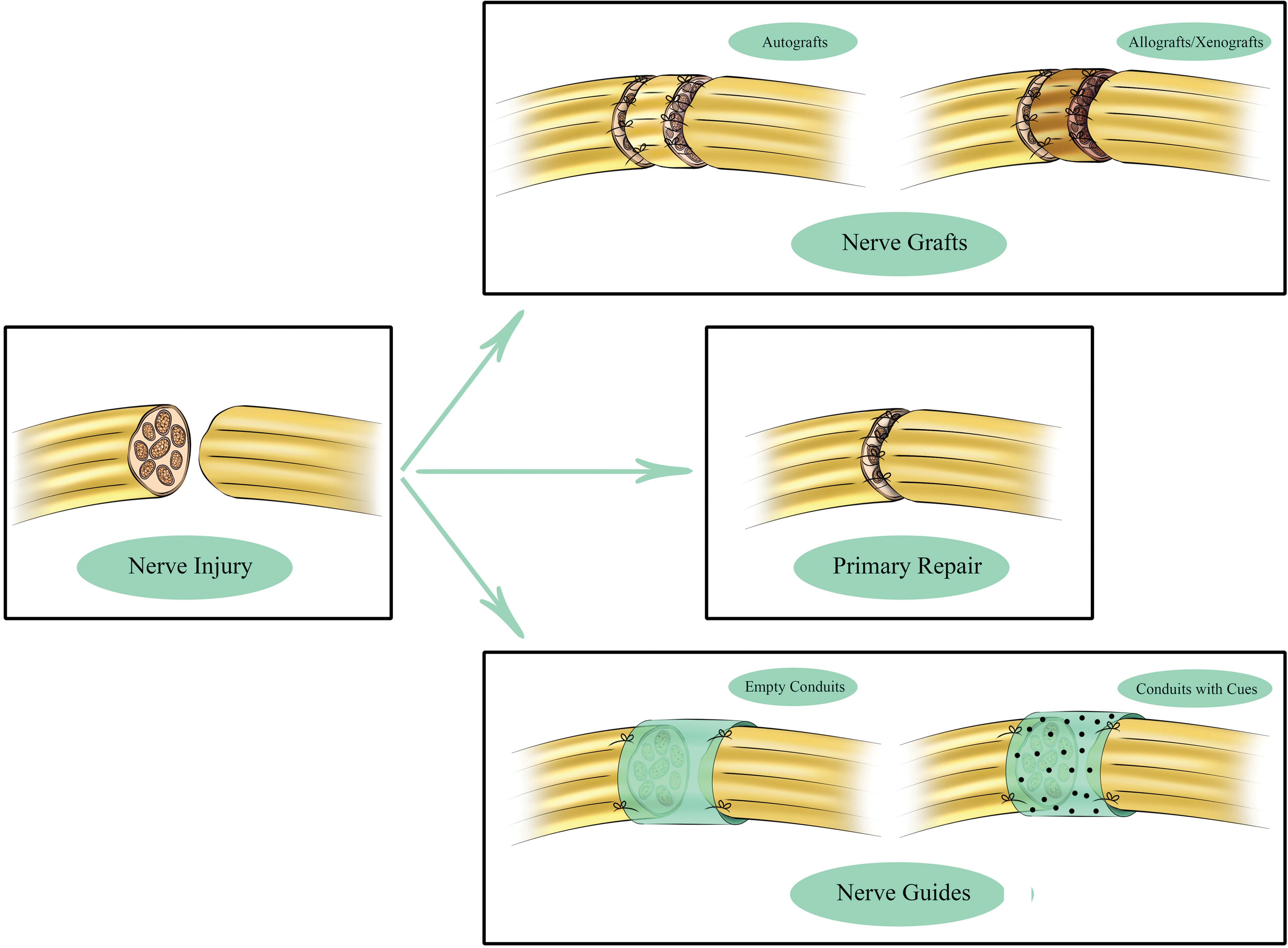
Various approaches for repairing facial nerves, such as primary healing and applying nerve grafts or conduits. Color images are available online.
When selecting the best animal model for studying nerve regeneration, it is essential to consider the species' innate neurobiology, including neural microstructure and composition, postinjury inflammatory responses, and nerve regeneration capacity. 75 Bioengineered conduit studies typically use relatively small nerve gaps, whereas nerve graft studies focus primarily on more significant gaps in larger animals. Rat models are used in the majority of preclinical investigations to examine facial nerve restoration. Large species with gaps up to 60 mm, including sheep, pigs, and monkeys, have been used to test synthetic neural scaffolds. Due to animal care costs, narrow assessment ranges, and complex training requirements for functional testing, studies in these large species are limited. 76 Figure 3 summarizes various craniofacial tissues and the role of tissue engineering in craniofacial regenerative medicine.

A schematic of different craniofacial tissues and primary contributions of tissue engineering to each tissue. Color images are available online.
Various animal models in craniofacial regenerative medicine and tissue engineering
Different studies and investigations relate animals to the evolution of human history. 10 It is often not practical, ethical, or scientific to study human biochemistry, physiology, or pharmacology in detail in humans. The term “animal model” refers to a study that uses animals to model what would happen in humans. Animal models have provided a large portion of our understanding of human biology, and animal models have been used in nearly all of our current medical approaches. Disease models simulate various diseases and conditions developed to mimic human disease. Based on the method of generation, these models have been classified.
In an induced disease model, a previously healthy animal is induced with the disease, or an aspect of it, to be investigated. Defect models are limited by their lack of chronic diseased states. 77 Spontaneous models refer to animals with diseases analogous to human conditions occurring naturally in the studied animal. They also involve mutant animals with genetic variants that make them suitable for studying a particular human condition. Due to their deficient immune systems, “nude” mice models are helpful for organ and tissue transplantation studies since they cannot reject a tissue graft from another species.
A “transgenic” animal has a foreign gene inserted deliberately into its genome, causing the translation of the matching messenger RNA (mRNA) to produce a new protein, resulting in a defective phenotype. Knock-in and knock-out models are now included in the definition of transgenic animals. A knock-in mouse is a mouse that has a mutated DNA sequence instead of a standard sequence. An inactivated gene is present in a knock-out mouse. An animal model that does not produce a particular disease is called a negative model. A model with a condition that naturally occurs in its species, but has not yet been detected in humans is referred to as an orphan animal model. 78 Experimental outcome can be confirmed by selecting the optimal animal assessment models.
Many models of large and small animals have been developed to translate basic research findings into clinical applications and confirm new strategies, ideas, and concepts.79,80 The healing potential of growth factors, different scaffolds, and stem cells has also been studied in smaller animals like rabbits. As a small animal with a bone mineral density compared to humans, rabbits are ideal models for studying healing over scarred dura mater because studies on them are clinically relevant.5,6,16,81 Small animal models are more amenable to high-throughput testing than large animal models.
The low cost and simplicity of genetic manipulation and handling cause rats and mice to be used as the most appropriate animal models. Their small size, however, makes it difficult to compare them with the biomechanics of humans. Therefore, large-scale animal models involving horses, goats, and sheep, similar to human size, with larger knee joints could be evaluated by performing magnetic resonance imaging (MRI) and arthroscopy. Large animal models that resemble human anatomy and physiology must be developed regardless of how animal models are used in a biomedical investigation to gain preclinical information and assess the clinical potential of vascular grafts, orthopedic bone plates, and implants.5,10 As in the case of humans, heterogeneity and complicated physiological and genetic interactions with the environment make large animals ideal for evaluating the efficacy and safety of novel treatments.
The concept “One Health, One Medicine” was developed when many diseases in animals occurred naturally (or spontaneously) and were comparable to those in humans, which infers that human and animal diseases (primarily mammals) share identical pathophysiology and etiology, and therefore need similar treatment approaches. A one-health approach can therefore benefit both veterinary and human medicine. It is possible to research not only novel treatment options but also disease progression and prevention at a pertinent age by using big animal models of naturally occurring diseases with comparable pathophysiology as in humans. However, it is challenging to standardize disease grade and recruit sufficient clinical cases for studies using naturally occurring disease models, even though this best represents the disease's complexity. 5
Rat (Rattus norvegicus) and mice (Mus musculus)
In many cases, the tiny size of teeth prevents small animal models from being used as orthotopic investigation models. On the other hand, endodontic therapies on their brachydont molars are considered possible. Nonetheless, access is limited due to the small size of the mouth. A substantial risk exists that endodontic instruments will perforate soft dentin walls, especially in curved roots. However, rodents can now be used for endodontic applications, thanks to magnifying devices like operative microscopes and micro-sized instruments. 82
Rats continue to be the most often utilized animal model in malocclusion and TMJ degeneration research. The rat has become well known due to the TMJ's fast remodeling and degeneration following loading perturbations. 7 However, the fact that the majority of studies exclusively assesses histologic changes is a significant drawback of rat models for investigating TMJ. Larger animals are typically required to connect these histologic results to the mechanical properties of joint compartments, as well as the resulting discomfort and degeneration. The inflammatory response and pain of the TMJ in rats were studied using mustard oil (allyl isothiocyanate) as an irritant. A study investigated how injecting mustard oil into rats' TMJs affected the muscular activity in their jaws and necks. TMJ pain was also induced with formalin or formaldehyde in the rat model. As a result, the cross-linking of proteins by these chemicals causes inflammation. 7
The interaction of dysbiotic oral microbiota and host immunity is the cause of periodontal diseases. Similar anatomical and histological characteristics with the human periodontium have led to using rats and mice as periodontitis-induced models. Despite this, differences exist in the size of the oral cavity, the anatomy of the teeth, the microbiota of the mouth, the inflammatory processes, and the progression of periodontal diseases. Rats appear to have a high resistance to periodontal diseases under natural conditions, which is remarkable compared to humans. The injection of specific bacteria or the fixation of ligatures surrounding targeted teeth may nevertheless result in the development of periodontitis in some strains.
Some advantages of the ligature-induced periodontitis model over other models include predictable bone loss, rapid disease induction, and the ability to investigate periodontal tissue and alveolar bone regeneration due to the model being developed inside the periodontal apparatus. Researchers can also use rats to study bone healing and periodontal tissue regeneration. Rats were used to evaluate whether enamel matrix derivatives heal supraosseous and intraosseous defects. 83 The mandibular ramus could also serve as a proper site for studying guided bone regeneration approaches. 84 Both the mandibular border and the edentulous alveolar crest between the incisor and the first molar serve as intriguing surgical models for guided bone regeneration.85,86 However, modeling and analyzing the results can be problematic due to continuous occlusal eruption and osseous apposition on dental roots. 87 Although mice are the most accessible and adaptable animal models for periodontal disease research, large animals have been employed more frequently for ligature-induced periodontitis. 88
Rodents and humans have significantly different bone microstructures. The advantages of these models include simplicity in terms of husbandry, administration of bioactive agents and genetic manipulation, and availability of tail discs for inducing mechanical trauma and asymmetrical compression; nevertheless, various regenerative treatments have been carried out using rodents as disc degeneration models. 89 Biological treatments are accomplished mainly based on rats rather than mice. Rats and humans differ significantly in terms of bone composition, density, and quality. The lamellar bone in rats has a profound cancellous structure, but with less cortical remodeling. 90 Mice, while lacking a Haversian canal system, are still attractive owing to the availability of genetic knockouts and the growing understanding of their genetic blueprint.
When evaluating bone replacement scaffolds on such a small scale, concerns have been raised concerning the size of these models and their applicability to the actual scenario.91,92 The lower effectiveness of rhBMP-2 in human orofacial bone regeneration, including socket preservation approaches, sinus lift procedures, or alveolar cleft reconstruction, is one recognized biological difference between rats and humans. 93 A mouse model is easy to handle, has a pure strain, and is highly resistant to infections, making mice an ideal research choice. Also extensively explored is the mouse genome, which enables molecular analysis of bone replacement materials. Since there are few differences between individuals when inbred line mice are used, the findings are more trustworthy.
Furthermore, nude mice are well-developed models with no immunological rejection to human cells incorporated in bone substitutes. Ye et al. 68 made a bond defect on mice calvaria. This model was used to determine the effect of the silk scaffold/induced pluripotent stem cells in promoting bone growth. These models have successfully assessed bone graft materials in vivo for osteoinductive and osteoconductive properties. 68 Nevertheless, establishing a large defect model on a mouse skeleton is impossible due to its limited size. In addition, some bone implants cannot be shrunk to fit the defect size to evaluate their clinical effectiveness. A rat model is also a better value for biomechanical testing when compared to a mouse model. 94
Since the anatomy, biology, and function of salivary glands in these species are rather well studied, rodent models are frequently employed in salivary gland research. Researchers are, however, limited in using rodents for studying disease mechanisms because of their small salivary glands, narrow gland ducts, and short life spans. Furthermore, compared to human glands, rodent salivary glands are relatively radioresistant; this is presumably because of anatomical differences from humans that make radiation-induced functional and structural changes to salivary glands almost impossible. 95 For ductal obstruction with clipping, rats were used more frequently than mice, probably due to their surgical feasibility. In contrast, because of their well-studied genetic backgrounds, experimental mice were thought to have a unique advantage.
The mini-clip, on the other hand, has enabled the mouse to be utilized more actively by getting over its size restriction. 96 An extraoral or cervical approach is used to obstruct the proximal ductal portion of the obstruction, and an intraoral approach is used to occlude the distal ductal portion. 97 A connective sheath surrounds the central duct and parasympathetic nerve on the proximal section of the salivary duct. Distal obstruction is typically used to reduce any aggravating effect that might result from the clipping or ligation of vital structures, including the parasympathetic nerve and the blood artery that supplies it (Fig. 4).41,98,99 The restricted size of mice and surgical accessibility, however, make it challenging to apply the distal obstruction in mice. High surgical skill and using a surgical stereoscope are necessary to avoid ligating nearby nerves and blood vessels to perform the surgical procedure in the mouse. 97
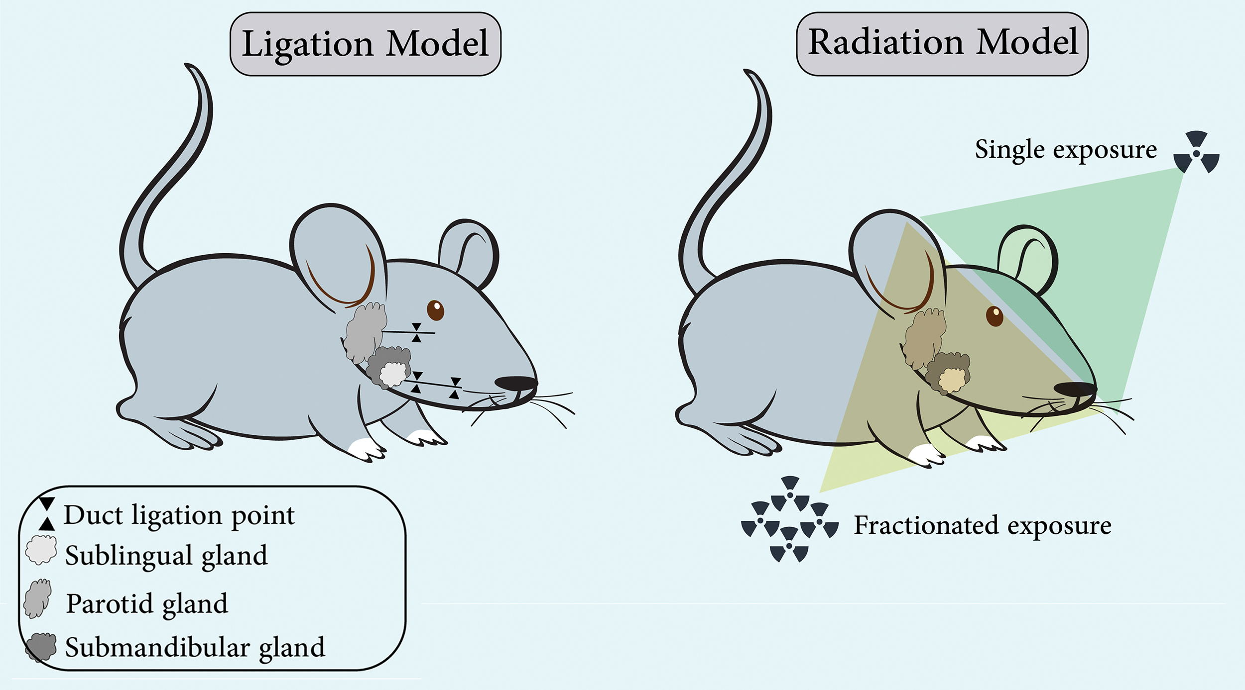
An overview of animal models that simulate salivary gland damage caused by radiotherapy induction and duct obstruction. Color images are available online.
The skin of rodents is thinner compared with humans, produces less scarring, and heals differently due to the rapid contraction of the wound.100,101 In rodent models, scar formation mechanisms can only be studied in the short term rather than the slow and long-term development of pathological scars in humans. When using mice, it is vital to consider the potential for variation in wound healing and inflammation due to different strains. 102 Despite these limitations, it is still viable to use excisional and incisional wound models in rodents to examine the process of typical scar development and the impact of interventions. 103
Rodents are the primary focus of the well-established preclinical models used today for facial nerve regeneration. Nerve crush, nerve transection, and nerve gap are the three main types of nerve injury models.104,105 For conduit investigations, the average size of the facial nerve defect or gap is <10 mm. With a handful of nerve transection-only investigations, the great majority of conduit studies are conducted in the 5–8 mm gap range, and there are just a few uses in gaps wider than this.
On the other hand, models for graft investigations (autografts and allografts), like cross-face neurotization, require gaps >10 mm. 76 Rat models can be used to study electrophysiology, measure functional recovery, analyze the morphology of muscles and nerves, and evaluate other aspects of nerve regeneration. Using relatively small gaps—the majority has employed ones of 10 mm or less—ranging in length from 1 to 50 mm is a drawback. Compared to target nerve lesions and nerve axotomies in humans, small gaps in rats undergo complete recovery. Furthermore, humans regenerate peripheral axons slower than rodents, further complicating the situation. 76
The use of silicone tubes in various studies has been demonstrated as an effective method for locally delivering and encapsulating factors that promote nerve regeneration. A silicone conduit was used to deliver FGF-2 embedded in microspheres into a 7 mm facial nerve defect by Matsumine et al. 106 A scaffold-free bioprinting method was used by Zhang et al. for creating constructs from gingiva-derived MSC spheroids that were transplanted into a 5 mm defect in the buccal branch of a rat facial nerve. The functional restoration was superior to that of the plain silicone tubes, but it fell short of that of autografts. However, the target muscle recovery obtained by electrophysiology and the histopathological organization of nerve fascicles was comparable to the autologous grafts, suggesting potential applications for improving nerve regeneration in the future. 107
Grafts made from autogenous veins and arteries enhance axonal regeneration by preventing scar tissue from growing inside the graft environment.108–111 The histology or functionality of the regenerated axons in a transection model was not improved by an autologous venous ensheathment to the facial nerve trunk. 112 Another study used a decellularized allogeneic artery graft enriched with adipose-derived stem cells in a 6 mm gap; results showed facilitated recovery, but at a lower extent than nerve autografts.113,114 The abdominal aorta was utilized as a Y-shaped conduit in a significant facial nerve defect. It improved axonal pathfinding, but did not improve functional outcomes.115,116 Table 2 summarizes some of the most essential benefits and drawbacks of utilizing rats as model animals.
Advantages/Disadvantages of a Variety of Animal Models Used in the Engineering of Craniofacial Tissues
Rabbit (Oryctolagus cuniculus)
Compared to pig, ovine, bovine, canine, and nonhuman primate models, rabbit models are more economical. 89 They are frequently used as TMJ anterior disc displacement models and disc perforations. 7 Rabbits provide large amounts of tissues for reliable mechanical testing for TMJ, such as friction, shear, tensile, and compression. 117 The rabbit model is an ideal small animal model for assessing cartilage repair due to its larger joints and convenient size for surgical practices and sample processing (Fig. 5). Even though some 1-year rabbit experiments have been conducted, rabbits have been widely used to evaluate cartilage restoration. 47 A rabbit randomized controlled trial conducted by Köhnke et al. evaluated the chondro-regenerative potential of intra-articular stem/stromal cell therapy. They followed a single intra-articular injection of a developed therapeutic medicinal product that complied with Good Manufacturing Practice (GMP) and contained stem cells as a treatment. 118
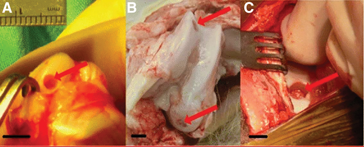
Macroscopic view of the distal femur (condyles of the distal epiphysis) of
For testing biomaterials or evaluating treatments for peri-implantitis, rabbits have been used in periodontal regeneration studies. 87 Periodontitis does not occur spontaneously in rabbits, so the disease must be induced. Furthermore, the periodontal pathogenesis model provided by rabbits is poorly standardized. 119 The bones of rabbits differ considerably from those of humans in both macrostructure and microstructure. Rabbit skeletal bones have a primary vascular longitudinal tissue structure rather than the secondary hierarchical structure of mature human bones. Despite this, rabbits have a more human-like bone structure than mice. 13 Rabbits have quicker bone remodeling than primates and rodents.
Therefore, the rabbit's findings should not be extrapolated to possible human clinical outcomes. Compared with rodents, rabbits have a larger bone size, which facilitates creating bone defects. Blood samples can also be obtained from the marginal ear vein of rabbits, which is advantageous for blood analysis. 94 Lee et al. 120 implanted four experimental groups into rabbit calvarial defects to conduct their research. A bony defect model for rabbits was used to study the osteogenic effects of bone marrow stromal cells and dental pulp stem cells (DPSCs) (Fig. 6). 120 Using a bone defect model, Chen et al. tested TCP/poly(lactic-co-glycolic acid) (PLGA)/icaritin scaffold on the ulnar area. 121 Delgado-Ruiz et al. 122 used a rabbit model for a tibia defect to evaluate titanium granules with and without membrane covering. According to their findings, when grafting more significant defects, the titanium particles needed to be protected by a barrier membrane. 122
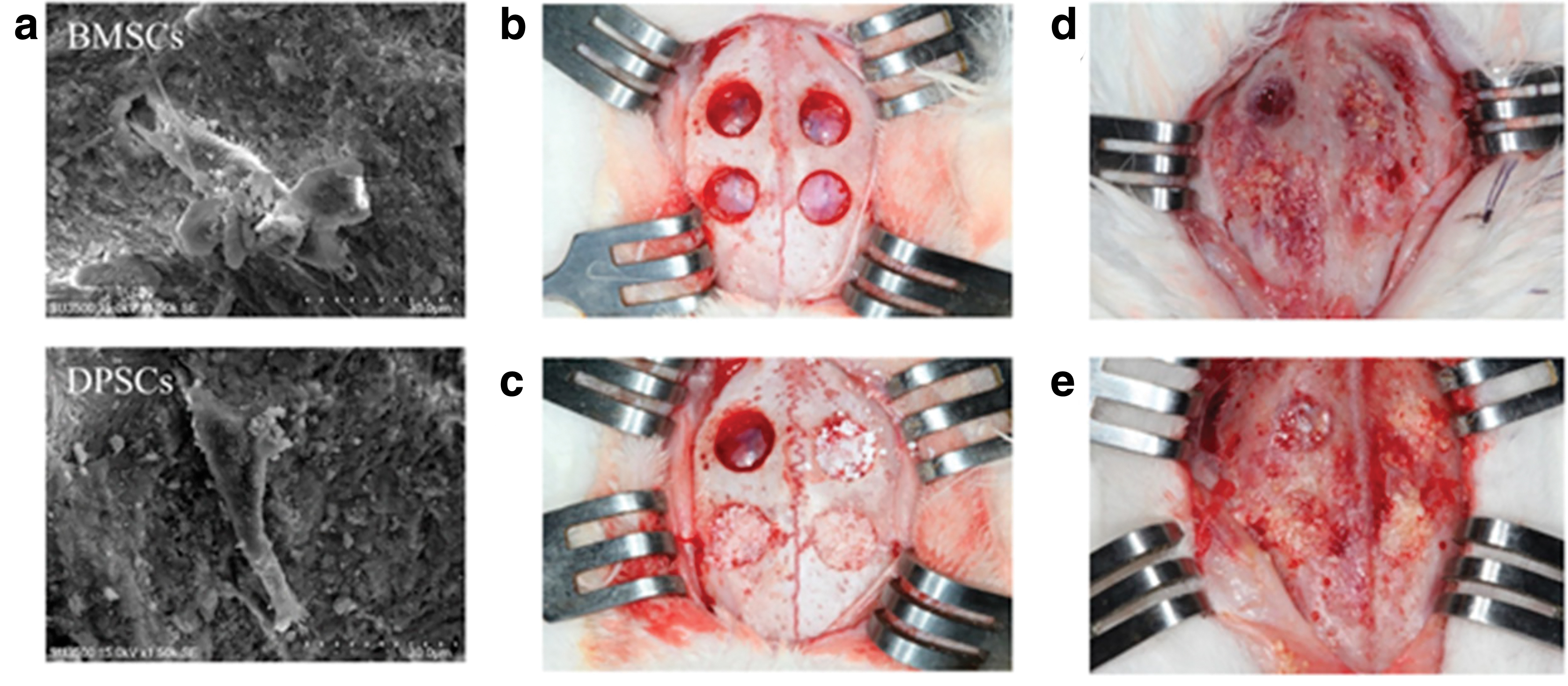
Microscopic examination of rabbit calvarial bone defects.
It is widely accepted that rabbit ears are a reliable model for hypertrophic scarring. There was also significant scar reduction in this experimental model after using silicone gel dressing, a widely used therapeutic approach for hypertrophic scars. 123 When epidermal hydration is maintained by occlusion, keratinocyte activation and proinflammatory mediators are inhibited, and dermal fibroblast activity is decreased, resulting in less scar tissue formation.124,125 Celecoxib alone, 126 or with captopril, 127 has also been shown to reduce scar elevation in the rabbit ear model. This model has been utilized in several investigations to uncover differentially expressed genes, alter biological processes, 128 and investigate the role of adipose-derived stem cells 129 in developing hypertrophic scars.
Rabbits are also frequently used to study peripheral and cranial nerves. Various nerve gaps have been used with rabbits, ranging from 2 to 50 mm. Neuromorphometric and electrophysiological analyses have usually been used to assess the regeneration capacity in this species. 76 Due to the animal's larger size and ample facial nerve length, rabbits are commonly used for nerve regeneration in more significant defects (>10 mm). On the other hand, face nerve repair rabbit models are limited in number compared to rat models.
Several studies have shown that silicone tubes can restore the facial nerve in rabbits. For instance, silicone tubing serving as conduits for nerve growth factor (NGF) solutions was employed in an 8 mm nerve gap.130,131 Compared to autologous nerve grafts, growth factor-filled conduits resulted in comparable functional regeneration with fewer collateral nerve sprouts. A 10 mm facial nerve defect was more efficiently recovered using neural stem cells combined with NGF in a chitosan conduit and protein sponge by Guo and Dong. 132
Several studies have used venous autografts for facial nerve repair in rabbits. Tang et al. compared inside-out vein grafts with autologous ones in a segmental nerve gap of 10 mm. 133 The former significantly facilitated axonal regeneration, although both benefited nerve regeneration. A 10 mm segmental nerve defect was also treated with vein grafts to deliver stem cells. 134 Table 2 summarizes some of the most significant benefits and drawbacks of utilizing rabbits as model animals.
Dog (Canis familiaris) and cat (Felis catus)
Companion animals have gained significant attention as models for human diseases as a result of the One Health initiative, which seeks to “break through species barriers” to connect medical and veterinary research for the benefit of both human and veterinary patients. 135 Joint disorders, particularly temporomandibular disorders (TMD) and osteoarthritis (OA), have been studied using dog and cat models for a long time. Dogs have received more attention for OA than sheep, horses, and goats. 5 The canine and feline TMJs serve primarily as hinge joints.
Laterotrusion is possible in dogs to a limited extent. Cats have a more dominant pure hinge movement since their TMJ anatomical characteristics are more constrained. Dogs and cats have thin TMJ discs due to their primarily rotary articular motion, which correlates with their structure-function correlations. 7 TMJ-OA is the most prevalent naturally occurring TMD in dogs, and it is also the second most pervasive TMD in cats, behind fractures. 136 Dogs and cats also show similar clinical manifestations of TMJ-OA. The severity and presence of computed tomography (CT) findings may not be related to clinical symptoms. 137 Ankylosis, luxations, fractures, and a range of neoplasms, which resemble comparable conditions in humans, are some more naturally occurring TMJ problems in dogs and cats. 137 Such conditions can be used in clinical trials as a clinically meaningful platform for translation, much like the scenario with TMJ-OA.
In contrast to dogs, cats are less frequently used in craniofacial tissue engineering experiments. The use of cats for hard and soft tissue research has nonetheless been widespread. According to research, cats are maybe the most human-like nonprimate model for investigations on nasal growth. 138 Studies on cats' soft tissues have focused on the temporalis muscle, which appears to be connected to their prominent temporalis muscle that is especially receptive to surgery.139,140 The regeneration of nerves has been studied in dogs and cats with gaps up to 90 mm. It has been found that neuromorphometric analysis is the most common method to determine regeneration ability in both species.
The assessment of muscular and spinal kinematics characteristics following experimentally induced spinal cord or cerebral cortex injuries is done using electromyography (EMG), force plates, treadmills, and biomechanical motion analyses. 5 In addition to studying the cat's inferior alveolar and lingual nerves, several NGFs have also been studied, which may aid in the reconstruction and regrowth of trigeminal nerve branches. This includes the reaction of these nerves to injury, as well as their spontaneous and surgically induced regeneration.141–144
Cat models have been utilized to evaluate nerve guidance and eyelid reflexes because the depiction of facial muscles in felines' facial nuclei is similar to that of human facial muscles. A collagenous nerve guide placed in a 5 mm facial nerve gap, for instance, was shown by Kitahara et al. to be related to improved healing. 145 A study using various surgical techniques evaluated the flexibility and plasticity of the facial motor system as well. 146
Most studies have used dog models in oral tissue regeneration. The growth patterns, anatomical structures, and pathophysiology of canine teeth are similar to those of human teeth. The size, pulpal structure, and periodontal ligament attachments of dog teeth are comparable to those of human teeth. 139 Because of their striking resemblance to human molars, canine premolars are the preferred choice in studies. 82 The significant differences between dogs and humans are their limited lateral movements, no occlusal contact for all premolars, and open contact between teeth. A further difference between dogs and humans regarding their periodontal conditions is the composition of bacterial plaque/calculus. 87 The evaluation of filling biomaterials and regeneration procedures also involves combined techniques, such as surgically creating periodontal defects and placing bindings.147,148 The treatment of periodontal diseases with MSCs has been reported in research on dogs.149–151
Guided bone regeneration,152,153 and implant surgery154–156 have also been performed on dogs. There is a crucial role for canines in implantology. 38 While dogs' jawbones are denser and more resistant than humans, the bones of dogs are most similar to those of humans in terms of turnover, composition, and mechanical properties.13,38 In the implant experiments, the ramic angle is also employed to create critical-sized flaws filled with different biomaterials. 157 Numerous research examined re-osteointegration models and peri-implant bone defects in dogs. Bindings around implants were used to induce peri-implantitis over 2 months.
In this way, osseous defects around implants appeared to be reasonably comparable to those found in humans. 158 There are many similarities between canine and human bone components. In the surrounding endosteum and periosteum, canine bone develops plexiform/laminar bone in the form of secondary osteonal structures. Large animals and children with rapid growth have plexiform bones. Thus, the canine bone represents the human bone structure in the best possible way. 94 Dogs' age-related bone changes are comparable to humans. 139 According to consensus, dogs have faster bone turnover than humans. Even though canine bone has a similar organic composition to human bone, implant-associated changes may not be as apparent in human cases as in canine models because humans have a lower remodeling rate. 159
Although the use of dogs as bone defect models in orthopedic research has declined due to public complaints, they were previously used as models in orthopedic research. 16 A canine mandible edentulous area was used to assess the effect of implant placement with the bone ring technique.160,161 The amount of mineralized bone was similar in the two groups based on micro-CT and histomorphometric analyses; the one-stage process achieved equal osseointegration. There was a more significant residual bone graft in the single-stage group, whereas more de novo bone development was observed in the two-stage group.160,161 Sawada et al. implanted three natural bone blocks into the beagle dogs' calvarial bone defects: β-TCP, deproteinized bovine bone mineral (DBBM), and α-TCP/hydroxyapatite (HA). All bone blocks retained their original shape, despite only limited bone growth in the bone substitute during healing. 162
There have also been studies conducted using dogs to examine facial growth. Dogs have furthermore been investigated for their potential to serve as spontaneous animal models mimicking human diseases. Maxillary brachygnathism, for instance, occurs naturally in many dogs. Infantile cortical hyperostosis may be investigated using these animals as pilot studies for humans. Orthognathic surgery and tooth viability studies have also been conducted with dogs. 139 DO, which involves gradually distracting the facial bones following an initial osteotomy, has also been investigated with dogs. A modified intraosseous distractor was used by Esposito et al. to perform DO on a dog's posterior mandible to augment its vertical bone length. In a separate study, 163
Terbish et al. investigated the effect of injecting rhBMP-2 into beagle dogs after a DO procedure. The alveolar ridge's width and height improved after receiving an rhBMP-2 injection, substantially enhancing bone volume. 164 Dogs and cats receive an unparalleled amount of affection and attention in our culture as companion animals and pets. Thus, the ethical discussion around studies using dogs and cats is more heated compared to experiments involving other animals. Veterinary hospitals and clinics, however, are the most common settings for these studies, underscoring the significance of this investigative resource. The most significant benefits and drawbacks of employing dogs and cats as model animals are listed in Table 2.
Pig (Sus scrofa domesticus)
The pig is one of the most common domesticated animals in the world. Pigs grow rapidly and have a shorter generation interval than other livestock and primates. Pigs have gradually become more prevalent as animal models for human diseases due to comparable body sizes, genetic backgrounds, anatomical and physiological attributes, and diets. 165 It is easier for pig experiments to be transferred to human conditions because pigs have physiological similarities to humans, as opposed to mice, rats, or rabbits.
The anatomical similarities between pigs and humans, the functional similarities, and the availability of disease models have made pigs and minipigs popular in medical and pharmacological research. 166 Conducting clinical trials on nonrodent species for pharmaceuticals intended for human use is essential. Dogs or, to a minor extent, primates are the most popular options. Due to their similar hematological and cardiovascular characteristics to humans, pigs and minipigs are also suitable for testing pharmaceutical products for toxicity. 17 Another advantage is that euthanizing pigs, considered livestock, is viewed as less critical. 82 Table 2 provides a summary of some of the most significant drawbacks and benefits of utilizing pigs as model animals.
Porcines have a narrow and long oral cavity, which does not differ among breeds, unlike humans, whose oral cavity is almost oval shaped. Compared to other species, pigs have short labia and are less moveable, so they have difficulty opening their oral cavities fully; this anatomical layout makes intubation for anesthesia challenging. It is possible to study scarless wound healing in the oral mucosa of the pig. The palatal mucosa's histological structure exhibits a pattern similar to that of humans. 167 It may be possible to develop new clinical techniques and find molecules suppressing scar formation after surgical procedures by understanding these scar-associated molecular pathways and the healing cascade of the oral pig mucosa without scarring.
The human tongue shares a similar histological structure with the swine tongue, which is long and tightly bound to the floor of the oral cavity by a double frenulum. Cold ablation (coblation) has been studied as a surgical technique to reduce tongue volume in the swine tongue model.168,169 The porcine tongue was used as a test bed for determining the lesion/scar formation process after this procedure and improving it further.
Studying salivary glands in pigs makes more sense because they share similar gland size and comparable physiological and morphological characteristics with human glands, including their structure and ductal system. 95 Since patients with ionizing radiation-treated head and neck carcinoma often have salivary glands affected, researchers and clinicians have paid attention to these glands. 170 In this regard, minipig salivary glands may prove helpful in preclinical gene transfer experiments, given their similar volume and morphology to human salivary glands.171,172
Pigs are also utilized in various research fields, particularly as surgical procedure models. This is due to their resemblance to humans regarding physiological characteristics, growth patterns, and head size. Due to their similar brachydont and bunodont dentition and enamel mineralization patterns, pigs have been used as dental models for a long time. 17 Because of their ease of access and suitability for dental pulp tissue engineering testing, porcine premolars were used for endodontic procedures. Significant consideration was also given to the potential for human tooth regeneration to restore missing or damaged teeth. 173 The advancements in dental implants, restoration of the periodontal system, or the regeneration of biological and functional teeth from embryonic/postnatal tooth buds are the main areas of this research.174,175 Root/periodontal complexes are successfully created in minipigs using periodontal ligament/human apical papilla-derived stem cells. 176
In numerous disciplines of research on the craniofacial bones, including nasal, calvarial, maxillary, or mandibular bone defects, 3D ridge augmentation, DO, implantology, management of peri-implant diseases, and sinus lift procedures, porcine breeds have been utilized as experimental animals. 30
Various types of dental implants, coatings, and osseointegration rates were tested in minipig maxilla to assess their stability and healing process.177–179 Growth factors and collagen have also been proven to assist osseointegration and healing following dental implant surgery.180–182 Dental implants are generally challenging to be tested on pig mandibles for two reasons: (1) The large inferior alveolar canal is superficially located. Therefore, they are prone to have the superior border of the inferior alveolar canal penetrated during implant drilling/placement and (2) the canine teeth occupy a significant portion of the mandibular bone. If these teeth have an issue, removing them without damaging the intricate bone structure would be difficult. This may affect the success rate of implanted teeth in the pig mandible since proper implantation is more complicated.
The primary complication of inflammatory periodontal diseases is the loss of periodontal supporting tissues. 183 Pigs' periodontal tissues undergo an inflammatory process similar to humans 95 ; therefore, it was tested in pigs whether periodontium/alveolar bone-derived cultured cells could develop new periodontal tissues. 184 It was discovered that cultured cells helped develop new bone, cementum, and attachment fibers, as well as helped wounds heal by preventing epithelial recession. 184 A porcine periodontitis model was also used to study the potential use of autologous periodontal ligament stem cells (PDLSCs) in treating periodontal defects. 185 Minipigs were used to obtain autologous PDLSCs. Alveolar bone and periodontal defects were treated using in vitro cultured cells. Periodontitis can therefore be treated with stem cells, as confirmed by regenerating periodontal tissues using PDLSCs.185–187
For creating and closing oral communications, pigs are also suitable animal models. 188 In addition, biodegradable materials were assessed for their ability to close these defects. 189 Although bone regeneration for dental implants or sinus augmentation is frequently employed in human beings, practitioners are unable to obtain histology samples to better understand the process. 190 In vivo models of these processes can be studied in greater detail in the pig, providing new potential study avenues. As applicable models for testing novel biomaterials in preclinical settings, pigs have been employed to study bone substitutes and their influence on de novo bone regeneration. 191 On the other hand, using pigs for the Caldwell-Luc approach is constrained by the excessive thickness of the cortical bone.
Thus, the swine model is not the best for practicing these concepts. 192 Various bone substitute materials were tested using a porcine craniofacial defect model.193,194 To investigate platelet-rich plasma (PRP) in bone regeneration surrounding dental implants, it has been established that the pig is an appropriate animal model. 195 According to Wehrhan et al., porcine craniofacial bone defect models and alternatives to these models are commonly used for evaluating replacement materials. A porcine craniofacial bone defect model was used to develop a gene delivery technique for bone regeneration. More new bone was formed when gene delivery was used at the defect site. 196
Pig orbits are also great experimental models for testing and developing alloplastic materials for reconstruction after trauma, tumors, or developmental difficulties. These materials would deteriorate gradually, and because of their osteoconductive qualities, osteogenic tissue could replace and repair them. Testing these properties in a pig model before applying them to humans will be necessary. 197
With current techniques, replacing bone and teeth in craniofacial reconstructions necessitates complicated procedures. Although pigs have been used in bone regeneration studies, their use is often discouraged due to the need for careful species management. 16 The porcine maxillary and mandibular tissues can be employed for integrated autologous tooth and bone regeneration.
It also serves as a foundation for advancing tissue engineering capabilities for potential clinical use in humans.194,198–201 Children with cleft palates, with upper teeth positioned behind the lower teeth, are typically treated with maxillary distraction. For researching the healing process during DO, the pig has long served as a helpful model using Le Fort I devices. 202 Minipig experiments may also aid in constructing internal biodegradable/nonbiodegradable distraction devices. Considering the thickness and mass of the maxillary bone, DO studies have used the frontal bone for easier access.194,203,204
The ideal timing and long-term consequences of fixation procedures on a developing skull in utero were assessed using temporal and parietal bone fragments. 205 The mandibular blood supply and design of osteosynthetic screws and plates can also be studied using pigs as experimental animals.206,207 Understanding the mechanical and molecular processes that control bone growth during DO also requires knowledge of the pig mandible.208–210 Testing endoscopic procedures such as resectioning mandibular angles and placement of distraction devices can also be conducted with a pig mandible.211,212
The pig can be a beneficial model for studying stem cell-based tissue engineering for bone regeneration. 213 Miniature pig stem cells were extracted from the ilium, bone marrow, or deciduous teeth and grafted onto the critical-sized bone lesions created in pig mandible models.199,200,214–217
Models of porcine joints and tissues, such as ligaments, bone, and cartilage, have long been used to study the biomechanics of these joints. 5 The pig has been utilized for direct measurements of TMJ tissue deformation and load during biting, making it the greatest nonprimate model for human TMJ diseases.218,219 The pig's disc can be used in tissue engineering46,220 to develop new management techniques for post-traumatic complications and degenerative TMJ problems 221 because it has topographical, biochemical, and biomechanical characteristics, which are comparable to those of the human disc.
In addition, performing invasive arthroscopic TMJ surgery is technically complex and necessitates the development of competent arthroscopic skills, which are difficult to acquire by treating patients alone. As a result, the arthroscopic surgery of the TMJ and its future development in pigs serve as a trustworthy training prototype. 222 The swine model's huge size also enables the evaluation of tissues that are typically challenging to evaluate.
Murphy et al. evaluated the response of six TMJ discal attachments to tensions of different orientations. They found that the superior-posterior attachment of the TMJ had the highest modulus values under mediolateral stress. All the attachments, with the exception of the anterior-inferior attachment, showed anisotropy when the collagen orientation was examined. 223 Also, TMJ discs in pigs show region-specific compressive characteristics, with higher rigidity in the tissue's medial and posterior portions. These directionally dependent and regionally specific characteristics of the TMJ disc serve as crucial design parameters for studies in TMJ tissue engineering. 224
The skin structure of pigs is rigid, and they have a thick epidermis, elastic fibers, Langerhans cells, and a collagen structure similar to human skin. Pig and human skin also exhibit similar blood vessel distribution/orientations, as well as papillae and dermal ridges. 52 Due to its similarities in skin structure and healing mechanisms to human skin, the porcine model has been widely used to study normal scarring.225,226 To determine whether moist wounds heal better than dry wounds, Reish et al. 227 studied different wound types in pigs, including full/partial-thickness wounds, meshed/sheet split-thickness skin grafts, incisional/excisional wounds, and minced skin. Additionally, findings from pig burn trials have demonstrated that surgical debridement, 228 ideal graft bed features, 229 or less severe injury 230 result in reduced scarring. These studies indicate that porcine models can be valuable preclinical scar study models.
In nerve experiments, large gaps in minipig models were used along with collagen nerve conduits to convey specific cues to the defects. Cui et al. also found that collagen scaffolds combined with recombinant proteins produced favorable results in a 35 mm gap. 231 Type I collagen and nanoscale β-TCP conduits were investigated and coupled with NGF in a comparable 35 mm gap model. 232
Sheep (Ovis aries)
Research with domestic sheep offers unique advantages due to its accessibility, affordability, and social acceptance as a research animal. Some anatomical and biomechanical characteristics of sheep, such as weight, joint structure, bone composition, and architecture, are very similar to those of humans, making them excellent models for studying osseous and cartilaginous tissue repair and remodeling. 5 Treatment of segmental bone defects is most commonly tested in sheep. The findings can be easily applied in a clinical setting because adult humans and mature sheep have similar body weights.
Due to their voluminous size, they can be repeatedly sampled over a long period from various anatomical locations. Compared to other large animal models, like the horse, their size is excellent for clinical imaging modalities like MRI and CT intended for humans. As a result, utilizing sheep eliminates the need for major investment in surgical facilities or specialized handling equipment. This human-sized model also enables the testing of surgical techniques and medical equipment. The well-documented mechanical loads of ovine (sheep) hind limbs, nearly half that of humans while frisking, is another factor facilitating the translation of research results.
Sheep bones have a different microscopic structure than humans, but their metabolism and bone remodeling rates are similar. 16 Thus, sheep bones can be reliably used to test bone substitutes. Yang et al. studied the osteogenesis and remodeling effects of a biphasic synthetic bone graft comprising calcium sulfate and TCP on a vertebral sheep defect model. 233 Kobayashi et al. also investigated TCP formulations for histological characteristics in the sheep vertebral bone defect model. 234 A 3D printed TCP/HA porous block was examined histomorphometrically using a sheep calvaria vertical guided bone regeneration model by Carrel et al. Various bone substitutes were implanted on sheep calvaria using titanium shells. These included Particulate Bio-Oss®, 3D printed blocks, and β-TCP. All other biomaterials showed inferior bone growth after 8 weeks, but the 3D printed block had superior bone growth. By 16 weeks, the new bone had almost filled all the spaces. 235
The use of scaffolds with/without MSCs has also been studied in sheep to treat critical-sized bone defects. A comparison of these treatment modalities with gold-standard methods such as autologous bone grafts showed that they enhanced bone formation and improved mechanical properties.16,236–240 There are little data on segmental mandibulectomy and maxillary sinus augmentation in ovines. The applicability of ovine models for follow-up investigations may be constrained by differences between ovine and human bones, including increased density and mechanical resistance in ovine bones and aging-related structural and remodeling changes. 12
In vivo, preclinical research in TMJ surgery frequently uses sheep as a large animal. In TMJ studies, sheep have been employed as an appropriate model to study postcondylectomy consequences and condylar reconstructive options, minimally invasive procedures like arthroscopy, TMJ ankylosis, and OA. 7 For the study of TMJ surgical methods, larger animals are preferred over laboratory models due to their larger size, which allows for better surgical access and larger tissue appropriation for mechanical testing. The condyle of the sheep has an elliptical form and is mediolaterally concave, with a longer axis in this orientation. A sheep model for TMJ surgery is suggested due to its anatomical similarities to humans and extensive in vivo experience in TMJ surgery. 241
Sheep are also rarely studied in periodontology. A constant cement apposition is characteristic of sheep periodontium as compensation for teeth attrition. Consequently, this periodontal physiology may impact regeneration mechanisms, representing a bias for clinical conditions. 242 It is also reasonable to consider sheep as animal models for dental regeneration, although they are not widely used. However, DPSCs have been used in various studies associated with bone grafting techniques.243–245 Using ring-shaped bone blocks of three different materials, Jinno et al. implanted the lateral margins of sheep mandibles with Bio-Oss, resorbable biphasic ceramic implants, or autogenous bone. They then used 3D imaging and histomorphometric analysis to evaluate the volume changes and osseointegration of implants in the bone rings. Even though the grafts in all groups were shrunk, all cylindrical shaped blocks were well stabilized around the implants. Dense cortical bone blocks outperformed the other materials regarding volume maintenance. 246
The ovine species is also a suitable and efficient model for studying nerve regeneration due to its comparable nerve size and regenerative characteristics to humans. 5 The similarities in nerve size and regenerative pathways between sheep and humans are the key benefits of using sheep as models for nerve regeneration. Histological investigations show that their nerves are likewise poly-fascicular, much like human nerves. 247 Several translational research projects have used sheep's hypoglossal or facial nerves in orofacial medicine. Niimi et al. analyzed the anatomy of these nerves using an ovine model. As a result of the resemblance in nerve structure and innervation, they contend that the ovine model can be employed for research on the facial nerve. 248
Sheep have not been frequently employed as cutaneous animal models for studies on wound healing. However, given their affable nature and the availability of superficial skin tissue for developing wound models of various shapes and sizes, they could serve as suitable skin models. Different gels, PRP, and other topical treatments have been used to investigate the regeneration potential of various skin wound healing modalities in this animal species.249–253 In addition, studies have been conducted linking MSCs to better outcomes in wound healing experiments. By assisting in the vascularization, reepithelialization, and extracellular remodeling of the skin, MSCs could promote wound regeneration and reduce the healing period.249,254,255 The most significant drawbacks and benefits of employing sheep as model animals are outlined in Table 2.
Horse (Equus caballus)
They could serve as a model for developing and testing novel therapeutics through naturally occurring disease models. 7 For studying musculoskeletal diseases, horses have long served as an approved, established, and clinically appropriate animal model. 5 The horse is also a good model for cartilage thickness and common morphological characteristics. The horse may also be suitable for modeling naturally occurring TMJ diseases. Horses chew in a cycle that includes an opening, closing, and power stroke, just like other herbivores do. Horses have a unimodal power stroke, and their mandibles travel mediolaterally. Previous kinematic studies also showed that the working side of a horse's TMJ moves lateroventrally during the opening stroke and mediodorsally during the power stroke. 256 Furthermore, equine age-related joint degeneration, known as articular disc mineralization, occurs in a manner comparable to humans. 257 Equine TMJ fractures, OA, and septic arthritis in horses are similar to those in dogs and humans.
The horse is a well-liked model for nonterminal studies, with comprehensive assessment and follow-up being feasible because sophisticated diagnostic approaches, such as arthroscopy, magnetic resonance imaging, ultrasound, radiographs, computed tomography, scintigraphy, serial sampling, and second-look arthroscopy, have been shown to be applicable in horses. 5 The voluminous size of horses also provides a high tissue quantity for sampling, further analyses, and creation of multiple/critical size defects, allowing for thorough studies that may not be achievable with laboratory animals.
It is then possible to compare diagnoses, follow-ups, and results for horses using these methods combined with well-established histologic and pain scores.258,259 Thus, in order to carry out equine surgeries, it is essential to have a highly specialized facility equipped with state-of-the-art equipment, staffed by qualified personnel, and situated within a specialized natural environment. All this adds up to significantly higher study-related costs. 47 The biomechanical strains, which are significantly greater than those considered physiologic in humans, may limit the rehabilitation of injured structures, the application of transplants, and suturing. Thus, horses do not provide an ideal model for bone healing.
Notably, ponies heal wounds more quickly and effectively than horses because they produce a more rapid and robust inflammatory response and have superior infection resistance. In human and equine clinical trials involving cartilage repair, the study duration must be at least 8–12 months since long-term failure is common, even when short-term results appear promising. 260 Table 2 overviews some of the critical drawbacks and benefits of applying horses as model animals. Figure 7 illustrates different animals used as investigative models in tissue engineering for craniofacial tissues.
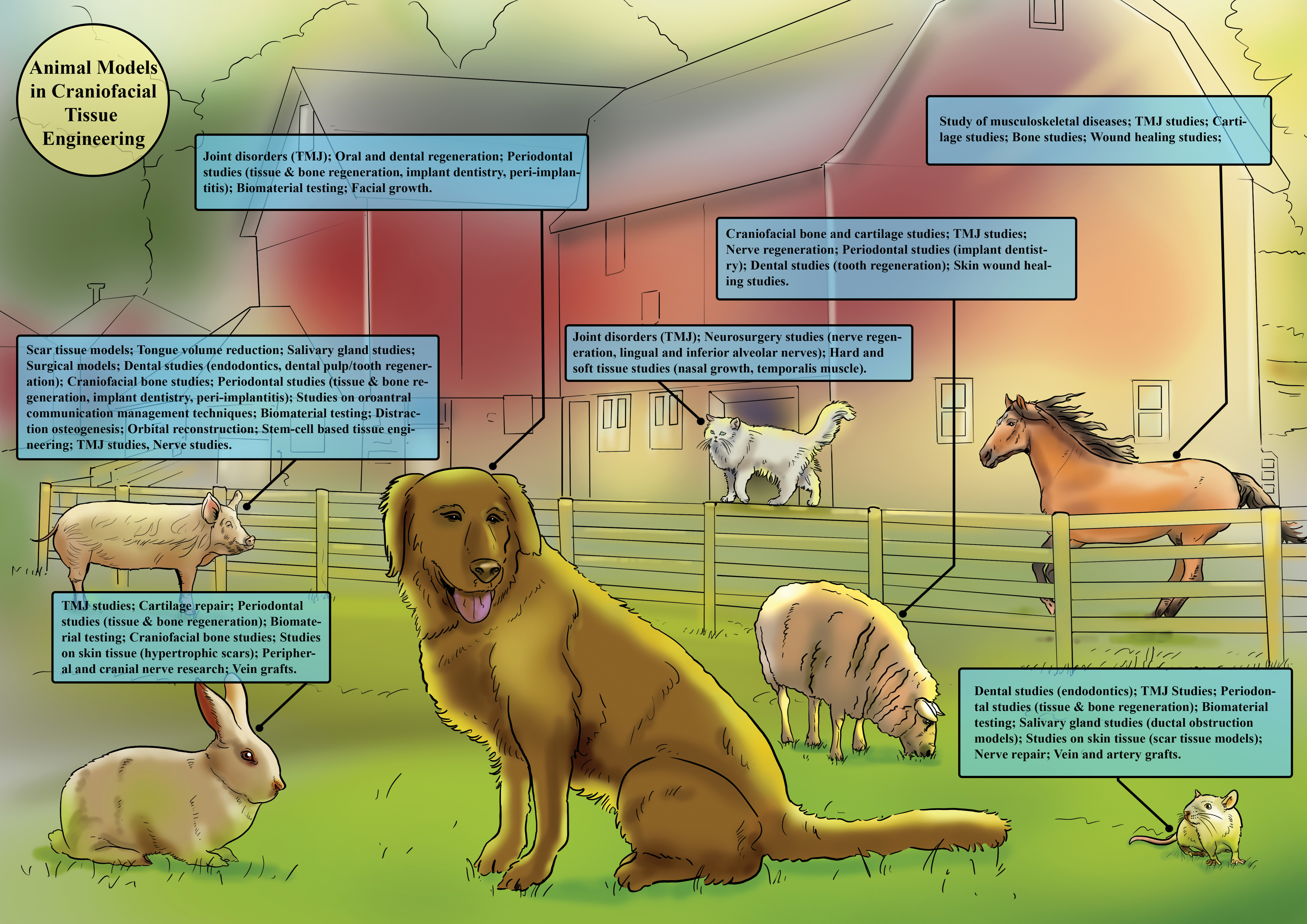
Frequent animal models used in biomedical investigations focusing on their potential contributions to craniofacial tissue engineering. Color images are available online.
The practical value of the discussed animal models in studies of craniofacial regenerative medicine is scaled down in Table 3 by two authors (S.A.M. and H.T.).
The Suitability of Common Animal Models in Craniofacial Tissue Engineering, Considering Their Clinical Translation
The information utilized to structure this table was adapted from included studies.5,7,12,13,16,17,30,32,34,38,41,46–49,54–56,66,67,76,82,87,88,91,92,95,100,101,104,105,119,123,138–140,152–156,158,168,169,171,172,218,219,225,226,242,248,257
++, Highly recommended; +, recommended; −, less recommended; −−, not recommended; N/A, not applicable; TMJ, temporomandibular joint.
Biological characteristics of craniofacial tissue regeneration in animal models
Despite evident differences (Table 4), the physiological and anatomical similarity between humans and animals, especially mammals, make the latter an appropriate model for drug development and biological research. 261 Given the physiological complexity of humans and mammals regarding their circulatory factors, cellular structures, tissues, and hormones, different pathophysiological dimensions, such as organ and cellular functions and molecular structures, should be evaluated. All animal models have restrictions that make them unsuitable for biomedical research, making their selection procedure particularly complex.
Physiological Parameters of Different Animal Models Compared to Humans
Optimal animal models should be selected based on limitations, physiological factors, and pathophysiological similarities between human and animal species. Detailed evaluations should also be performed when applying medicines, molecules, and medical devices to measure their capacity to regenerate the condition or pathology at the same level as humans. The other influential factors include the availability, size, and life span of animal species, as well as the duration of the study. For instance, a large animal population can provide several subspecies.10,261 Further complicating matters is that the experimental outcome is also highly variable depending on which animal species/strain is selected, where the defect is located, its size and shape, which control groups are selected, and the time points assessed.
The animal model's age should also be considered regardless of the species. The use of skeletally mature animals to simulate adult physiology and healing capability is a crucial factor in the design of a definitive study. 262 It is also important to note that preclinical models often use young, healthy animals, which do not represent human patients' aging and potential age-associated comorbidities. 263
A bone defect model should have a size exceeding the body's self-repair capacity, leading to nonunion without immediate treatments. Nonunion is caused by a critical size defect, a bone defect that cannot recover in a spontaneous manner or that will not regenerate more than 10% within the experimental period. 264 Given the confirmed regeneration of critical size defects by guided bone regeneration, it is recommended that experimental models be developed to evaluate the impact of diverse treatment modalities on bone regeneration. 36 The models of these defects, involving healing orthotopic bone sites such as the calvarium and mandible, can provide “proof-of-principle” through discriminative and standardized evaluations of bone regenerative modalities and bone grafts.
The extent of the defect varies depending on the animal model, age, weight, the location of the lesion, and other disease variables. 94 The calvarial bone defect model is frequently used by researchers to create a standardized lesion that can be examined using histology and radiography. Biomaterials can also be implanted without external fixation with sufficient surgical access because of the support that the dura mater and the skin provide. In some clinical situations where the bone tissue must support mechanical stresses, it is required to evaluate the performance of biomaterial under physiological mechanical loads; this cannot be done using a calvarial model. 265 Preclinical animal models commonly used for calvarial defects include rats, rabbits, and mice. 2
Continuous and noncontinuous defects of the mandible can be distinguished. There is no need for an additional mechanical fixation in noncontinuity defects because their geometry is circular or rectangular without compromising the mandibular unity. Small animal models are more frequently utilized to study these defects, which can shed light on the biocompatibility and effectiveness of tested constructions, but often fall short of accurately simulating clinical conditions like size and load-bearing, as was the case with calvarial defects. 266 Post-tooth extraction bone remodeling is a clinical example of employing noncontinuity defects in a preclinical model. Continuity defects are often caused by segmental resections resulting in mandibular continuity loss following invasive procedures such as tumor resection. As a result, internal fixation is necessary to give sufficient mechanical stability. 267 Microbial infections, while employing intra-oral techniques, 268 as well as plate failure in continuity defects 267 are complications related to mandibular defects.
Other craniofacial reconstruction approaches include combined dental implant placement with sinus and 3D ridge augmentation. Employing human subjects in clinical studies and animal models in a preclinical setting helps acquire knowledge about peri-implant hard and soft tissue healing. Surgical interventions trigger a complicated cascade of biological events that heal the defects. The temporal sequence of healing events that leads to osseointegration has been reported in humans and animals. After inserting an implant into the alveolar bone, the healing steps include clot formation followed by bone maturation in direct contact with the implant surface. 36
The handling simplicity, ease of rearing in laboratories, and similar biological features to those of humans cause rodents to be frequently used in these trials. According to updated regulations, the trials should be conducted simultaneously with rodents on nonrodents, including rabbits, cats, dogs, and primates. 10 Salvi et al. reported the associations of osseointegration with reductions in inflammation and increases in angiogenesis-, neurogenesis-, and osteogenesis-associated gene expression during the initial stages of wound healing. The soft tissue maturation and its attachment to implants, that is, the connective tissue and epithelium, were observed 6–8 weeks after the surgery. 269 Araújo et al. found histologically described wound healing stages in dogs to resemble those in humans significantly. However, they found bone modeling and re-modeling to be 3–5 times faster during socket healing. 270
Availability of antibodies for different animal models
Immunohistochemistry (IHC) constitutes a popular tool in medical and biological studies for identifying organisms, structures, and materials secreted by cells through localizing and identifying amino acids, peptides, proteins, and other macromolecules in tissue samples. IHC also contributes to comprehending the role of proteins in different disorders, malignancies, and physiological processes. IHC can identify tissue constituents (the antigens) and visualize them through staining by using specific antibodies. 271 Animal models can also be used in IHC studies to elucidate the mechanism of diseases and develop novel medicines. To serve this purpose, animal-based IHC requires antibodies for identifying and localizing proteins in animal tissues.271,272 Obtaining accurate and reproducible findings heavily depends on the sensitivity and specificity of antibodies as essential reagents in IHC.
Polyclonal antibodies and monoclonal antibodies (mAbs) make up the primary antibodies. A polyclonal antibody (pAb) is produced by immunizing animals (such as rabbits, goats, monkeys, rats, mice, ewes, etc.) that recognize many epitopes of the same antigen, thus generating higher detection sensitivity. On the other hand, mAbs are produced from hybrids and are more specific because they target only one epitope of the antigen. 271
There are some commercially available rabbit mAbs, but mouse mAb is the most common. 273 mAbs have significantly improved the sensitivity and specificity of findings based on IHC. mAbs help detect proteins in the tissue by identifying a single epitope on the protein. A pAb contains antibodies against multiple antigens, causing more noise in the background and being less specific than a mAb. The specificity of pAbs can be increased by subjecting them to numerous adsorption protocols for different antigens, despite their lower specificity than mAb. 274 An antibody's diagnostic specificity is determined by whether immunostaining is present or absent in certain cell types or tissues in a multitissue control block. 274
Few cellular antigens in animals—aside from CD (cluster of differentiation) markers—are recognized by species-specific antibodies, and even fewer are visible in formalin-fixed, paraffin-embedded (FFPE) tissues. In veterinary IHC laboratories, antibodies are usually developed against human or rodent antigens, and their characterization should be available. The widely available commercial antibodies for animal models have significantly facilitated the application of IHC to research.274,275 Polyclonal and mAbs are used as commercial antibodies in mice IHC studies.
Using antibodies against different antigens has dramatically facilitated research on the function and localization of proteins in mice tissues.276–278 Diverse polyclonal and mAbs against antigens in mice are offered by Santa Cruz Biotechnology, Cell Signaling Technology, and Abcam for IHC studies. Databases such as Antibodypedia also provide data on the sensitivity and specificity of antibodies against different antigens in mice.279,280 Despite their lower frequency of application in biomedical research compared with mice, rats constitute a standard animal model with a more developed immune system. In addition, commercial antibodies for rats are not as readily available as those for mice. Vendors that offer antibodies against antigens in rats include Abcam and Novus Biologicals.
The application of these antibodies to IHC studies has been approved, and their specificity and sensitivity are provided on vendors' websites.281,282 Compared to rodents, the larger size and generation of high-quality polyclonal antibodies have promoted the application of rabbits in research. The manufacturers that provide antibodies against antigens in rabbits include Dako, Bio-Rad, and Thermo Fisher Scientific.283–285 The creation of equine-specific antibodies, molecular tools, and markers is constrained by a number of criteria, including a stringent annotation status. Although commercially available, molecular tools (antibodies) for ovines are becoming more readily available, they are still relatively limited compared to those for rodents.
Future directions
Using many models to assure the reliability of data and new genetic and metagenomic methods to create and improve “humanized models” that more closely resemble human settings will lead to animal models that replicate human conditions. New models will likely be developed for diseases/specified defects that small models have not adequately replicated due to advances in genetic engineering in nonlaboratory species. In addition, large species can be studied for their naturally occurring diseases that are optimally matched to human diseases. A growing trend is using humanized models, such as transgenic animals expressing human genes.
Furthermore, significant findings from big animal models would aid in developing small animal models that can more accurately reproduce essential disease facets for analyzing etiology and generating potent treatments. Animal models will thus probably continue to be crucial for translational research and improving human and animal health. As a result, acknowledging resemblances between human and veterinary medicine can help both fields. There are many alternatives to in vivo animal models, such as biomimetic and animal-saving in vitro models, organ, tissue, and cell culture, DNA/microfluidic chips, microbiological systems (from bacteria or fungi), and recreated enzyme systems could be considered for future studies regarding their efficacy to provide satisfactory outcomes applicable to clinical situations.
Conclusions
Various tissues (soft tissues, bone, cartilage, nerves, and blood vessels) can be recreated using modern regenerative medicine and tissue engineering resources in facial esthetic surgery and maxillofacial reconstruction. In addition, choosing appropriate animal models for preclinical testing is a fundamental challenge in translating experimental therapies into clinical practice. Although recent advancements have been made, the success of tissue regeneration concepts from the bench to the bedside is rooted in the standardization and validation of preclinical models.
Experimental models that are reproducible, easy to manipulate, low cost, and can be used to translate theory into clinical practice are essential for developing new therapeutic approaches in craniofacial regeneration. A staged approach using laboratory models for early-phase testing, followed by studies on larger animal models with a more realistic perspective toward decisive long-term results, would be more promising to successfully translate from animal models to regulatory authorization and clinical use. Animal models must be optimized to the fullest degree feasible to produce the best results while adhering to the three R's approach (replacement, reduction, and refinement) of employing the fewest animals possible. It is crucial to consider species, anatomical, physiological, and biomechanical factors, and how they apply to clinical settings.
Footnotes
Consent for Publication
Not applicable.
Data Availability Statement
Not applicable.
Ethical Approval and Consent to Participate
This article is a review and contains no study with humans or animals performed by the authors.
Acknowledgments
The authors would like to acknowledge colleagues who gave valuable comments.
Authors' Contributions
Conceptualization: A.H. and H.T.; methodology: S.A.M. and H.T.; software: S.A.M. and H.T.; validation: A.H.; formal analysis: S.A.M. and H.T.; investigation: A.H., H.T., and S.A.M.; resources: A.H., H.T., and S.A.M.; data curation: A.H., H.T., and S.A.M.; writing—original draft preparation: A.H., H.T., and S.A.M.; writing—review and editing: A.H., H.T., and S.A.M.; visualization: A.H., S.A.M., and H.T.; supervision: A.H. and H.T.; project administration: A.H. All authors have reviewed and accepted the final draft and are in charge of the content and similarity index of the article.
Author Confirmation Statement
Dr. Tebyaniyan is from Islimic Azade University (Tehran, Iran), where education and research are the primary functions.
Disclosure Statement
The authors declare that they have no competing interests.
Funding Information
There was no financial support.