




Abstract
Three-dimensional (3D) bioprinting, or additive manufacturing, is a rapid fabrication technique with the foremost objective of creating biomimetic tissue and organ replacements in hopes of restoring normal tissue function and structure. Generating the engineered organs with an infrastructure that is similar to that of the real organs can be beneficial to simulate the functional organs that work inside our bodies. Photopolymerization-based 3D bioprinting, or photocuring, has emerged as a promising method in engineering biomimetic tissues due to its simplicity, and noninvasive and spatially controllable approach. In this review, we investigated types of 3D printers, mainstream materials, photoinitiators, phototoxicity, and selected tissue engineering applications of 3D photopolymerization bioprinting.
Impact statement
The main goal of this review article is to present recent developments in utilizing light-based or light-assisted three-dimensional (3D) bioprinting for tissue engineering applications. In this study, we discuss several 3D printing technologies, materials (photopolymerizable hydrogels), and photoinitiators, which are key factors to consider when utilizing photoinduced polymerization to engineer tissues or organs in vitro.
Introduction
Over 40
Three-dimensional printing enables the formation of complicated structures in a controllable manner. For TE purposes, the concept of using 3D bioprinting is shown in Figure 1. The 3D bioprinting process is defined as the precise layer-by-layer deposition of a liquid solution, known as “bioink,” composed of biocompatible materials and viable cells to fabricate biomimetic tissues in vitro. 10 To maintain the integrity and architecture of the 3D construct during bioprinting, a key procedure called crosslinking is necessary to form chemical bonds between polymer chains to influence the thermal, rheological, mechanical, morphological, and physiochemical properties of 3D printed constructs.11–13 Crosslinking can either be initiated by heat, reagents, or light.14–16
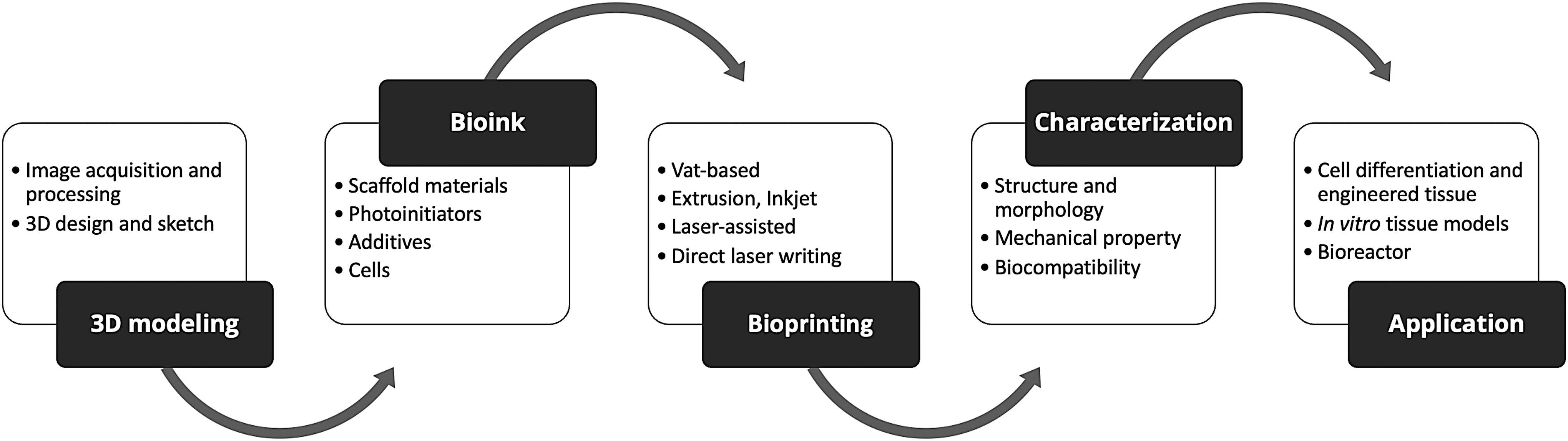
The fundamental of light-assisted 3D bioprinting. The first step of light-assisted 3D bioprinting is 3D modeling, which consists of (1) image acquisition and processing and (2) 3D design and sketch. Next, components of bioink should be determined to be compatible with each specific system and application. The currently available bioprinting processes are stereolithography, digital light processing, extrusion, and laser-assisted printing. After the printing process, characterization of the printed structure, including structure and morphology, mechanical property, and biocompatibility, should be performed to confirm the characteristics of the printed construct. Finally, the bioprinted material can be utilized in tissue engineering applications.
Among these, photocrosslinking and the use of photosensitive hydrogels have been receiving precedence because of mild temperature and pH conditions, tunability, and noninvasive means by which they can be executed. 17 In addition, photocrosslinking can be spatially controlled in 3D printing. Herein, we reviewed the 3D printing strategies used for photocrosslinkable materials and their applications to TE.
Three-Dimensional Bioprinting Technologies
Vat polymerization bioprinting
Vat polymerization 3D printing, commonly known as resin-based 3D printing, relies on the use of visible or ultraviolet light to photopolymerize or cure the liquid photopolymer resins. 18 The process of vat polymerization involves the use of a liquid polymer resin tank and a build platform that actuates to expose a designated path at the top layer of the liquid polymer resin bath with UV light to create a solid object in a layer-by-layer manner. 19 Common 3D bioprinting technologies that utilize the concept of vat polymerization are stereolithography (SLA) and digital light processing (DLP).
SLA bioprinting (Fig. 2A) is a solid free-form fabrication method that converts liquid monomer solutions into solid polymeric materials by utilizing a scanning mirror that controls the ∼355 nm laser beam in the x-y axis, a reservoir containing the bioink, and a build platform controlled in the z-axis. 20 Before starting the proper bioprinting process, a 3D model design and a bioink solution should be prearranged.

Schematic diagrams of photo-assisted additive manufacturing techniques. Bottom-up
In SLA bioprinting, depending on the position of the build platform, the light source precisely targets the desired X-Y path to solidify and encapsulate cells only in the illuminated area, while leaving the nonilluminated areas liquid. 21 This process continues by incrementally raising (bottom-up printer) or lowering (top-down printer) the position of the build platform until the patterned slices project the 3D scaffold design. Conventional SLA bioprinting offers several advantages such as having a nozzle-free approach to creating scaffolds, while maintaining a high spatial resolution. However, the use of UV light could cause cytotoxicity and limit its capabilities. 22
Similar to SLA, DLP bioprinting utilizes a light source to cure bioinks to form solid 3D structures from liquid polymeric materials (Fig. 2B). However, DLP bioprinting employs an array of digital micromirrors to illuminate light on top of the vat to cure an entire cross-section at one time. 23 This results in DLP bioprinting offering faster processing times when compared to the point-by-point scanning mechanism of an SLA. Moreover, instead of employing UV light, conventional DLP bioprinting uses near-UV light (∼405 nm) to cure the bioink, which eliminates the risk of damaging the cells in the process. 24 Nevertheless, DLP has its drawbacks such as still requiring photoinitiators and having lower spatial resolution compared to SLA.
Extrusion bioprinting
Extrusion bioprinting (Fig. 2C) is a bottom-up fabrication approach that extrudes bioink in a predefined plane through a nozzle driven by a mechanical actuator (pneumatic, piston, or rotary screw). 25 Although extrusion bioprinting heavily relies on mechanical mechanisms, they have been utilized to print photosensitive polymers by exposing the extruded bioink to UV or visible light. 26 Zhuang et al. demonstrate the use of a RegenHU extrusion bioprinter to fabricate gelatin methacryloyl (GelMA)/gellan gum-based hydrogels encapsulated with immortalized C2C12 rat myoblasts using a UV-assisted (365 nm) layer-by-layer curing approach to increase printing resolution, maintain cell survival rate, and optimize material microstructure and stiffness. 27
While layer-by-layer printing coupled with a slower UV scanning speed would result in a higher degree of crosslinking, its overall effects on the printing resolution, structural stability, and cell viability were shown to have negligible effects. Conversely, another method is to first finish printing the 3D construct before UV irradiation. A research group from the Mayo Clinic, for example, first printed the construct of a bone scaffold made of a photocrosslinkable polymer called oligo(poly(ethylene glycol) fumarate with impregnated MC3T3 preosteoblast cells before immediately exposing it to the built-in UV lamp of the Cellink Bio-X bioprinter. 28
Inkjet bioprinting
Inkjet bioprinting (Fig. 2D), inspired by commercial inkjet printing technology, deposits droplets of the bioink in a predefined 2D path through thermal or acoustic actuators to create a 3D construct. 18 Thermal-based inkjet bioprinter produces droplets by subjecting the bioink to high temperatures using a heating element without affecting cell viability. 29 Meanwhile, an acoustic-based inkjet bioprinter utilizes a piezoelectric crystal to generate acoustic waves within the bioink cartridge to intricately expel droplets out of the printing nozzle. 30 Both methods offer several advantages such as ease of use, high-throughput manufacturing capability, and high spatial resolution. 31 The laboratories of Gao and Cui have repeatedly demonstrated the capability of inkjet bioprinting to organize photosensitive PEG-based hydrogels to create functional cartilage tissues by simultaneously depositing the bioink, while photopolymerizing.32,33
Laser-assisted bioprinting
Laser-assisted bioprinting (LAB) is a bioprinting technique that is based on the laser-induced forward transfer effect and is composed of a laser source, a ribbon, and a receiver substrate for the printed material. 34 A typical LAB setup works by focusing a pulsed near-infrared (NIR) laser beam onto a donor substrate containing a gold-coated transparent quartz slide and a thin layer of cell-laden bioink to propel microdroplets toward the receiver substrate (Fig. 2E). 35 Sorkio et al. demonstrated the use of LAB to create a 3D corneal tissue from a bioink composed of human embryonic stem cell-derived limbal epithelial stem cells to mimic the epithelium and human adipose-derived stem cells to mimic the lamellar corneal stroma. 36
Direct laser writing
Direct laser writing (DLW) (or two-photon lithography) is an additive manufacturing technique that utilizes ultrafast NIR laser pulses to initiate photopolymerization in an extremely localized focal volume (Fig. 2F). 37 The laser beam generated from DLW systems is capable of creating constructs with a high resolution by introducing enough light on a defined spatial region to induce photopolymerization microscale/nanoscale deep into the material without initiating crosslinking outside the target focal volume.38,39
Early utilization of DLW printing in TE was demonstrated by Marino and coworkers wherein they investigated the topographical effects of patterned submicron surface on neuronal differentiation and outgrowth. 40 A more recent study demonstrated by Valente et al. shows the ability of DLW to selectively crosslink silk fibroin (SF) hydrogels without the need to modify the bioink composition. 41 In this study, they were able to show that the process of DLW printing can enhance the printing resolution, mechanical properties, resistance to enzymatic degradation, and cell viability of SF hydrogels.
Common Photopolymerizable Materials
Photocrosslinkable polymers paired with mild photocrosslinking conditions to create biomimetic 3D constructs have attracted significant attention within the scientific community. 42 Natural polymers have been widely utilized as a primary component for creating 3D constructs due to their inherent capability to improve biocompatibility, biodegradability, and bioinductivity. 43
To enable the photocrosslinking process, a functional group that is susceptible to photocrosslinking should be present in the polymer structure. An example of functional group modification is the methacrylation at the amino side chain of gelatin, which yields GelMA.44,44–53 The methacrylate forms covalent bonds induced by photoinitiators. Similarly, the modification of other natural materials like hyaluronic acid,54–58 extracellular matrix (ECM)/decellularized ECM (dECM),45–48 SF,49–52 collagen,53,54 alginate,55–58 chitosan,59–62 and pullulan63–66 through methacrylation has produced photocrosslinkable polymers used in engineered bone and neural, ocular, and vascular tissues.
Conversely, synthetic polymers such as poly(ethylene glycol),67–70 polyvinyl alcohol,71,72 and poly(ɛ-caprolactone), 73 due to their nature, largely possess lower levels of biocompatibility as they lack cell adhesion sites compared to natural polymers. 74 Nonetheless, synthetic materials have proven to be outstanding alternatives as they generally offer several advantages, including a controllable biodegradation, molecular weight, mechanical property, and purity of the material. 75 Table 1 is a summary of frequently used natural and synthetic biomaterials and their corresponding functionalized modifications in photocrosslinking and 3D bioprinting applications.
Tissue Engineering Applications of Photocrosslinkable Polymers
HUVEC, human umbilical vein endothelial cell; hESC-CM, human embryonic stem cell-derived cardiomyocyte; hPDLC, human periodontal ligament cell; mBMSC, mouse bone mesenchymal stem cells; mUVEC, mouse umbilical vein endothelial cell; hfRPC, human fetal retinal progenitor cells; hADSC, human adipose-derived stem cell; hCAEC, human coronary artery endothelial cell; hMSC, human mesenchymal stem cell; ATII, alveola epithelial type II cells; PEG, polyethylene glycol; PEGDA, polyethylene glycol diacrylate; PEGDMA, polyethylene glycol dimethacrylate; PEGdiPDA, polyethylene glycol diphotodegradable acrylate; PVA, polyvinyl alcohol; PCL, poly(ɛ-caprolactone).
Several studies have also shown the advantages of complementing synthetic polymers with natural polymers to improve scaffold characteristics. For example, Hu et al. showed that an interpenetrating polymer network hydrogel composed of GelMa and polyvinyl alcohol exhibited improved degradation and mechanical properties, while being as capable of promoting angiogenesis as single network GelMA. 76 Similarly, Wang et al. demonstrated that the addition of polyethylene glycol diacrylate (PEGDA) in GelMA hydrogel produced a more robust hydrogel that exhibited higher rates of cell viability and proliferation of mouse osteoblasts MC3T3–E1 when compared to solely GelMA. 77
Moreover, the PEGDA/GelMA hydrogel was shown to have a longer degradation and lower swelling rate. Alternatively, studies have also shown that composites of natural polymers are also capable of improving the biological and mechanical properties of the materials when compared to their individual components. 78 Osi et al. demonstrated this when they were unsuccessful in bioprinting methacrylated chitosan in its pure form due to poor structural integrity, but when reinforced with GelMA, the researchers found success and were able to bioprint and encapsulate the rat BMSCs into the composite scaffold. 79
Photoinitiators
Photoinitiators are often used to produce reactive species needed for initiating polymerization reactions. In this process, the photoinitiators absorb light in the 250–450 nm range and convert it into a reactive species that commences polymerization to form crosslinked hydrogels. 95 However, these reactions may vary depending on the photoinitiator implemented, irradiance, radiant flux, and photon source. For instance, Xu and coworkers systematically compared the effects of Irgacure2959 and lithium phenyl-2,4,6-trimethyl-benzoyl phosphinate 96 on the physical properties, microarchitecture, and cell viability of the 3T3 fibroblasts embedded in GelMA hydrogel. 97 The group found that constructs with lower concentrations (0.3% and 0.5%w/v) of both photoinitiators displayed similar cell viability after 3D printing. However, at higher concentrations (0.7% and 0.9%w/v), LAP-cured constructs displayed higher biocompatibility with slightly smaller pore size, slower degradation rate, and lower swelling ratio compared to Irgacure2959 constructs.
In contrast, Krishnamoorthy et al. reported that an increase in 3T3 fibroblast density in Irgacure2959-GelMA hydrogels leads to an increase in pore size and biodegradation, but a significant decrease in overall mechanical property. 98 A similar report by Pahoff and group on engineering cartilage tissues employing Irgacure2959 in crosslinking 3D printed chondrocyte-laden GelMA/HAMA hydrogels lead to exhibiting lower cell viability compared to using LAP; however, the Irgacure2959 constructs were shown to be better in mimicking native articular cartilage tissues. 99 Therefore, the selection of a photoinitiator requires careful consideration when constructing engineered tissues. Table 2 summarizes commonly used photoinitiators in TE scaffolds.100–110
List of Photoinitiators Commonly Used in Three-Dimensional Bioprinting
Irgacure 2959, 2-hydroxy-1-(4-(hydroxyethoxy)phenyl)-2-methyl-1-propanone; HaCaTs, immortalized human keratinocytes; FMN, flavin mononucleotide; UC MSC, Umbilical cord MSC; BMSCs, bone marrow stromal cells; Gel-GMA, gelatin glycidyl methacrylate; HCEp, human corneal epithelial cells; HCF, human corneal fibroblasts; HCEn, human corneal endothelial cells; NDC, hybrid neuroblastoma cells; Irgacure 1173, 2-hydroxy-2-methylpropiophenone; Clay, laponite XLG nanoclay.
Visible-light photoinitiators can be classified as either free radicals or cationic photoinitiators based on the polymerization of their active species. However, cationic photoinitiators produce protonic acid when excited and thus cannot be used for biomedical applications. 111 Free radical photoinitiators, based on their intermediate steps for photocrosslinking, can be classified as either cleavable photoinitiators (type-I) or bimolecular photoinitiating systems (type-II). 112 Type-I photoinitiators, such as Irgacure2959 and LAP, do not require complex procedures and operate by absorbing suitable incident photons to produce two free radicals to initiate photocrosslinking. Meanwhile, type-II photoinitiators (ex. eosin-Y based) require a multistep mechanism that involves generating radicals in the presence of a co-initiator to initialize crosslinking. 113 Since type-II photoinitiators involve more steps, the reaction times are generally slower than those of type-I photoinitiators. However, utilizing type-II photoinitiators has shown better optical absorption in the near-UV range. 114
While established photoinitiators have a minute effect on the biocompatibility of bioinks, researchers have continuously sought out possible photoinitiators that may be utilized for TE applications. For instance, Zeng and associates investigated seven commercially available visible-light photoinitiators by evaluating their cytocompatibility and corresponding reaction to irradiation. 115 It was revealed that hydrogels handled with photoinitiators ethyl (2,4,6-trimethylbenzoyl) phenylphosphinate (TPOL) and methyl benzoylformate (MBF) displayed the least cytotoxicity in HEK293T, LO2, and human umbilical vein endothelial cells (HUVECs).
However, MBFs were incapable of deep curing, showed noticeable discoloration, and are less cytocompatible when compared with TPOL. Subsequently, implementing TPOL as a photoinitiator for DLP printing urethane dimethacrylate/bisphenol-A-ethoxy dimethacrylate/triethyleneglycol dimethacrylate dental resins has also shown to have superior biocompatibility, color stability, and dimensional accuracy, suggesting that TPOL may be utilized in other TE applications. 116 Several reports have also proposed implementing co-initiator photocrosslinking systems to generate secondary free radicals. For example, Wang et al. implemented a photoinitiator system composed of eosin-Y, triethanolamine, and 1-vinyl-2-pyrrolidinone to SLA print 3T3 fibroblast-encapsulated GelMA-based hydrogels.106,117 The developed photoinitiator system was later successfully adopted in engineering human corneal stroma equivalent tissues. 82
Phototoxicity
In the curing process, light excites the photopolymer causing it to harden; however, extended exposure to light, which ultimately amounts to a longer printing time, runs the risk of losing its biocompatibility. 97 In addition, other parameters such as luminous intensity, polymer concentration, and layer thickness should be considered to maintain cell viability during vat printing. 118 With short exposure times and low luminous intensity, the near-UV wavelength (λ) of 315–405 nm has consistently expressed good cytocompatibility and degree of crosslinking.
Although the difference between utilizing UVA (315–400 nm) and visible light (400–700 nm) seems minute, Khoshakhlagh et al. found that longer exposure times in UVA display increased apoptotic and cell damage to neuronal cocultures compared to visible light. 119 For applications that entail harder materials like bone, a longer curing time is deemed necessary to meet physiological demands. Parthiban and coworkers illustrated this by DLP printing methacrylated demineralized and decellularized human bones (BoneMA) with encapsulated human dental pulp stem cells (HDPSCs) and HUVECs. 120 Interestingly, the length of light exposure had minimal effects on the viability of HDPSCs. However, vascular network formation was more prominent in softer, highly porous, and less crosslinked BoneMA hydrogels.
TE Applications of 3D Photopolymerization Bioprinting
Cardiovascular TE
Mortality attributed to cardiovascular diseases (CVD) has been increasing worldwide due to the difficulty in treating CVD owing to the lack of regenerative properties of cardiomyocytes.96,121 Thus, many have been trying to apply TE to propose alternative methods in CVD treatment. Human adipose-derived mesenchymal stem cells (hADMSCs) encapsulated in GelMA hydrogel modified with nanocrystalline cellulose 3D bioprinted heart valve with phenotypes found in the spongiosa were successfully done by Ma et al. 122
Meanwhile, Izadifar et al. proposed an alternative approach by introducing functional carbon nanotubes, which they found to have greatly improved the mechanical, biological, and electroconductive properties of 3D printed human coronary artery endothelial cell-laden alginate/ColMA scaffolds, indicating its potential utilization in myocardial regeneration. 53 The work by Liu and group using a DLP-based bioprinting system called microscale continuous optical bioprinting (μCOB) to develop an aligned ventricular myocardium composed of neonatal mouse ventricular cardiomyocyte GelMA constructs, which exhibited almost two times the force equivalent and having higher cell compaction compared to 2D seeded samples. 123
In Zhu's research, a μCOB system was used to print prevascularized tissues without a sacrificial material using HUVEC-encapsulated GelMA-HAMA-based bioinks (Fig. 3A). 80 The grafted prevascularized GelMA-HAMA tissues demonstrated significant endothelial network formation with the host circulation and the dorsal skin of mice, while the non-prevascularized tissues showed very limited vascularization and were only visible in the periphery. Although GelMA has arguably been the benchmark for creating vascularized tissues through photocrosslinking, Pien and coworkers argued that photocrosslinked ColMA was revealed to have superior mechanical properties and cell-biomaterial interaction in HUVECs. 124
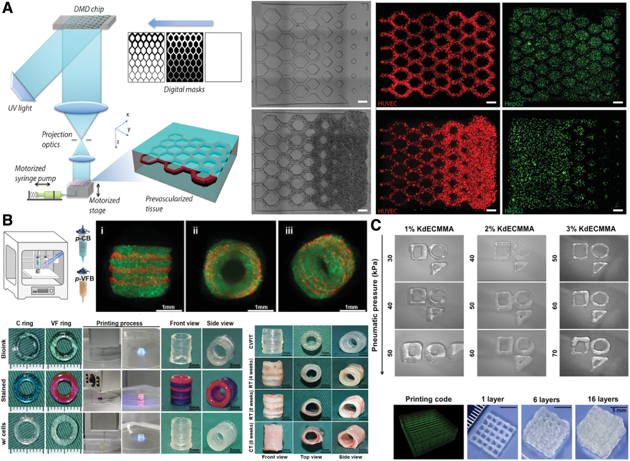
Applications of light-assisted 3D Bioprinting.
Pulmonary TE
The intricate and complex hierarchical architecture of the human lungs have made it extremely challenging to bioengineer 3D printed lung tissue models in vitro. Several studies have also recommended the approach of recellularizing acellular lung ECM to preserve the vascular network, but were received with additional complications such as reperfusion, leakage, and scarcity of ECM source.125–127 Nevertheless, attempts to bioengineer lung mimetic segments have made significant strides in recent years.
Grigoryan et al. utilized a custom SLA printer to develop a multivascular alveoli model from a hybrid PEGDA-GelMA hydrogel with encapsulated human lung fibroblasts and A549 epithelium-like cells in the interstitial space and airway lumen to study biological phenomena happening in the lung. 70 There has also been growing interest in utilizing 3D printing technology as a clinically viable option for tracheal reconstruction of long segmental airway disorders. 128 Huo and associates (Fig. 3B) took advantage of layer-by-layer crosslinking hybrid photocrosslinkable materials to create a 3D printed trachea with alternant stiff-to-soft cartilage and vascularized fibrous layers with GCC (GelMA, chondroitin-sulfate-methacrylate, and methacrylated-acellular-porcine-ear-cartilage) and HPD (HAMA, 8-arm-Polyethylene-glycol-succinic-acid-ester and methacrylated-acellular-Derm-matrix). 129
The alternate stiffness arrangement of the engineered tissue resembled that of a native trachea as it exhibited a similar longitudinal and lateral flexibility compared to a cartilage-only scaffold. Moreover, angiogenesis in the fibrous layers was also observed. Alternatively, while no blood vessel formation was detected in the cartilage layers, the adjacent fibrous layers in the engineered stiff-to-soft trachea displayed superior cartilage regeneration as it is still able to supply sufficient nutrients to the tissue.
Urinary TE
One of the first clinical accomplishments of 3D bioprinting was the development of autologous collagen-PGA-based scaffolds in treating patients needing bladder cystoplasty. 130 Since then, there has been growing interest in applying 3D bioprinting technology to fabricate scaffolds that could treat urological diseases, which require tissue/organ transplantation such as end-stage renal disease, bladder incontinence, and urethral strictures. 131 Ali et al. (Fig. 3C) reported a photocrosslinkable bioink composed of gelatin, HA, glycerol, and methacrylated porcine kidney dECM (dKECM), which could mimic kidney microenvironments. 132 The UV irradiation and tuning of methacrylated dKECM concentration provided prominent structural integrity and rheological characteristics of the native kidneys. Above all, the bioprinted methacrylated dKECM bioink showed excellent biocompatibility and biological activity of human kidney cells.
For tissue grafts, Gu et al. studied the use of mouse fibroblast L929 cell-seeded poly(2-hydroxyethyl methacrylate) (p-HEMA) hydrogel for 3D printing urethra tissue. 133 Interestingly, it was found that since p-HEMA hydrogels have poor mechanical strength, the addition of sodium alginate drastically improved the material's overall mechanical properties, while showing better cellular adhesion, growth, and proliferation. Furthermore, FTIR analysis showed that adding the sodium alginate also greatly increased the blue light curing mass and depth of the hydrogel.
Hepatic TE
The great challenge of engineering liver models in vitro lies in its intricate microarchitecture and multicellular configuration, which allow it to play an important role in plasma protein synthesis, xenobiotic and allogeneic metabolism, detoxification, and enzyme activation. 134 Regardless of the strong regenerative properties of the organ, the only current option for treating end-stage liver disease is either liver or hepatocyte transplantation. 135 Thus, the current scientific outlook involves the use of 3D printing technology as one of the modes of meeting organ donor demand. Mao et al. developed a liver microtissue by utilizing DLP to print encapsulated human-induced hepatocytes in hybrid liver dECM-GelMA bioink. 47
The incorporation of dECM was shown to improve not only the printability of the construct but also the cell viability, proliferation, and spreading of hepatocytes in the microtissue. Ravichandran et al. demonstrated that methacrylation of solubilized liver dECM (dLECM) alone, capable of encapsulating immortalized human hepatocytes, is a viable candidate for TE liver. 136 Moreover, adjustments in dLECM concentration and photocrosslinking time demonstrate the prospect of tuning the mechanical properties of methacrylated ECM-based hydrogels. Although the use of dECM is recognized as a suitable candidate for hepatic TE, concerns on ethical issues, xenopathogenic transmission, and immune rejection should be fully addressed. 137
Skeletal TE
Engineering bone tissues has remained a challenging feat owing to the complex mechanical property and anisotropic architecture of native bone tissues. 138 Currently, the main focuses are on developing new materials and new methods for creating bone implants with sufficient mechanical and osteoinductivity requirements, while reducing complications. 139 3D printing has been an attractive method for engineering bone tissues due to its ability to control scaffold architecture, tune mechanical strength, and promote bioactivity. 140 Shen et al. reported that the in situ vascularized tissue-engineered bones made of bioprinted BMSCs and aortic vascular endothelial cell-laden GelMA hydrogels exhibited excellent osteogenesis and angiogenesis formation for healing rats with calvarial defects. 141 To improve GelMA's printability and mechanical properties, Liu et al. detailed that the incorporation of nano-attapulgite (nano-ATP) in 3D printing cell-laden GelMA hydrogels would significantly improve its printability and mechanical strength, while maintaining similar cytocompatibility with that of pure GelMA. 84
Moreover, the encapsulated rat BMSCs and mouse umbilical endothelial cells (MUVEC) in their nano-ATP/GelMA constructs could promote osteogenic differentiation and angiogenesis activity in vivo. Unlike bone, cartilage TE possesses a unique obstacle in spontaneous healing due to its lack of vascularization and low metabolic rate. 142 Cao et al. devised an FDM-printed trilayer stratified scaffold from PCL hydrogel impregnated with methacrylated alginate for blue light photocrosslinking. 143 The group found that a gradient scaffold design with different filament gaps, laydown patterns, and pore morphology showed better collagen II deposition, BMSC proliferation, and chondrogenic differentiation when compared to homogenously printed scaffolds.
Neural TE
Injury in the central and peripheral nervous system has been proven to be an enduring challenge due to their inability to regenerate, which has led to various neurodegenerative diseases. 144 To address this, a common approach is utilizing nerve guidance conduits (NGCs), which are devices that guide axonal growth, that lost the capacity to establish synaptic connections due to injury or degeneration. 145 Ye et al. propose the use of DLP-printed multichannel NGCs made from GelMA to aid regenerate peripheral nerves. 146 In brief, the fabricated NGCs were able to support the long-term survival, proliferation, and migration of PC-12 cells and induce neuronal differentiation from neural crest stem cells in vitro. A similar study published earlier also shows the effectivity of DLP-printed GelMA/PEGDA composite NGCs capable of guided sciatic nerve regeneration in mouse models. 147
Ocular TE
The intention to engineer ocular constructs is to treat major optical diseases such as corneal diseases, retinal degeneration, and glaucoma by repairing or replacing the damaged cornea or retina. 148 Engineering corneal tissues present an interesting task of needing to be optically transparent on top of being biocompatible and mechanically stable for it to properly refract light into the lens, and eventually the retina. 149
Bektas and Hasirci studied the transparency of corneal stroma equivalent by investigating the light transmittance of 3D printed human corneal keratinocyte-encapsulated GelMA hydrogels. 86 They found that, while the pattern in the 3D construct scattered light, the transparency still closely resembled the native cornea. Conversely, the retina is a complex multicellular region that typically requires translational cellular/biomaterial-based therapies to promote retinal repair. 150 Wang et al. developed a DLP-based bioprinting system capable of printing a multilayered retina-like structure composed of hyaluronic acid and glycidyl methacrylate (HA-GM) with a coculture of human fetal retinal progenitor cells and retinal pigment epithelium. 151 In this study, the researchers found that the HA-GM hydrogels that exhibited closer mechanical properties and morphology to the native retina had a better cell survival rate.
Skin TE
The objective of skin TE is to generate scaffolds capable of repairing damaged skin with the goal of accelerating wound closure, while simultaneously minimizing scar formation. 152 Yang and coworkers proposed that introducing recombinant human type III (rhCol3) collagen in GelMA would help promote epidermis-dermis formation. 153 In vitro cell viability assay of encapsulated HACATs and HDFs revealed over 90% cell viability after extrusion printing, regardless of rhCol3 concentration. Furthermore, while type III collagen is only the second most abundant collagen type in skin, the presence of rhCol3 in GelMA would reveal faster wound closure and freshly regenerated hair follicles.
Meanwhile, Urciuolo et al. developed an in situ 3D bioprinting approach wherein they directly fabricated cell-laden NIR photosensitive hydrogel in mice. 154 They claim that this approach could minimize damage in surrounding tissue by accurately positioning the bioprinted structure in the site of interest to support cell function and generate new myofiber bundles that are integrated with the host vascular network.
Conclusion and Future Perspective
Photocrosslinking-based 3D bioprinting technology has demonstrated great promise in our mission of addressing human donor demands for replacement tissues and organs. Among these, SLA and DLP techniques have shown to be capable of assembling highly precise, intricate, and complex constructs that recapitulate natural tissues. Despite the rapidly growing interest in photocrosslinking-based bioprinting, there have been limited studies to assess the biocompatibility, printability, degradability, mechanical property, biomimicry, and potential clinical application of photoresponsive bioinks and photoinitiators.
Moreover, the selection of appropriate bioprinting methods and optimization of printing parameters such as print speed, curing wavelength, and exposure time are equally important to minimize disparity in evaluating the biocompatibility of 3D printed constructs. Therefore, these factors should be considered and optimized specifically for each experiment setup. In addition, the more thorough biomolecular assays, including transcriptomics, proteomics, and drug responses, could provide very useful information to ensure that the 3D bioprinted tissue construct can represent the original tissue biological functions for applications such as drug screening.
Footnotes
Disclosure Statement
The authors declare no conflicts of interest.
Acknowledgment
The authors are grateful for the Taiwan Ministry of Education scholarship, and the use of resources of Taipei Medical University.
Funding Information
The work was supported by the Ministry of Science and Technology, grant number MOST110-2222-E-038-001-MY3, and Taipei Medical University, TMU108-AE1–B48.