




Abstract
Polyurethane (PU) and PU ceramic scaffolds are the principal materials investigated for developing synthetic bone materials due to their excellent biocompatibility and biodegradability. PU has been combined with calcium phosphate (such as hydroxyapatite [HA] and tricalcium phosphate) to prepare scaffolds with enhanced mechanical properties and biocompatibility. This article reviews the latest progress in the design, synthesis, modification, and biological attributes of HA/PU scaffolds for bone tissue engineering. Diverse HA/PU scaffolds have been proposed and discussed in terms of their osteogenic, antimicrobial, biocompatibility, and bioactivities. The application progress of HA/PU scaffolds in bone tissue engineering is predominantly introduced, including bone repair, bone defect filling, drug delivery, and long-term implants.
Impact statement
Development of biomaterials with enhanced mechanical attributes, excellent bioactivity, and biocompatibility to meet the requirements of human bone tissue repair or replacement has become popular in biomedical engineering. In this review, Polyurethane (PU) combined with hydroxyapatite (HA) is discussed as a composite material for bone tissue engineering. Several performance analyses proved that the HA/PU nanocomposite scaffold material could be used for bone tissue engineering repair.
Introduction
Bone defects caused by accidents or diseases are a common and serious problem in orthopedic surgery. 1 Typically, these defects can be repaired by autologous bone transplantation, allogeneic bone transplantation, or synthetic material filling. Autologous and allogeneic bone grafts have the disadvantages of few donors, high risk of infections, and easy disease transmission. 2 Therefore, the use of composite materials with good biocompatibility, pore structure, and mechanical strength has become popular for treating bone defects.3,4
The scaffolds should be biocompatible and porous. The purpose of a scaffold is to fill defects and act as a load. It also provides a three-dimensional (3D) environment for the growth of cells and new bone tissue growth. 5 An ideal tissue engineering scaffold must have good biocompatibility, surface activity, easy processability, mechanical properties sufficient to meet the support requirements, biodegradability, and a highly porous 3D structure. 6
The human bone tissue predominantly comprises 65% (w/w) hydroxyapatite (HA) crystals and 35% (w/w) collagen matrices. HA [Ca10(PO4)6(OH)2] is a bioceramic with a composition, structure, and mechanical strength similar to those of natural bone tissue. HA crystals are biocompatible and are vastly used for bony tissue regeneration. 7 It can form strong bonds with bone tissues and is often used as an adsorbent due to its high affinity for diverse proteins.8–11
Polyurethane (PU) is a novel polymer material with unique properties and multiple applications. Its application in the medical field began in the late 1950s, and it was first used as a fracture repair material in 1958. In the early 1980s, artificial heart transplantation using PU elastomers was successful, further promoting the application of PU materials in biomedicine. 12 Since then, PU medical materials have demonstrated good blood compatibility, histocompatibility, excellent physical and mechanical properties, adjustable performance, and easy processing; thus, they can be vastly used in several medical treatment fields, including artificial organs, medical catheters, family planning supplies, controlled-release drugs, medical adhesives, medical devices, and medical auxiliary materials.
PU is a linear block copolymer with a changeable structure that can be designed. 13 The molecular weight of polyester or polyether diols comprising PU soft segments is 500–4000, and the most used molecular weight is 1000–2000. 14 Increasing the soft segment content is an important structural parameter for adjusting the physical properties of PU. General PU soft segment content range was very vast and can reach 30–90%. 15 The hard segment content determines the level of phase separation of the soft and hard segments and the level of order and crystallization of the hard segment phase, thus affecting the physical properties of PU. In general, the hardness, modulus, and tear strength of PU can be improved by increasing the hard segment content. 15 By changing these blocks, the mechanical strength of the polymer could be changed within a certain range. In fact, its variable range is vast, from soft to hard plastics.
Synthesis of HA/PU composite scaffolds has been studied in bone tissue engineering. A scaffold combining the excellent properties of the two materials is used for bone repair, drug loading, implantation, and other bone tissue engineering applications (Fig. 1). It was verified that the composite scaffold fabricated from reusable polyols exhibited good bone repair properties. This demonstrates that HA/PU scaffold improved cell proliferation compared with the scaffold without HA crystals.

Properties and applications for HA/PU scaffolds. HA, hydroxyapatite; PU, polyurethane. Color images are available online.
Fabrication of HA/PU Scaffolds
HA preparation
HA can be prepared by diverse calcium-containing chemicals, such as Ca(OH)2, Ca(NO3)2, and CaCO3 or by using extraction from natural materials such as limestone and bioinorganic materials, including bone, shellfish, coral, or eggshells.16–19 HA preparation methods have been classified into five groups: dry methods (mechanochemical and solid-state methods), wet methods (chemical precipitation, hydrolysis, sol–gel, hydrothermal, emulsion, and sonochemical methods), high-temperature processes (pyrolysis and combustion), synthesis methods based on biogenic sources, and combination procedures. Some of the most common techniques are sol–gel, hydrothermal, ultrasonic, etc. 8 Summary as Figure 2. 20
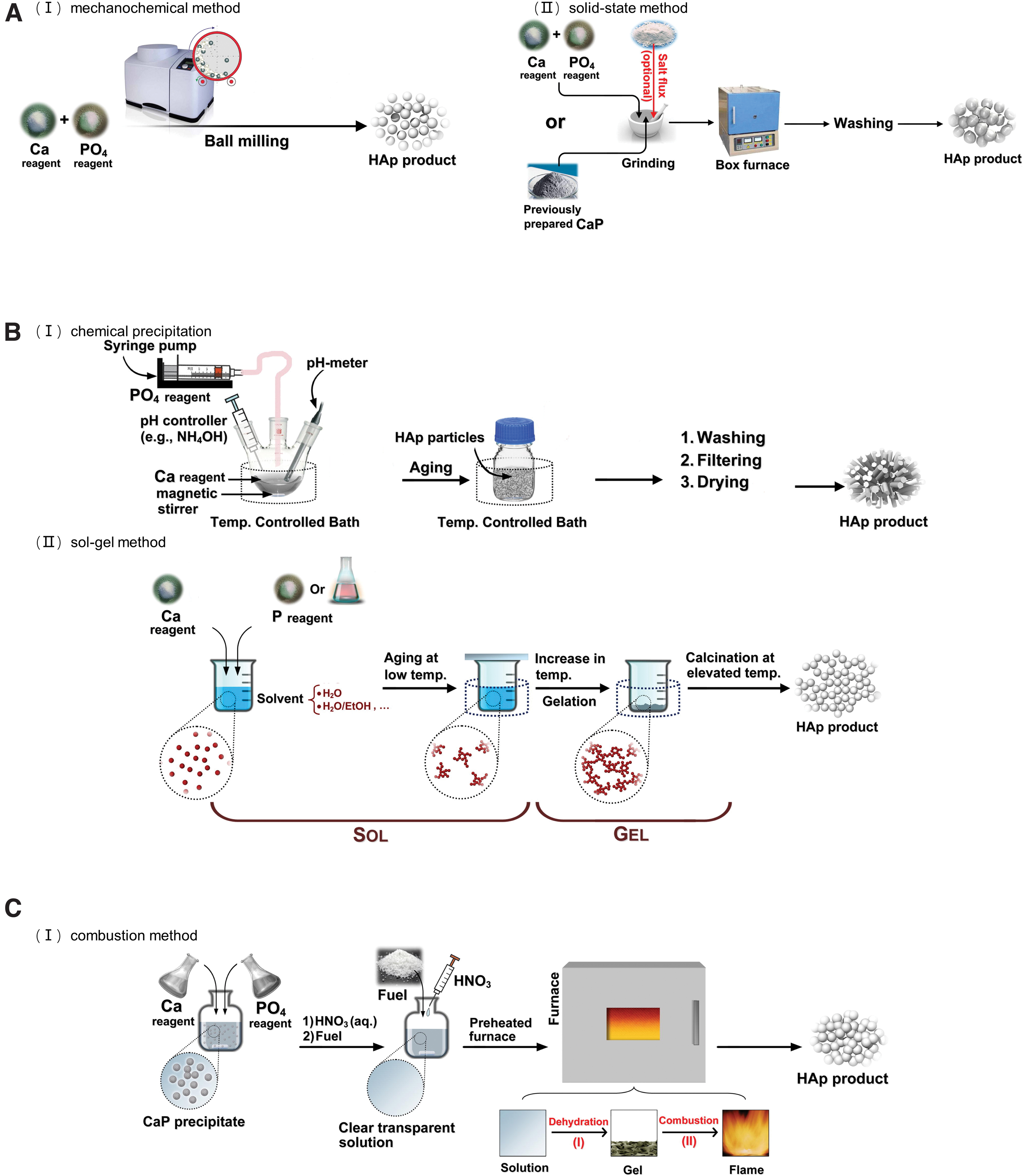
Different methods for the preparation of HA.
HA, in combination with other polymers,21,22 including polyethylene glycol (PEG), polycaprolactones (PCL), polyamides, and polylactic acid (PLA), is the most common and effective way to address the brittleness and fatigue susceptibility of HA. Among several polymers, PU is vastly used owing to its ease of processing, thermal stability, and unique pore structure. This demonstrates strong promise for HA/PU composites as bone tissue engineering scaffolds. 23
PU preparation
PU is typically prepared by the stepwise polymerization of polyisocyanates, oligomer polyols, polyols, or aromatic diamines, which are typical block copolymers (Fig. 3). In addition to carbamate groups, urea, biuret, and other groups were generated. PU has an obvious microstructure for microphase separation owing to the incompatibility of the soft and hard segments. The soft segments provide elasticity, and the hard segment plays a role in enhancing filling and crosslinking. 24 This multiphase polymer material exhibited high strength, hardness, and wear/corrosion resistance.
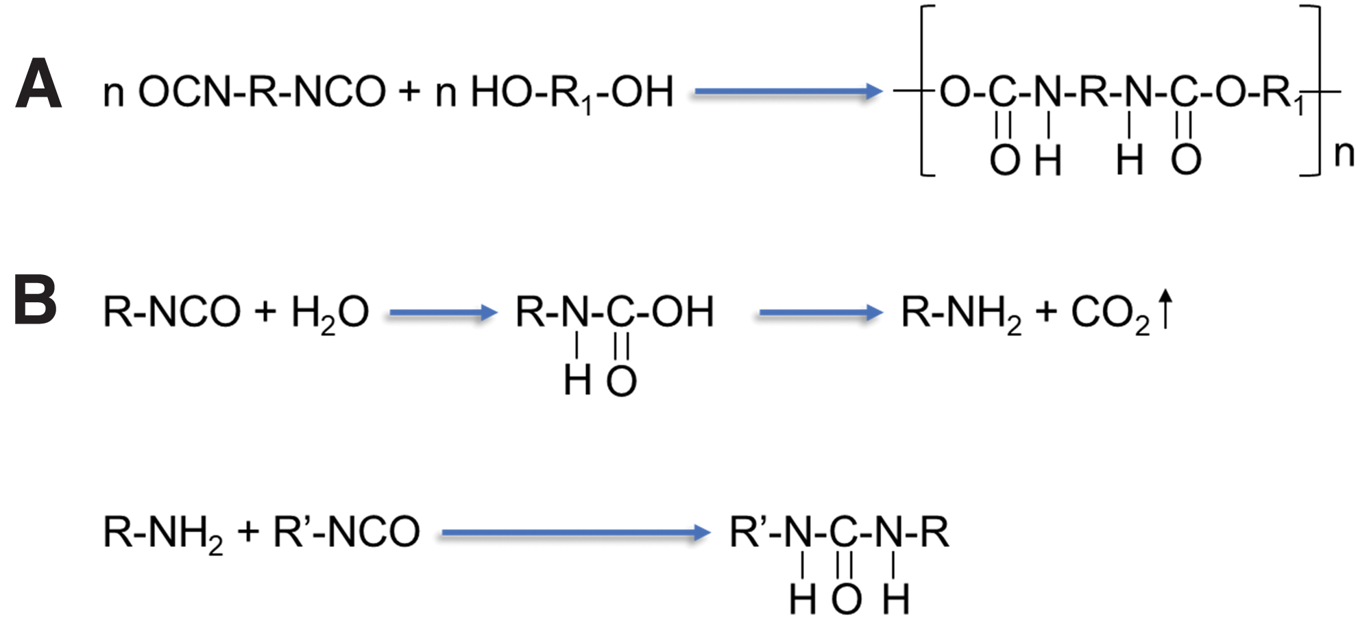
The chemical and physical properties of PU can be designed over a vast range by using different isocyanate/polyol ratios or by selecting diverse types and amounts of chain extenders and/or additives. However, petrochemical-based polyols and isocyanates are toxic and nonbiodegradable. Therefore, the use of sustainable raw materials as synthetic components of PU can effectively improve their adverse effects on the environment, for example biorenewable polyols, such as PEG and polyoxypropylene diols, or polyols obtained from renewable sources, including unpurified sustainable poly hydroxyls from biomass fermentation, 25 rapeseed or palm oils, 26 tannins, 27 and sunflower oils 26 have been largely used in PU composition. Moreover, nanofillers, such as maltodextrin, 28 cellulose, 29 talc, and calcium carbonate 30 have often been compounded into PU to design the final properties of the composites, contributing to enhanced sustainability. In foamed materials, nanofillers can change PU morphology and structure, which can enhance certain mechanical properties or thermal stability compared with materials that do not carry nanofillers. 31
HA/PU scaffold preparation
HA/PU composite scaffolds can be prepared using diverse methods. Cakmak et al. 32 used a 3D printing approach to manufacture PCL/gelatin/bacterial cellulose/HA composite bone scaffolds with diverse HA contents and found that the simultaneous incorporation of bacterial cellulose and HA can increase cell proliferation and adhesion. Nahanmoghadam et al. 33 produced a novel composite scaffold based on PCL and poly (hydroxybutyrate-co-hydroxyvalerate) complexed with HA nanoparticles. Cao et al. 34 and Liu et al. 35 prepared zirconium dioxide/HA scaffolds using a digital light processing (DLP) technique. Zhao et al. 36 and Askari et al. 37 introduced methods and techniques for preparing diverse scaffolds.
The different compositions and preparation methods8,38–43 of the obtained HA/PUs are depicted in Table 1.
Composition of Polyurethane, Methods for Preparation of Polyurethane, Hydroxyapatite, Particle Size of Hydroxyapatite, and Method for Preparation of Hydroxyapatite/Polyurethane Scaffold
BDO, 1,4-butanediol; HA, hydroxyapatite; HDI, hexamethylene diisocyanate; HMDI, 1,6-hexamethylene diisocyanate; IPDI, isophorone diisocyanate, mixture of stereo isomers; ISO, 1,4,3,6-dianhydro-D-sorbito; PCL, polycaprolactones; PEG, polyethylene glycol; PU, polyurethane.
Properties of HA/PU Scaffold
Mechanical properties
Bone–tissue-engineered scaffold materials exhibit excellent mechanical properties. However, the mechanical properties of existing polymers and polymer composites do not meet the application requirements. Conventional polymer composite porous foam material exhibits compressive stresses and moduli in the range of 0.1 and 10 MPa, respectively. 44 Therefore, improving the mechanical strength of the scaffolds is important in bone tissue engineering. It is generally believed that increasing the concentration of HA, 45 adds fillers to PU45–47 as depicted in Figure 4A, and selection of different PU soft and hard segment materials 48 is available.
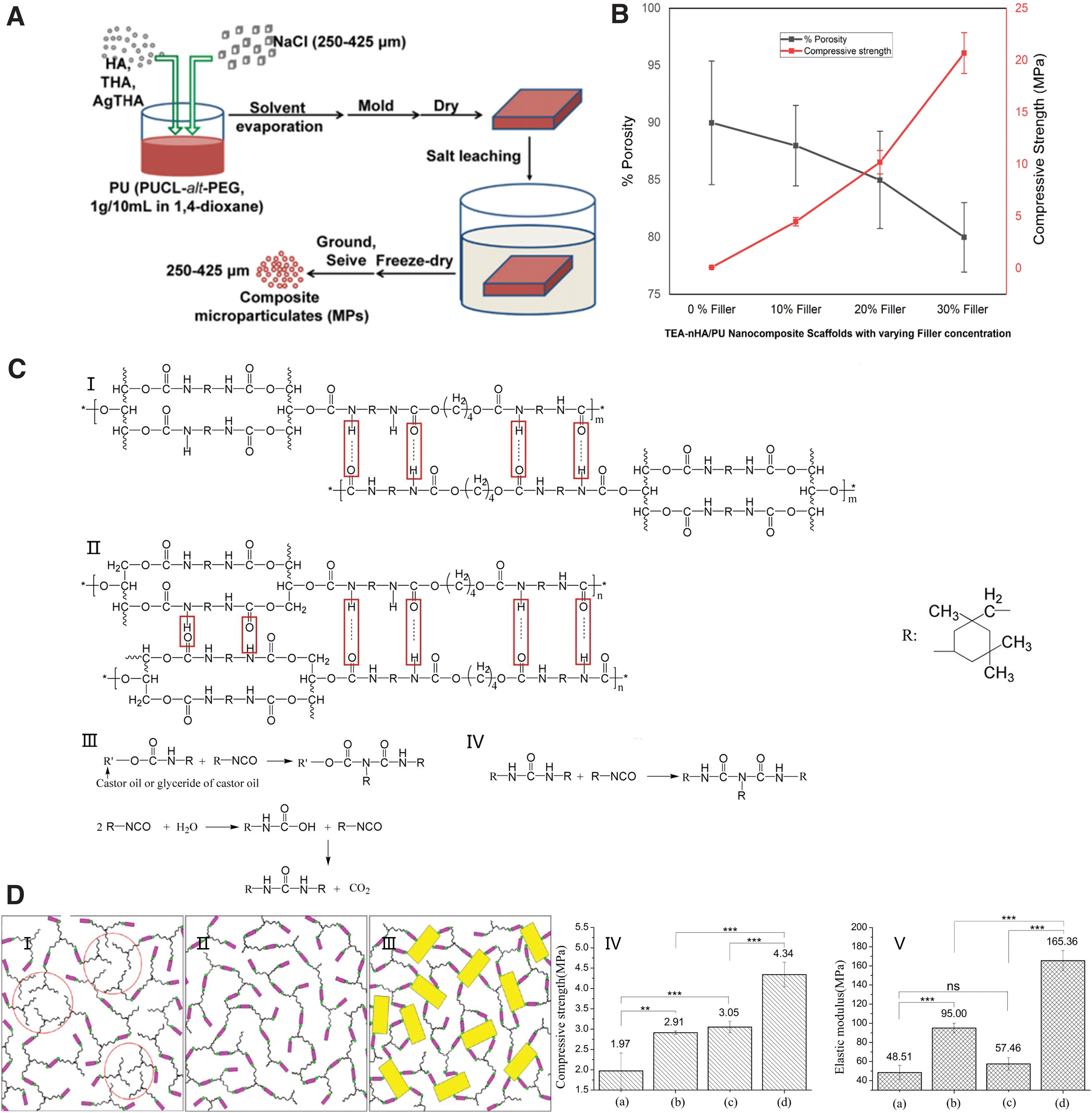
HA/PU scaffolds modified with triethanolamine (TEA) were fabricated by in situ polymerization, wherein HA nanoplates were grafted with TEA. 45 As depicted in Figure 4B, as the filler content increased, the compressive strength increased and the porosity decreased.
To achieve bone tissue regeneration, a newly designed porous HA/PU scaffold was prepared through in situ polymerization. Alcoholization of castor oil (CO) was conducted, and the glyceride in castor oil (GCO) was synthesized. 49 Figure 4C depicted CO-PU and the chemical structures of CO–PU and GCO–PU. More intermolecular hydrogen bonds could be formed in the GCO–PU matrix owing to the modification with polar GCO. A schematic of the three mechanisms that determine the mechanical properties is depicted in Figure 4D(I–III), and the compressive strengths and elastic moduli of the scaffolds are depicted in Figure 4D(IV–V). The results demonstrate that GCO was formed post GO transesterification, which improved the mechanical attributes of the material to a certain extent. When nano-HA (n-HA) particles were continuously mixed into the matrix, the compressive strength and elastic modulus were further improved, increasing by 49 and 74%, respectively.
The uniformly dispersed n-HA particles not only improved their interfacial binding to the GCO-PU matrix, but also provided superior binding to bone tissue. The hierarchical structure and mechanical attributes of the n-HA/GCO–PU composite scaffold were suitable for cell growth and bone tissue regeneration, demonstrating a good prospect for the application of this material in the field of bone repair and regeneration. 48
Etidronic acid was grafted onto the surfaces of HA particles and mixed into PU scaffolds prepared by foaming to develop HA/PU nanocomposite scaffolds for bone tissue engineering. The obtained HA/PU nanocomposites had ∼200 times higher compressive strength and good biocompatibility. 46
Biological properties
HA/PU scaffolds with a 3D structure and high porosity and HA contents of 0, 15, 25, and 32wt% were prepared by a simple and rapid casting technique using the schematic principle in Figure 5A, B. The cell survival rate increased monotonously with prolonged incubation. Post 72 h, the cell survival rate gradually increased with increasing HA content. The trend observed in Figure 5C may be related to the biological activity of HA and the high surface area of the HA particles.50,51 Additionally, combined with the FESEM images in Figure 5D of the PU-HA scaffolds, the results demonstrated that the increase in HA content in the composite scaffold was accompanied by an increase in porosity and pore connectivity, which may contribute to cell migration and nutrient transfer.
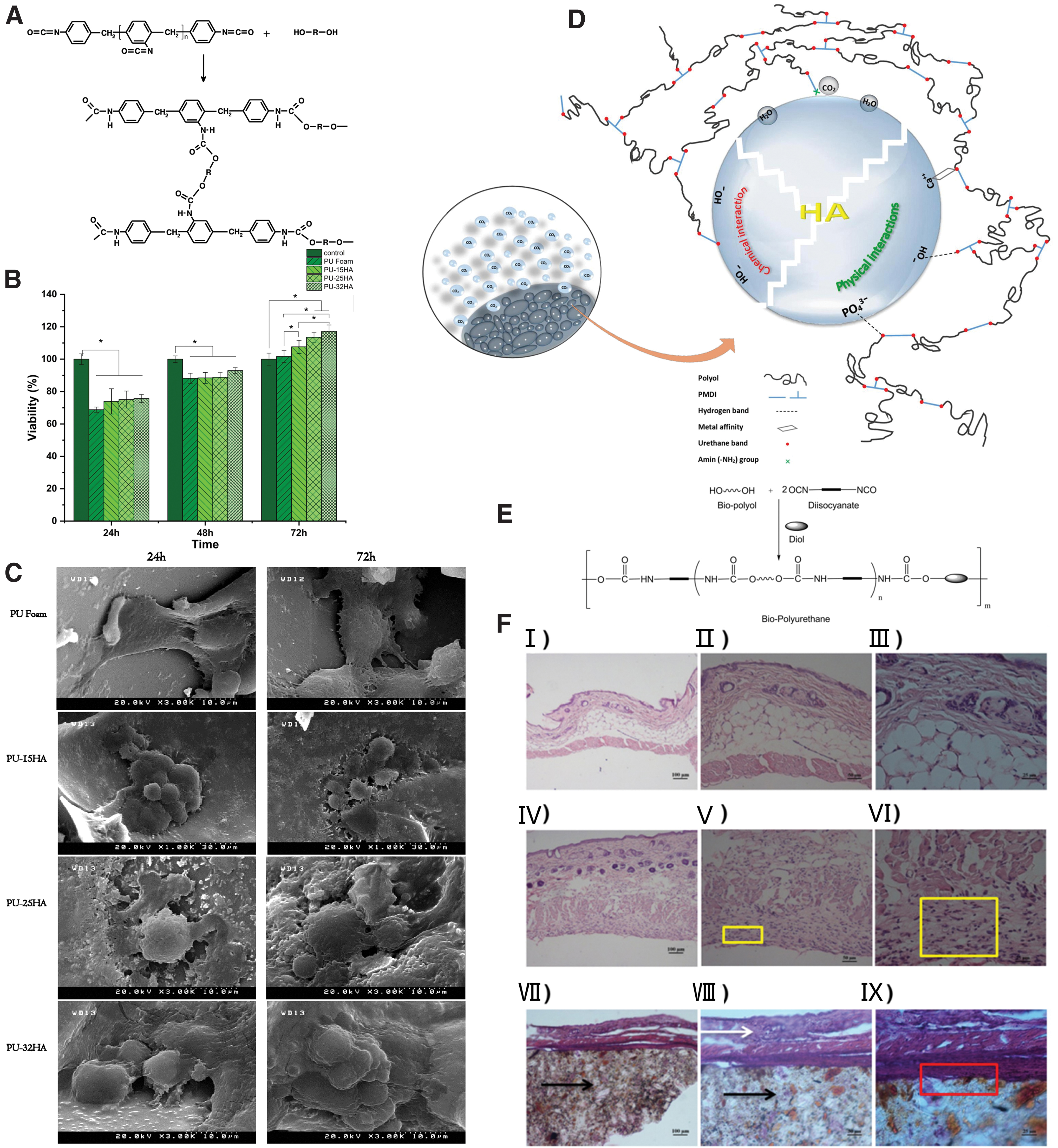
The biological and cellular activities of the scaffold surfaces were improved by increasing the HA content of the HA/PU composite scaffolds. Additionally, an increase in HA content promotes cell proliferation and improves the adhesion between cells, and the scaffold surface was better. 52 The study found that the incorporation of HA nanoparticles to the synthesized PU by the method depicted in Figure 5E can achieve better binding. As depicted in Figure 5F, the incorporation of HA promotes fibroblasts adhesion, while histological in vivo studies verified that the composite promotes adhesion of connective tissue and does not cause inflammation. 53
These studies demonstrate that HA/PU scaffolds have good biocompatibility and are vastly used in bone tissue engineering. To obtain better biocompatibility and cell compatibility, follow-up studies should also directly mix bioactive factors, such as quaternary ammonium salts, metal derivatives, or chloramphenicol as raw materials in the scaffold, or biological macromolecules, such as PEG and ester heparin mimics, grafted on the surface of the scaffold, adding nanofillers, or further modifying HA to obtain better biocompatibility.
In bone–tissue engineering, TEA can entrap cells and accelerate their growth. They can remove toxic metals while providing nutrients. 54 Kumar and Ahuja 55 developed a novel HA/phosphate-based 3D biodegradable composite. The 3D structures provide designable mechanical attributes and connected pore structures to the material and provide a way for biodegradable nanocomposites to be compatible with natural bone. 56
Antimicrobial activity
It would be promising if bone tissue scaffolds could inhibit the activity of diverse human pathogens without compromising bone conductivity. The inhibitory activities of the bone engineering scaffolds were examined against diverse human pathogenic bacteria, such as Escherichia coli, Salmonella typhimurium, Vibrio cholerae, Pseudomonas aeruginosa, Rhodococcus rhodochrous, Aeromonas hydrophila, and Bacillus cereus, which may cause bone infections (such as osteomyelitis). The results demonstrated that the PU/Aloe vera-wrapped mesoporous HA nanorods displayed moderate inhibitory behavior against all tested human pathogens. 57
HA modified with tannin (THA) and HA modified with Ag and tannin (Ag-THA) exhibited good in vitro biocompatibility. After combination with PU, the antibacterial activity, bone conductivity, and bone inductivity of the Ag-THA/PU composite scaffolds were studied in an infected rat model of femoral condylar defect. Ag-THA/PU exhibited excellent antibacterial activity in vivo, and the bacterial concentration decreased to <3% at 12 weeks postsurgery. 46 Based on clinical requirements and previous research, HA/PU materials were compounded with silver phosphate as an antibacterial agent and calcium hydrogen phosphate crystal hydrate (DCPD) as a foaming agent. Under certain conditions, DCPD released crystal water to achieve homogeneous foaming of PU-based composite materials and prepared antibacterial composite bone repair scaffolds with high porosity, uniform pores, and good interconnectivity. Ag3PO4-nHA/DCPD/PU scaffold can effectively inhibit the adhesion of bacteria to the surface of the material, and the bacteriostatic rate can reach 95% after 24 h of contact with bacteria.
Biodegradability
Biodegradable medical materials can be degraded into safe substances after they play their due role. They can be rapidly excreted without toxic side effects and are not rejected by organisms, which greatly improves the safety and convenience of clinical medical materials. Therefore, this has become an important direction for international development in recent years.
Natural biodegradation of the material occurring in vivo can prevent secondary damage during surgery and provide space for new bone growth. However, an uncontrolled degradation rate is detrimental to bone repair. This indicates that it is essential to manufacture materials with degradation rates that match the bone repair rate. Research has demonstrated that incorporation of HA to the PU matrix can decelerate the scaffold degradation process. Phosphate-buffered saline has been used to investigate scaffold degradation behaviors.2,58 Scaffold degradation was also observed by soaking it in a simulated body fluid solution. 59
The long molecular chains of PU decomposed into small molecules.
59
The degradation of HA/PU composites may have been caused by reactions (1)–(3).
The degradation behavior of a scaffold can be regulated by water absorption or the presence of acid, 50 oxidant, or special enzymes in the body.
The weight of HA/PU scaffolds decreased rapidly in the first week, decreased by 13.8%. At 4 weeks, the weight loss rate decelerated, and at 8 weeks, the weight loss was 9.4%. The deposition rate of Ca2+ and PO43− may be faster than the degradation rate of PU, resulting in a decrease in weight loss rate. 41
Osteogenic ability
HA incorporation enhances the adsorption capacity of the scaffold for proteins and increases the surface oxygen content, thereby increasing the surface energy and improving the adhesion and proliferation of osteoblasts on the scaffold, which is beneficial for bone tissue engineering. 60 ALP activity can be used as a marker for the osteogenic differentiation of cells. In the early stages of cell culture, the PU scaffolds demonstrated higher ALP activity than the HA/PU composites. However, with increasing culture time, the 40 HA/PU scaffolds gradually exhibited a higher ALP activity. At the RNA level, polymerase chain reaction results also demonstrated that the HA/PU composites promoted Bsp and Runx2 expression, including Oc expression, compared with PU scaffolds. 61
HA/PU prepared with either l-glutamine or ascorbic acid as a chain extender exhibited good degradation under alkaline and acidic conditions. The 24-h eluents were exposed to cells for 24 h, and after 72 h were not cellulotoxic to human alveolar osteoblasts and allowed to proliferate for up to 14 days. Therefore, it is a promising candidate for bone tissue regeneration. 50 Compared with the HA/PU and blank control groups, the THA/PU- and Ag-THA/PU-treated skull defect sites had a higher bone mineral density. Moreover, coating HA with tannic acid enhances composite osteogenesis and angiogenesis. 62
Applications of HA/PU Scaffolds in Bone Tissue Engineering
Bone tissue regeneration
HA nanoparticles were introduced into the PU matrix to improve bioactivity. Compared with the control group, the PU scaffold with 40wt% HA clearly promoted its biomineralization ability and accelerated mesenchymal stem cell proliferation and osteogenic differentiation. Eight weeks after the in vivo implantation, a large amount of vascularized bone tissue was generated surrounding both the PU and 40HA/PU scaffolds, accompanied by bone marrow interstitial development. Thus, HA/PU composites are potential scaffolds for bone regeneration.61,63 Additionally, studies have demonstrated that the shape memory properties of PU can play an important role in the minimally invasive surgery of irregular bone defects to promote bone regeneration.
Shape memory polyurethanes (SMPUs) are smart materials that can recover their original shape from a deformed shape under external stimuli such as heat, 43 moisture, 64 ultraviolet (UV) light, 65 and magnetic field 66 (Fig. 6A). Compared with metal shape memory alloys (SMA), shape memory polymers (SMPs) have several advantages, especially in biomedical applications. 67 Among others, the most attractive advantage may that the SMPs still have a large recoverable strain at 100% deformation level, which is much larger than that of the SMA (<10%). 68 This feature allows SMPU foams to be implanted in a small size or a specific shape through minimally invasive surgery. When exposed to stimuli, such as body temperature, the stent returns to its original shape owing to shape memory effects.69,70 The working principle is illustrated in Figure 6B.
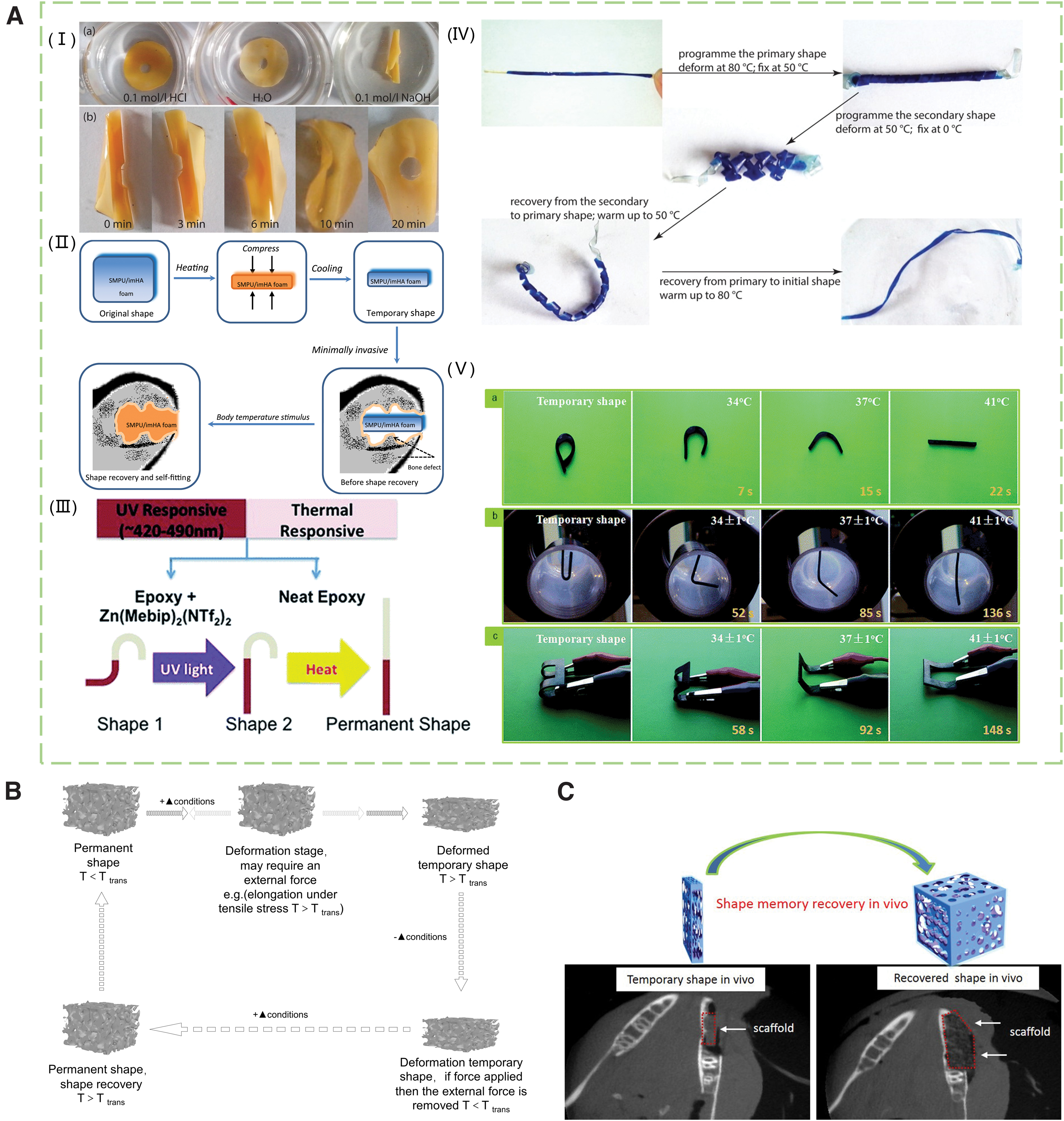
Bone tissue engineering is an important tool for treating bone defects in the craniomaxillofacial (CMF) region. Biological or bone grafts are required as functional supports to induce bone tissue growth. Autologous transplantation has certain limitations: the transplantation process is complex, and the incidence of infection is high. Additionally, shape remodeling, autologous transplantation, fixation, and complete adaptation to the defect boundary are difficult to achieve. Tissue-engineered scaffold materials are potential alternatives for critical-size CMF bone defects. 71 This effect is illustrated in Figure 6C. Accurately matching irregular size and shape of bone defects while promoting bone tissue regeneration. 66 In vivo experiments, microcomputed tomography, and histomorphometry demonstrated that the SMP scaffold not only exhibited good shape-restoration properties, but also promoted new bone production. 72
Therefore, the use of SMPU foam as a bone scaffold is expected to treat irregular bone defects. SMPU foam combined with HA nanoparticles is a bone scaffold material that can be used for minimally invasive bone regeneration.
The HA/SMPU foam has the advantages of matching the properties of trabecular bone, such as: (i) Pore size (500–800 μm), porosity (50–60%), and high pore interconnectivity (95–99%); (ii) Compressive stress (∼13 MPa) and modulus (∼700 MPa) at body temperature; (iii) Responsive shape memory performance at body temperature; and (iv) Noncytotoxic, good biocompatibility. 43
Alternatively, bone regeneration can be promoted by incorporating active factors or other additives to scaffolds. Carbonated HA (CHA) ceramics were fabricated at low sintering temperatures using the PU foam replication technique. Mg2+, Co2+, and Sr2+-doped CHA scaffolds exhibited improved architectural and mechanical properties. The presence of Mg2+ in the ternary-doped CHA improves its biocompatibility. The effect of the release of these ions on osteocytes and their surrounding cells, especially endothelial and immune cells, will provide new ideas for the design of new bone immunoregulatory scaffolds. 73
Bone defect filling drug delivery
PUs are vastly utilized in drug carriers and drug delivery systems due to their several processable possibilities. 74 PU scaffolds prepared using soft and hard segment materials with diverse proportions, hydrophilicity, crystallinity, and crosslinking degrees have diverse properties and morphologies, which allow us to control the speed and amount of drug release.
Currently, the development of local drug delivery systems that can prevent infection and inflammation postimplantation in vivo, while allowing vascularization and new bone formation, has attracted much attention. Li et al. 75 studied PU synthesized from 70% ε-caprolactone, 20% glycolide and 10% D,L-lactide, lysine triisocyanate, and Tegoamin 33 as a catalyst, and water as a foaming agent for the sustained and quantitative release of vancomycin to prevent implant-triggered infections. The implantation of PU scaffolds into infected bone defect sites in rats resulted in significantly less infection than in the controls. Kolmas et al. 76 investigated an implantation drug delivery system to inhibit bone tumor growth and cell invasion in the extracellular matrix. They studied the control effect of HA/PU composites on the sustained release of bisphosphonates. The results demonstrate that the amount of drug released is adjustable within a certain range, which provides a variety of possibilities for controlling the release kinetics. Nair et al. 77 fabricated reactive scaffolds from PU and PLA through a thermally induced phase separation.
The PLA/PU system can be used as a drug test substrate for diverse chemotherapeutic drugs on cell adhesion-mediated drug-resistant cells and can be used to characterize the clinical significance of diverse drugs.
Implant
Biomedical science of implant science and engineering require different magnitudes and properties of materials to address the design of implant structures, use, biocompatibility, and biodegradability. It is difficult to achieve a balance between the in vivo degradation and tissue regeneration of the implanted scaffold. This depends on diverse variables in clinical conditions, such as the shape and size of the bone defect site, which may lead to the release of acidic degradation products of the nonphysiological inflammatory response and functional load. These factors may affect bone regeneration and remodeling. 51 In the recent year, as a kind of 3D cell carrier, biodegradable PU scaffolds have been introduced into tissue engineering.59,78,79 Unlike bioceramic implants, which are prone to secondary damage owing to the viscoelastic attributes of PU, they can be almost frictionlessly implanted into damaged tissues. 80 Additionally, they provide a porous and interconnected environment for cell growth and proliferation. 81 They are noncytotoxic in vitro, exhibit good biocompatibility in vivo postimplantation, and exclude a strong leukocytic inflammatory host tissue response. 82
Compared with conventional biodegradable polymers, scaffolds can effectively control the degradation rate and can be used for long-term implantation to repair large-area defects. The prevention of tissue collapse, and maintenance of newly formed tissues are the major directions for future research. From this perspective, HA/PU exhibits a series of mechanical and morphological properties that are significantly stronger than those of other medical-grade biodegradable polymers, and can be degraded slowly.
Laschke et al. prepared a 3D HA/PU scaffold for evaluating vascularization. The scaffold was not implanted into a bone defect, but onto the intraluminal transverse muscle tissue, ensuring permanent tissue contact with the implant for continuous in vivo analysis. The results demonstrated that the scaffold exhibited excellent biocompatibility. 60 HA nanoparticles were modified with a polydopamine coating. Subsequently, a 3D porous thermoplastic PU (TPU) with a two-stage pHA modification was fabricated using ultrasonication-assisted solvent casting methods. The pHA/TPU-pHA composites exhibited interconnected porous structures. The hydrophilicity and mechanical properties of the pHA/TPU-pHA composites were significantly better than those of TPU scaffold. 83
Conclusions
HA/PU scaffolds have properties favorable for application in bone tissue engineering, including proper mechanical strength, biodegradability, good biocompatibility, cell adhesion mechanisms, tissue repair induction, and sustained drug release owing to the imitation of the structure and composition of mineralized tissues. The results of both in vivo and in vitro experiments verified that the HA/PU composites had satisfactory biocompatibility.
However, the degradability of PU materials needs to be further explored as most PUs exhibit poor degradability and long degradation periods. The properties of diverse tissue engineering scaffolds differ, and the properties of PU can be adjusted by changing the soft and hard segment materials, proportions, and mixing different additives.
Footnotes
Authors' Contributions
Conceptualization: T. Zhang; investigation: J. Li, Y. Wang, and W. Han; writing—original draft preparation: T. Zhang; writing—review and editing: D. Huang, Y. Wei, Z. Liang, X. Lian, and Y. Hu; and funding acquisition: D. Huang. All authors have read and agreed to the published version of the article.
Disclosure Statement
No competing financial interests exist.
Funding Information
This work was supported by the National Natural Science Foundation of China (Grant No. 12272253), Shanxi-Zheda Institute of Advanced Materials and Chemical Engineering (Grant No. 2021SX-AT008, 2021SX-AT009), and the Scientific Activities of Selected Returned Overseas Professionals in Shanxi Province (Grant No. 20220006).