




Abstract
Respiratory infections caused by coronaviruses (CoVs) have become a major public health concern in the past two decades as revealed by the emergence of SARS-CoV in 2002, MERS-CoV in 2012, and SARS-CoV-2 in 2019. The most severe clinical phenotypes commonly arise from exacerbation of immune response following the infection of alveolar epithelial cells localized at the pulmonary blood-air barrier. Preclinical rodent models do not adequately represent the essential genetic properties of the barrier, thus necessitating the use of humanized transgenic models. However, existing monolayer cell culture models have so far been unable to mimic the complex lung microenvironment. In this respect, air–liquid interface models, tissue engineered models, and organ-on-a-chip systems, which aim to better imitate the infection site microenvironment and microphysiology, are being developed to replace the commonly used monolayer cell culture models, and their use is becoming more widespread every day. On the contrary, studies on the development of nanoparticles (NPs) that mimic respiratory viruses, and those NPs used in therapy are progressing rapidly. The first part of this review describes in vitro models that mimic the blood-air barrier, the tissue interface that plays a central role in COVID-19 progression. In the second part of the review, NPs mimicking the virus and/or designed to carry therapeutic agents are explained and exemplified.
Impact statement
COVID-19 pandemic highlighted the urgent need to gain a deeper understanding of viral infection pathogenesis and develop approaches for rapid translation of research findings as vaccines or therapeutics that will protect the public from future pandemics. Numerous studies are in progress worldwide but advanced in vitro models that accurately mimic the natural microenvironment of the infection site are still under development. This review aims to introduce a brief summary of the features of the models currently used and that need to be improved, especially for researchers working in the field of regenerative medicine and nanotechnology.
Introduction
In diseases of distal airways caused by viral infections, including Severe Acute Respiratory Syndrome Coronavirus 2 (SARS-CoV-2), loss of the integrity of the pulmonary blood-air barrier is one of the leading factors of disease progression due to impaired gas exchange across the barrier. In vitro models that mimic this barrier have become even more important because of the significance of the research outputs and their usefulness in further studies. The models available at present have great superiority over the conventional monolayer cultures as they are closer to mimicking the essential physical, mechanical, and biochemical characteristics of the blood-air barrier.1–3
However, there are several key factors4,5 that need to be improved further to better mimic the native barrier. These include the micro/nano structures, which should accurately represent the composition; mechanical features that preserve their elasticity and strength for extended periods of breathing; the type and the proportion of the cells, and the microenviroment. This encompasses the protective constituents of the surfactant layer, which guard against the entry of particles into the alveoli; and the specialized cells that facilitate efficient gas exchange and respond to any possible damage in this critical interface. In addition to individual differences, 6 each of the factors mentioned affects the research findings related to the events that happen at the barrier following SARS-CoV-2 infection and the progression of the disease afterward. In this respect, it is vital to develop and use accurate, rapid, and functional models that can recapitulate the essential features of the barrier.
Given the limitations of existing models and the rapid progress in the development of advanced in vitro systems and nanoparticles (NPs), this review provides an overview of in vitro models of the blood-air barrier and the potential of NPs to mimic viruses and deliver therapeutic agents in the context of Coronavirus Disease (COVID-19) treatment.
SARS-CoV-2 Infection Mechanism and COVID-19
SARS-CoV-2, the causative agent of the COVID-19, is an enveloped, positive-sense, and nonsegmented ss-RNA virus belonging to the SARS-related coronavirus species and Betacoronavirus genus of Coronaviridae family.7,8 When a person encounters SARS-CoV-2, the interaction starts by infection of Angiotensin-Converting Enzyme II (ACE-2) presenting cells in the nasal epithelium. The virus enters the host cell by binding of its Spike (S) protein to its receptor ACE-2, and subsequent proteolytic cleavage by host cellular proteases such as TMPRSS2 and cathepsin L9,10; the two major proteases involved in S protein activation. The mechanism of the viral entry is one of the key processes targeted in the development of vaccines and potential therapeutics against SARS-CoV-2 infection, and already reviewed in detail elsewhere. 10 In most patients, when the virus reaches the upper respiratory tract, the local immune defense mechanisms limit the spread of infection by viral clearance. 11
The virus, on the contrary, possesses various mechanisms that delay the expression of cytokines and recruitment of immune cells; it thereby evades immune response and travels down to the distal airways. 12 Progression of the infection into alveoli, more specifically to the pulmonary blood-air barrier across which oxygen (O2)/carbon dioxide (CO2) exchange occurs, is the leading cause of increased severity and morbidity of the disease. In the alveoli, the virus primarily infects alveolar epithelial type II cells (AETII) and hijacks the cells to produce its copies and to release them into the alveolus. Infected AETII cells also release inflammatory signals to recruit alveolar macrophages to the site of infection. Macrophages release cytokines and chemokines that cause vasodilation inducing the recruitment of more immune cells to the infection area. Recruited CD8+ cytotoxic T cells and neutrophils destroy infected cells to fight off the virus but this causes increased inflammation and alveolar tissue injury.
As a result of vasodilation, and loss of barrier integrity, fluid accumulates in the alveolus and increases the thickness of the barrier, which in return decreases the rate of gas exchange and triggers the onset of alveolar collapse. Increased inflammation potentially progresses to acute respiratory distress syndrome (ARDS) which requires intensive care treatment to prevent further progression toward systemic inflammation, multiorgan failure, and eventually death.13,14 Today, a large number of studies are being conducted to gain a deeper understanding of infection pathogenesis and disease progression. As the loss of barrier integrity is one of the key pathophysiologic processes leading to the progression of viral pneumonia to ARDS,13,15 it is vital to regain essential constituents and microphysiology of the barrier in vitro.
Characteristics of the Blood-Air Barrier
The pulmonary blood-air barrier constitute the interface between the alveoli and the capillaries that surround them. On the alveolus side, the barrier consists of two types of alveolar epithelial cells (AETI and AETII) and on the capillary side, it consists of pulmonary microvascular endothelial cells (hPMEC) along with an adherent basement membrane and lung interstitial components. 16 AETI cells are large, squamous cells that cover about 95% of the alveolar surface, enabling efficient gas transfer across the barrier. AETII cells, in contrast, are cuboidal cells that are also progenitors of AETI and serve in the renewal and repair of the alveolar epithelium upon injury. 17 AETII cells have characteristic secretory organelles called lamellar bodies, which produce the surfactant layer, a thin, continuous lining layer that covers the alveolar epithelium.
Surfactant mainly consists of saturated phospholipids that reduce surface tension at air–liquid interface (ALI), and plays a vital role in the protection of the alveoli from collapsing during breathing. The surfactant layer also has immunomodulatory functions owing to the surfactant proteins (SP; SP-A, SP-B, SP-C, and SP-D) that bind and opsonize pathogens and interact with innate and adaptive immune system cells. 18 Any particle that reaches the alveoli is confronted with the surfactant layer. They may be opsonized by the surfactant proteins, leading to their being phagocytosed by alveolar macrophages or removal through mucociliary clearance. Alternatively, they may form aggregates with surfactant molecules and deposit on the alveolar epithelial cells. In the latter case, they may enter the alveolar cells or be introduced between the cells if the barrier integrity is compromised. 19
The barrier is very thin (0.2–2.4 μm) and allows effective gas transfer. It is also very soft (Young's Modulus: 1–2 kPa) and resists volume change during breathing.20–22 Barrier integrity is mainly maintained by intercellular tight junctions between the epithelial cells formed by multiprotein junction complexes, zonula occludens and claudins. As a result, only small molecules (e.g., O2 and CO2) can freely diffuse across the barrier. 23 Viral infections in distal airways are known to damage the integrity of this barrier by inducing epithelial cell death and/or decreasing the expression of junction proteins by the various virulence factors. 24
In Vitro Models Used in COVID-19 Research
In vitro approaches are rapid and sophisticated strategies used in developing disease models and to understand how a bioactive agent such as a drug reacts in the body at cellular level. In vitro models also enable a reduction in the number of test animals and allow the investigation of processes such as toxicity on human cells that cannot be done in studies with animals. A number of in vitro models recently developed, such as ALI, tissue engineered, and organ-on-a-chip systems, constitute highly controlled and reproducible experimental environments for COVID-19 studies, and are being used widely (Fig. 1).
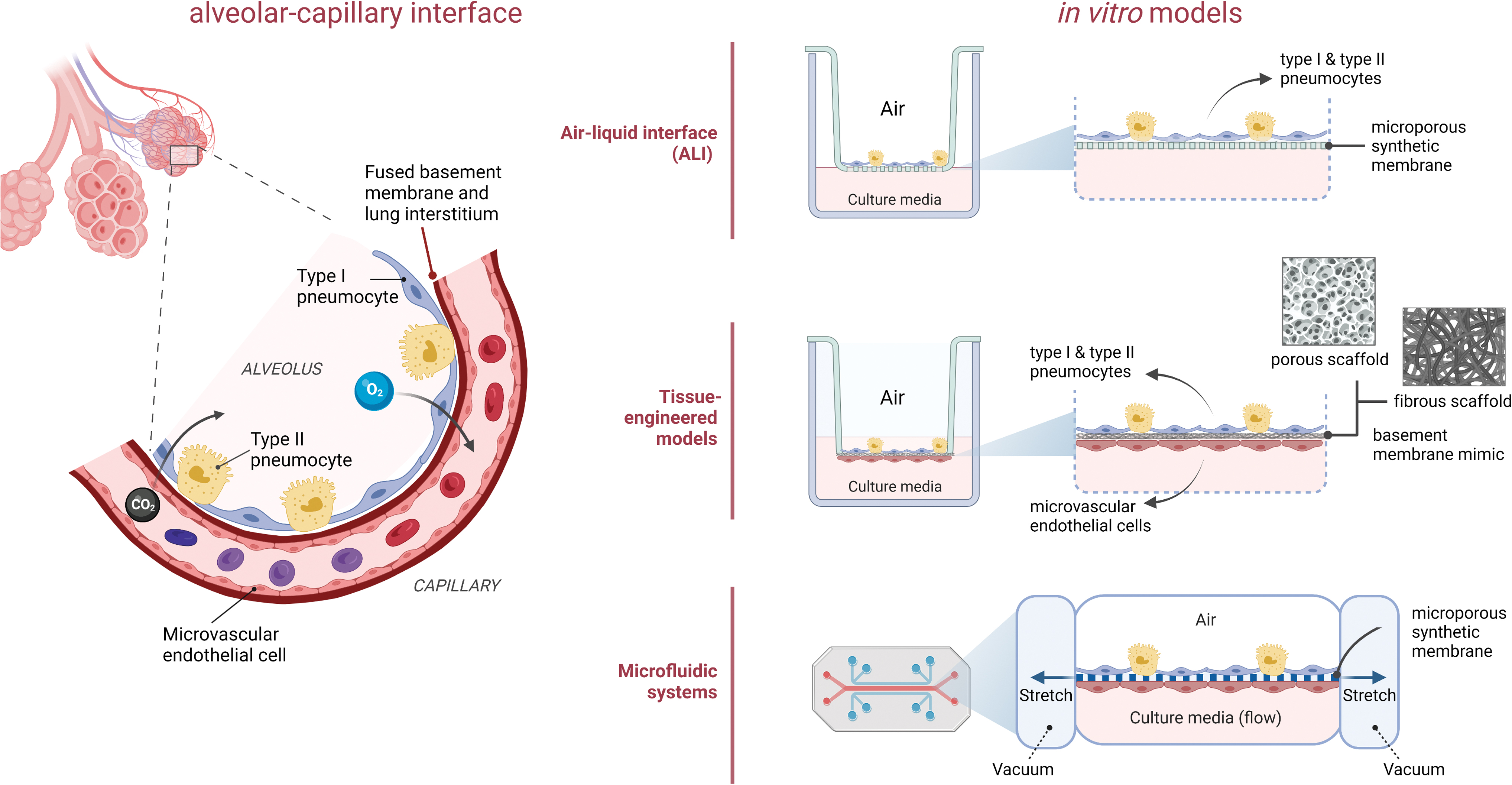
The blood-air barrier (alveolar-capillary interface) and in vitro models used in COVID-19 studies: ALI models, Tissue Engineered models and Lung-on-a-Chip systems. ALI, air–liquid interface. Color images are available online.
ALI models
The airway epithelium which is in contact with humid air on one side and blood on the other, acts as a physical and immunological barrier. 25 The defects in this unique barrier are implicated in the pathogenesis of respiratory diseases. ALI models that mimic the airway epithelium were developed to gain information on the physiology of the respiratory tract and allow us to investigate diseases affecting the lung alveoli and the introduction of antigens/reagents across them. The challenges in ALI models are the requirement for the provision of an adequate number of functional lung epithelial cells and miniaturization for use in high-throughput systems. There is need for in vitro ALI models especially to study the SARS-CoV-2 infection mechanism on its main target, the airway epithelium, and to develop relevant therapeutic approaches.
ALI models, are mainly composed of epithelial cells seeded on a porous membrane, where the epithelial cells are in contact with air on the apical side and with the culture medium on the basal side. 26 In this way, ALI models represent the interfacial function of airway epithelium on the barrier. The membrane serving as a cell support acts as a tightly regulated barrier upon polarization of epithelial cells, and this should result in the secretion of protective liquid layers, mucus and/or alveolar lining fluid, and thinning of epithelium on these membranes and also allows improved transfer of gases and aerosols. In vitro ALI conditions were primarily established with static Transwell inserts and culturing cells of alveolar and airway epithelium on porous synthetic membranes typically made of polyethylene terephthalate.25,27,28
In one of the COVID-19 disease models, ALI system with Transwell insert was used to investigate the expression levels of SARS-CoV-2 host factors in comparing induced pluripotent stem cell (iPSC)-derived alveolar and airway epithelial cells with primary lung epithelial cells. 27 In another study, Sasaki et al. demonstrated that in vitro ALI culture made alveolar epithelial cells susceptible to SARS-CoV-2 infection. 28 There are also in vitro ALI COVID-19 disease models designed to assess the efficacy of antiviral agents such as remdesivir, resveratrol, pterostilbene, and hydroxychloroquine.29–31
Transwell systems and tissue engineered barrier models
The first attempt to recapitulate the blood-air barrier under in vitro conditions had a simple design: it involved seeding the underside of a porous transwell membrane with endothelial cells (ECs) and the opposite side with alveolar epithelial cells and the culture of both these cells until confluency. This approach was followed by more complicated tissue engineered models where the basal lamina was mimicked by electrospun membranes made of biodegradable polymers or was bioprinted using extracellular matrix (ECM)-like hydrogels. Both these models allowed intermittent transepithelial electrical resistance (TEER) measurements to assess barrier integrity and visualization of the cultured cells, but without mimicking the alveolar ventilatory mechanics or flow.
Transwell systems
Hermanns et al. established an in vitro blood-air barrier in a transwell system for the first time by using human primary hPMEC and NCI H441 human lung epithelial cell line. 32 A polarized epithelial cell monolayer was generated on a permeable poly(carbonate) membrane, which possessed both native tight and adherence junctional structures, and an average TEER value of 500 Ωcm2 was obtained. Such a coculture system has been utilized since then as an in vitro model to study toxicity-related lung injury,33–37 acute lung injury pathogenesis, 38 distal lung infections, 39 and preclinical drug development. The addition of macrophages to this system created an in vitro model that is more relevant to the native alveolar microenvironment in terms of cellular composition. This made determination of immune response to the tested particulate material possible and allowed interplay among the different cell types.40–42
A high-throughput, transwell-based, blood-air barrier model was formed in a 96-well plate through density-driven underside epithelium seeding, which enabled rapid physiologic screening and was used to show the inflammatory response upon viral infection. 39
Tissue engineered models
The undesirable properties of the etched polycarbonate membrane in comparison to the natural basement membrane, such as greater thickness, lack of interconnected pores which restricts direct cell–cell interactions, and bioresorbability, encouraged the development of better basement membrane mimics. Nanofibrous meshes with interconnected pores were generated from bioresorbable polymers through electrospinning as a typical tissue engineering approach to mimic the natural basement membrane, while more sophisticated approaches like bioprinting served as an appropriate tool to deposit a hydrogel as an ECM mimic of the natural basement membrane between the endothelial and epithelial cell layers.
Basement membrane mimics
An electrospun poly(ɛ-caprolactone) (PCL) nanofibrous mesh with interconnected micropores was proposed for the first time by Nishiguchi et al. as a basement membrane mimic. 43 The interconnected pores enabled a direct contact between the cells on the opposite sides of the mesh.43,44 In this way, while the cellular penetration across the mesh is prevented, the transfer of growth factors and signals from one cell type to the other was still possible. The triple culture of epithelial, endothelial and immune cells on this 10 μm thick basement membrane mimic enabled the adequate maintenance of the barrier properties, and thus made this a promising in vitro model for further studies. An electrospun ultrathin (2 μm) PCL nanofibrous mesh was recently used to establish a blood-air barrier model, and TEER and permeability studies performed on this barrier in comparison to the conventional dense membranes with etched pores revealed integral tight junctions and improved mass transport.
Despite its high porosity, no barrier disruption was observed, and this thin nanofibrous mesh demonstrated a physiological relevance by allowing neutrophil transmigration. 45 A reconstituted basement membrane with 100 nm thickness was constructed by He et al. of biopolymers on a patterned honeycomb microframe; type IV collagen and laminin self assembled with a layer of crosslinked nanofibrous gelatin. Use of alveolar epithelial cells and ECs derived from human iPSCs enabled generation of a blood-air barrier model, where two distinct cell layers were grown successfully with no cellular migration to the opposite side of the membrane. 46
Three-dimensional bioprinted models
An endothelium–epithelium double layer separated by a basement membrane was fabricated using a layer-by-layer bioprinting approach to generate a blood-air barrier analog. 47 Tissue models prepared by three-dimensional (3D) bioprinting of cell-laden bioinks were also used to study influenza infection in the lung. 48 A three-layered blood-air barrier model with a total thickness of ca. 10 μm was fabricated by deposition of alveolar epithelial cells (NCI-H1703 and NCI-H441), lung fibroblasts (LFs; MRC5), and lung microvascular endothelial cells (HULEC-5a) through drop-on-demand inkjet printing. 49 This thin model was shown to better mimic the morphologies, structure, and functions of the lung tissue in comparison to two-dimensional cell culture model and to 3D nonstructured model, and to induce tissue-level responses to influenza infection.
Although these examples clearly show the potential of biofabrication in the engineering of a functional barrier model, many challenges, such as the development of materials and techniques for mechanically tuned and anatomically shaped constructs that mimic the native lung tissue, still exist.
Lung-on-chip systems
The development of research in the direction of organ-on-chip models is increasingly drawing interest within the scientific community. In comparison to the conventional in vitro methods, organ-on-chip models enable a better understanding of the interaction between the agents of interest and organs, tissues, and biological interfaces. The development of human organ-on-chip models of complex diseases and use as alternatives to animal testing, and commercially available chips were reviewed in detail elsewhere. 5 Here, a short overview of the recent activities in the development of lung-on-a-chip model and examples of successful application is presented. This article focuses on the functional elements of lung-on-chip systems and their fabrication technologies; especially production and functionality of membrane interfaces, the mechanical “breath mimicking” mechanism, channel configuration, system limitations, and other important aspects are considered.
Microfluidic settings used in lung-on-chip systems
The lung-on-chip model developed by Huh et al. at the Wyss Institute is the most widely used and well-known lung-on-chip model.50–53 It was based on poly(dimethylsiloxane) (PDMS) and fabricated by soft lithography. The model consisted of upper and lower microfluidic chambers with channels and a 10 μm thick, porous membrane separating them, with lung epithelium on the air side and endothelium on the basal side. To mimic the stretching of the alveolar membrane by breathing, cyclic pressure pulses can be applied to the sides of the membrane. The application of cyclic strain to the alveolar barrier was also demonstrated with a demountable alveolus-on-chip model by Stucki et al. 54 The model was designed to recreate the microenvironment of the pulmonary parenchyma, and a micro diaphragm system was used to simulate the native diaphragm.
Another model was reported by Zamprogno et al., called “second generation lung-on-a-chip” and it aimed to better mimic the geometrical dimensions of the in vivo alveoli. 55 The model used a gel membrane made by a collagen-elastin mixture and framed in a gold mesh with an array of 40 regular hexagons (Fig. 2). The membrane was integrated with a two compartment microfluidic setup made of PDMS and polycarbonate, and cyclic negative pressure was applied to mimic breathing motions.
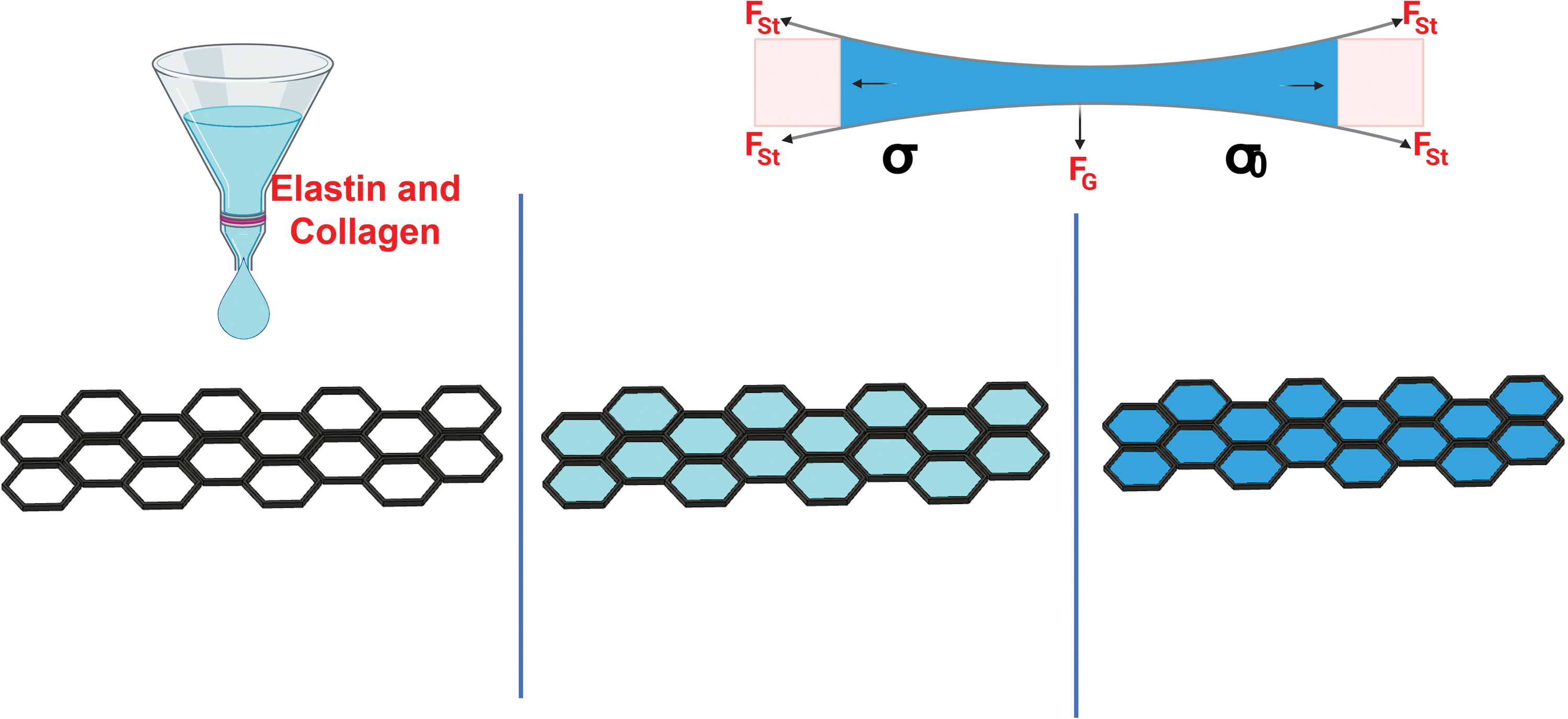
The Lung-on-Chip model developed by Zamprogno et al. 54 that was constituted of CE mixture gel membrane and framed in a gold mesh with array of 40 regular hexagons. CE, collagen-elastin. Color images are available online.
Another model comprises of three vertically stacked PDMS microfluidic compartments separated by nanoporous membranes to culture the airway epithelium, primary fibroblasts, and microvascular ECs. 56 A different three layer model containing a hydrogel segment was used to study the interaction between epithelial cells, smooth muscle cells, and a supporting ECM (collagen, Matrigel, or combination). 57 The microfluidic device was made of poly(methyl methacrylate) (PMMA), fabricated by micro milling and solvent-assisted thermal bonding methods to facilitate mass manufacturing and to enable the retrieval of the hydrogel for experiments by disassembling the device.
An airway-on-a-chip model was produced by 3D bioprinting a vascular network of PCL, bioinks of LF and EC, and PDMS. 58 The platform consisted of a central reservoir for EC and two side reservoirs of LF bioink separated by PCL microchannels for medium to flow. This method led to an airway model that created a functional interface with the vascular network.
Membranes used in lung-on-chip systems
PDMS membranes fabricated by soft lithography (Fig. 3) have been widely used in lung-on-chip systems despite the disadvantage that PDMS can absorb small hydrophobic molecules.59,60 To create membranes with pores, a solution of PDMS and crosslinker (10:1, v/v) is poured into a silicon mold containing pillars with predefined diameters. The poured PDMS was compressed and thermally treated to crosslink the PDMS. After crosslinking (ca. 24 h, 60°C), the PDMS membrane was removed from the master structure creating an interface for cell culture pretreatment (bioadhesion agents) and cell seeding preparation.
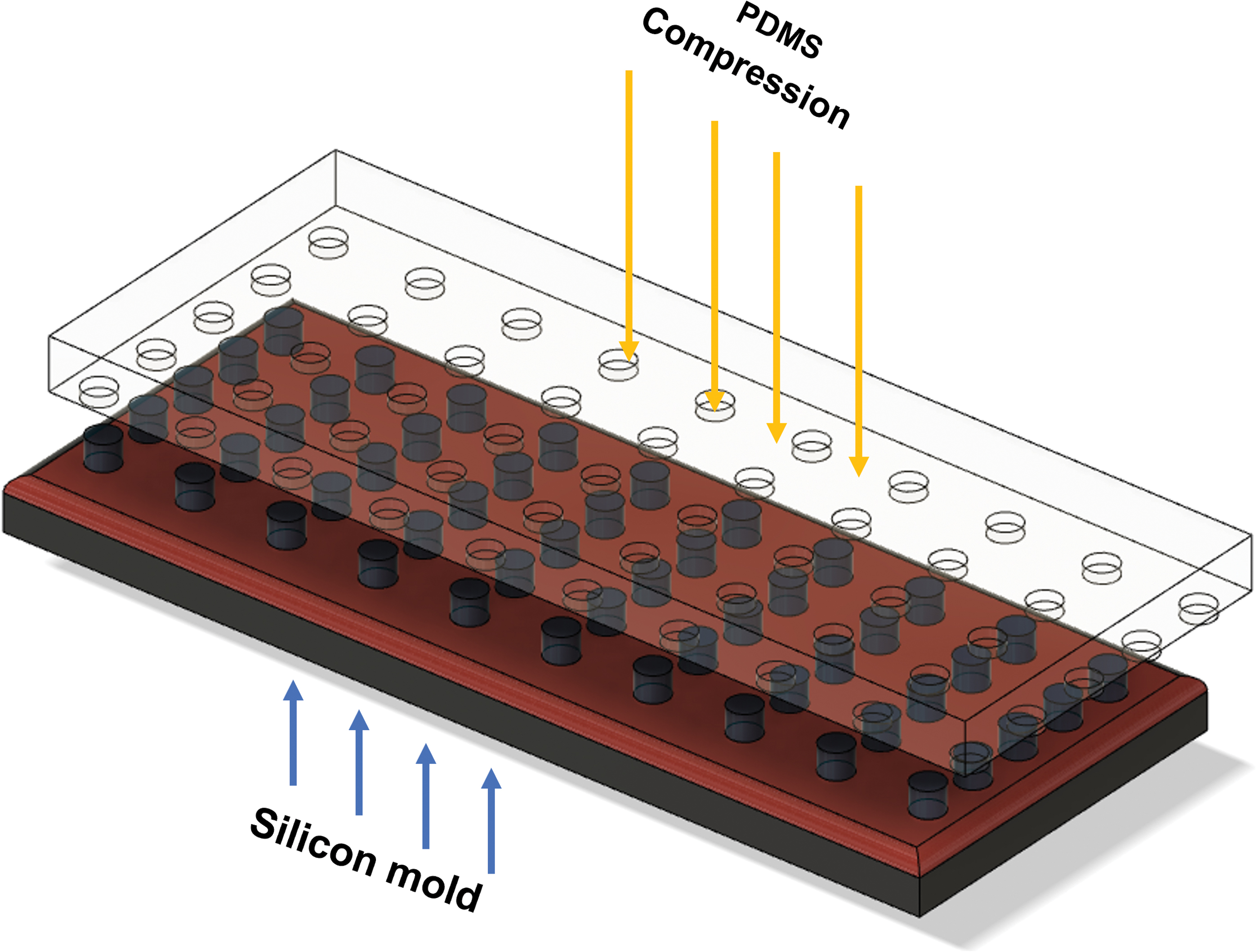
PDMS membrane fabrication by soft lithography. PDMS, poly(dimethylsiloxane). Color images are available online.
A micro curved culture membrane model integrated in a microfluidic system was developed by Di Huang et al., emphasizing the effect of microscale curved surfaces on cellular behavior. 61 The membrane fabrication involves assembling alginate microbeads in a closed lattice at the closest distance to each other.62–66 The free space between the microbeads was filled with GelMA solution. After crosslinking GelMA, the alginate microbeads were removed leaving behind an alveoli-mimicking hydrogel with uniform pore size with windows between the pores. Thus, effective airflow and ventilation within the hydrogel were achieved. The authors reported successful cell attachment, proliferation, and spread attributed to the presence of Arg–Gly–Asp sequences on GelMA and matrix metalloproteinase responsive peptide motifs in the model.
The introduction of 3D alveoli-mimicking structures to the lung on a chip model development is a necessary but a very challenging step in the direction of enhancing in vitro diagnostic and research tools. Table 1 demonstrates a comparison of the most important parameters of the membranes fabricated by means of the described technological approaches along with their advantages and disadvantages.
Comparison of Certain Parameters of Membranes Fabricated by Different Technological Approaches
3D, three-dimensional; CE, collagen-elastin; ECM, extracellular matrix; PDMS, poly(dimethylsiloxane).
NPs in the study of COVID-19
NPs of a variety of types and materials have been tested in the studies toward finding a therapeutic approach or cure against COVID-19. Most of these were synthetic polymer, lipid or carbon-based, and also metallic NPs. As subgroups of this list one can state dendrimers and their derivates, the dendrimersomes (Fig. 4). These differ from each other in terms of their biodegradability, which determines their effective life span; their surface chemical properties, which affect their distribution within the biological system; and their mechanical properties, which affect their transport properties. The preparation conditions of these particles are well established but their interaction with the nasal and the lung tissue necessitates the incorporation of certain targeting and interaction molecules, which significantly affect their efficacy. This diverse field as a whole is a topic for another study, while in this article, improvement of their interaction with COVID-19-related tissues and molecules are of specific interest.
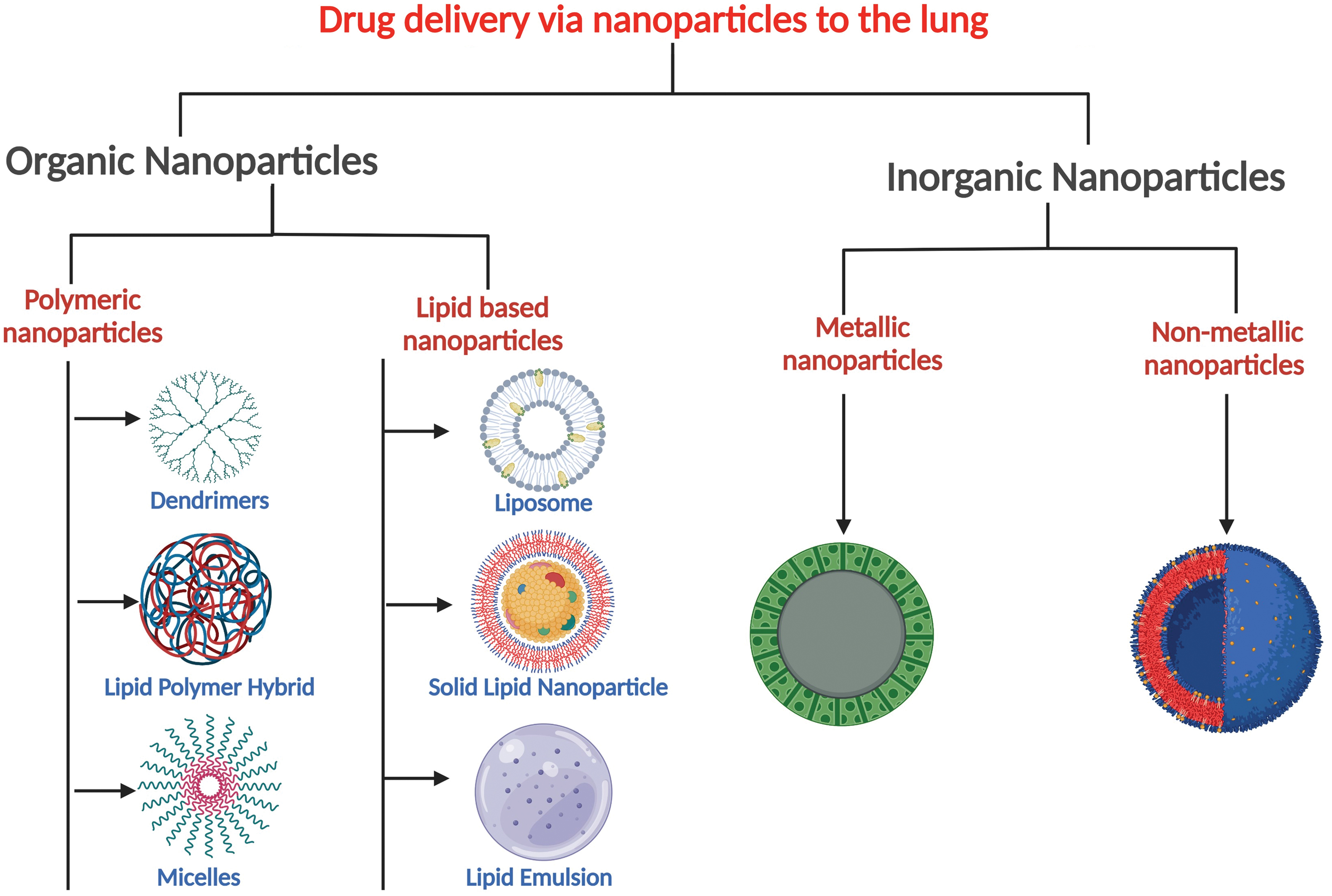
A variety of nanoparticles that can be used for the delivery of therapeutic agents to the lungs and thus can be used in COVID 19 modeling and therapy. Color images are available online.
The following sections present the involvement of NPs in COVID-19 research and applications based on their function.
Virus-mimicking NPs
A virus-mimicking NP can potentially be used to fight SARS-CoV-2 in various ways: It can display the viral antigen to compete with the virus or act as a vaccine by inducing the host's neutralizing activities; or it can target the delivery of therapeutics into the primary infection sites. Such NPs can be used to study the molecular mechanisms of host–virus interactions, enabling investigation into how the immune system recognizes and responds to viral infections, how viruses enter host cells, how they replicate and produce new viruses, and how they evade the host immune response. This information contributes to the understanding of the molecular mechanisms involved in viral infections and helps identify potential targets for antiviral therapies.
Mimicking the natural infection characteristics mainly relies on recapitulating the structural and antigenic properties of the S protein. This protein is responsible for initiating the primary immune response to the virus and facilitating the virus to attach and enter host cells. 67 Even though it is challenging to incorporate the antigen in its properly folded form into or onto nonprotein-based NPs without altering the accessibility of the epitope, synthetic polymeric NPs have been engineered to mimic various viral features. These features include morphology, multivalent antigen display, colocalization of adjuvant/antigen for enhanced immune response, as well as the breakdown of the antigen to facilitate its presentation on antigen-presenting cells through antigen processing.
The receptor-binding domain (RBD) in S1 subunit of S protein is an appealing antigen due to its capability of being reliably reproduced in a stable form.67,68 A virus-like hollow polymeric nanocapsule displaying RBD of MERS-CoV S protein was developed by Lin and colleagues. 69 The NP consisted of a poly(lactic acid-co-glycolic acid) (PLGA) shell and an aqueous core containing a soluble adjuvant for antigen pairing. The resulting vaccine was effective against MERS-CoV in mouse models. In another study, dendrimers were used, consisting of polymeric motifs with symmetrical tree-like branching units built around a center.70,71
Donalisio et al. developed a dendrimeric peptide consisting of a lysine peptidyl branching core covalently attached to the surface peptide that mimicked the heparin-binding domain of respiratory syncytial virus (RSV) envelope glycoproteins. This dendrimer binds to heparan sulfate proteoglycans on the surface of lung epithelial cells (A549), blocking the receptors and preventing attachment of RSV, thus preventing virus infectivity. 72
Pan coronavirus peptides generated by modifying certain spanning residues of the S protein S2 subunit helical bundle fusion core, were shown to inhibit viral membrane fusion in CoVs, including SARS-CoV-2.73–76 These antiviral peptides were displayed on NPs and found to enhance the immune response, partly due to the immunogenic properties of the NPs themselves. Carbon-based NPs, particularly nanodiamonds (ND), are known to trigger immune response and can be captured by neutrophil extracellular traps.77,78 It was demonstrated that surface-oxidized NDs displaying influenza virus trimeric hemagglutinin (H7) protein have both immunogenic and adjuvant properties in mice. 79 In another study, a pan coronavirus peptide heptad repeat 2 was covalently attached to the surface of NDs. The authors demonstrated that the displayed peptide exhibited reactivity with sera from convalescent COVID-19 patients, indicating its ability to recognize and bind to antibodies that are specific to SARS-CoV-2. NDs induced neutrophil-driven inflammation in mice and rabbits. 80
Effective protection against COVID-19 requires a vibrant and long-lasting immune response. This can be stimulated by highly immunogenic substances, which induce the production of antibodies and activate immune cells. One approach to achieve this is multivalent display of the S protein, as it has been demonstrated that the presentation of antigen as a repetitive array increases the potency of humoral immune response.81–85 For this purpose, protein-based NP platforms have been extensively used as multimerization frameworks for multivalent antigen display.86–88 24-subunit ferritin is one of the widely studied examples.89–91
Joyce et al. designed a ferritin NP conjugated with a modified S protein for its stable repetitive display on the protein scaffold and demonstrated immunogenicity against SARS-CoV-2 infection both in mouse models 89 and nonhuman primates. 92 Two-component I53–50 protein,93,94 encapsulin, 95 and lumazine synthase88,96 are other examples of protein-based NPs to display S protein RBD as antigens. In addition, as emerging variants of the virus possess mutations extensively in the S protein, recent studies also focused on displaying S or RBD of different viral variants of concern on a single NP. 97
One of the limitations of using virus-mimicking NPs for vaccination purposes is that most NPs are rapidly eliminated by body phagocytosis systems before they reach their target. One approach to solve this issue is to functionalize erythrocytes. This can be done by anchoring the S protein to the cytoplasmic membrane of erythrocytes 98 or decorating them with polydopamine NPs displaying the S protein S1 subunit and the Toll-like receptor 7/8 agonist R848. 99 This led to erythrocyte-mediated systemic antiviral immunity with both cellular and humoral immune responses in mice.
Study of high-risk infectious respiratory viruses such as SARS-CoV-2 in vitro is challenging as it requires biosafety Level 3 containment. Several pseudoviral systems have been developed to avoid this limitation, but generating a functional and safe system is still challenging. NPs mimicking the virus could be an alternative tool to study viral infection in vitro (Fig. 5).

Construction of a synthetic SARS-CoV-2 model using liposome-based nanoparticles. SARS-CoV-2, severe acute respiratory syndrome coronavirus 2. Color images are available online.
NPs as drug carriers
The use of NPs as drug carriers has a number of advantages: they can achieve release of drugs in a controlled manner, enhance and accelerate adsorption of the drugs carried, and prolong their in vivo half-life; or they can be made into intelligent delivery systems when triggered by certain stimuli or used for targeted delivery100–103 (Fig. 6).
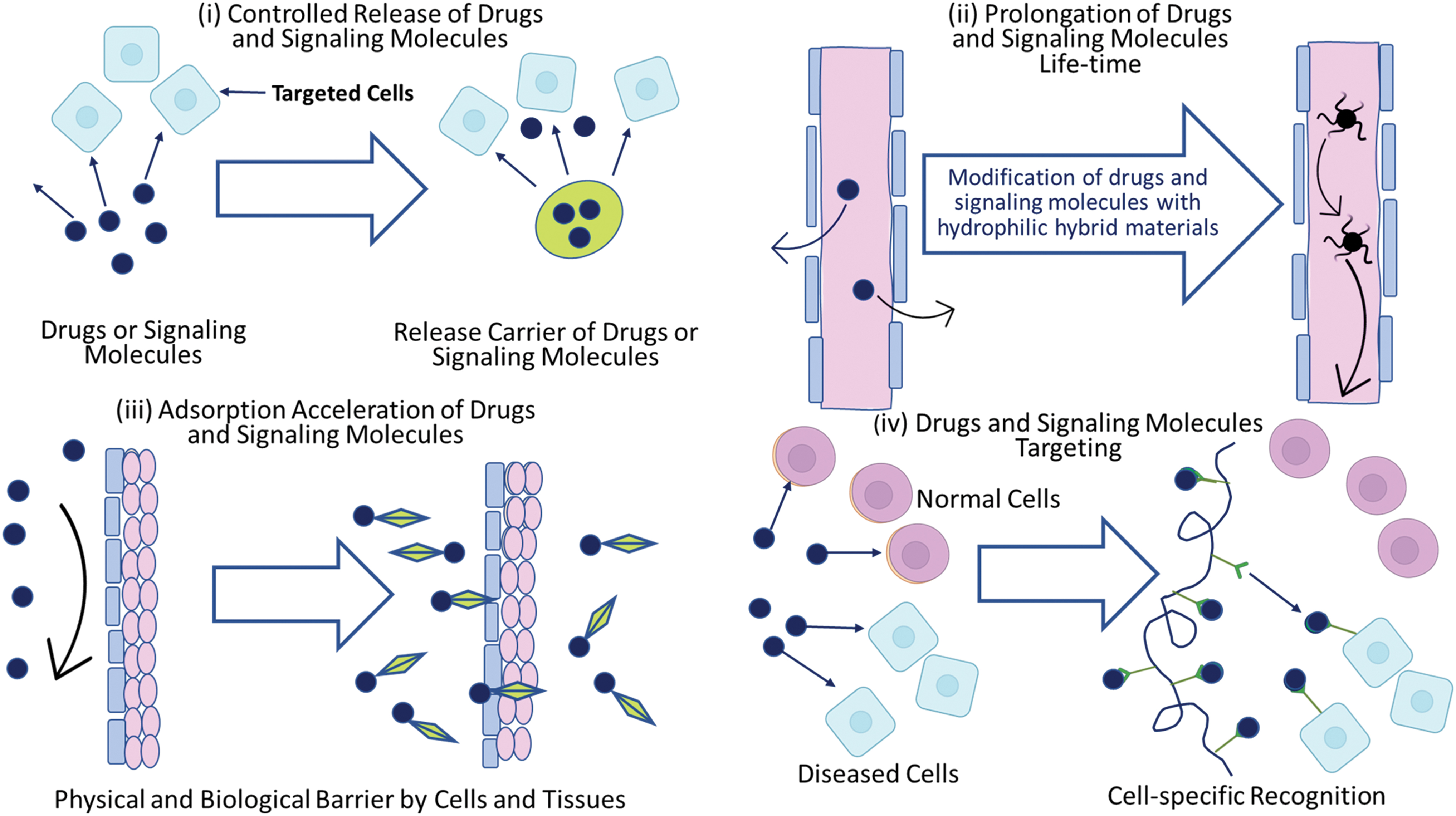
Properties and roles of nanoparticles or nanocarriers in bioactive agent delivery systems. The use of hybrid systems enables in vivo therapeutic activity of drugs and signaling or biomolecules to be intensified. There are four objectives of using drug and biomolecule delivery systems:
Toward the end of 2020, lipid nanoparticle (LNP) formulated vaccines BNT162b2 by Pfizer and BioNTech and mRNA-1273 by Moderna were accepted as effective and safe for use in the protection of public from the infection, and they still are in widespread use in the world.104–106 Both formulations contain a cationic LNP, which has a high encapsulation efficiency especially for negatively charged nucleic acids due to ionic interactions. The LNPs carry a modified mRNA encoding the full-length S protein that induces the neutralizing antibody production, CD4+ and CD8+ T cell activation, and favorable immunomodulatory cytokine responses. 107 The effectiveness of this technology in preventing disease has led to an interest in LNPs for the development of vaccines with an adjuvant effect and targeted drug delivery capability.
Using PLGA-based NPs to treat COVID-19 is another widely used drug delivery approach. An example is the delivery of a clinically approved antihelmintic agent ivermectin by oral administration. This drug acts by downregulating ACE2 receptor expression. The nanocarrier was constructed using poly(lactide-co-glycolide)-b-poly(ethylene glycol)-maleimide (PLGA-b-PEG-Mal) and tagged with Fc immunoglobulin, which enables the NPs to reach the bloodstream by crossing the gut epithelial barrier by binding to FcRn transcytosis receptor. 108 Another study used a similar approach where they delivered the antiviral prodrug oseltamivir phosphate labeled with spike-binding peptide , enabling it to bind to the S protein. 109
Metallic NPs such as gold (AuNPs) were coated with influenza membrane matrix protein 2 (M2e) using gold-thiol chemistry to produce a universal vaccine against influenza A viruses. The conjugation of the M2e peptide to AuNPs and using oligodeoxynucleotides (CpG) as a soluble adjuvant boosted immunogenicity against various types of influenza A viruses (H1N1, H3N2, H5N1) when administered intranasally to BALB/c mice. 110 Iron oxide nanoparticles (IONPs) have previously been shown to have antiviral activity against Dengue, 111 H1N1 influenza virus, 112 and rotavirus. 113 The antiviral activity was attributed to their interaction with viral surface proteins, which potentially prevented the binding of the virus to host cells. The affinity of IONPs to the S protein RBD was demonstrated with a docking model. 114
Another study demonstrated the potential use of meso-2,3-dimercaptosuccinic acid (DMSA)-coated IONPs as a drug delivery platform to treat COVID-19. DMSA serves as a shell for the IONPs, which helps to protect the IONPs and the tethered drug from degradation and improves their stability. The authors demonstrated that the NPs have no toxicity against the patients with COVID-19. 115
Another multifunctional use of NPs has been demonstrated with a polymeric NP containing a PEG shell carrying monoclonal neutralizing antibody against the S protein. 116 The NPs selectively captured the virus and effectively blocked its entry into host cells, resulting in an improved therapeutic effect compared to soluble neutralizing antibodies. The NP possesses a semiconductive polymer core, which has the ability to absorb light and convert it into electrical energy, thereby generating heat. This photothermal activity was used to generate local heat induced by 650-nm light-emitting diode to further inactivate the virus. The authors demonstrated the biosafety of the NPs both in vitro and in vivo, with satisfactory delivery to the lungs in mice.
Use of the Barrier Models and NPs in COVID-19 Research
The use of in vitro models of the blood-air barrier has significantly improved our understanding of COVID-19. Using an alveolus-on-chip system, researchers demonstrated that epithelial and endothelial cells of the barrier react distinctly to the infection, alveolar epithelial cells exhibited higher viral load and a broader innate immune response compared to pulmonary microvascular endothelial cells. Integrating circulating immune cells into the system resulted in elevated cytokine secretion and recruitment of immune cells, leading to the detachment of endothelial cells, and disruption of intercellular junctions in both epithelial and endothelial layers. 117 The models have also enabled researchers to test antiviral drugs and explore opportunities for drug repurposing. Notably, the results regarding the efficacy of remdesivir as an antiviral agent in the alveolus chip model aligned with another study which used an ALI model with stem cell-derived alveolar epithelial cells on a transwell system.
Both studies demonstrated a reduction in viral titers after treatment with remdesivir, as well as an improvement in the integrity of the on-chip barrier.117,118 In another study, researchers simulated the viral infection with SARS-CoV-2 pseudovirus on an alveolus-on-chip model, and demonstrated that monoclonal antibodies targeting the S protein RBD effectively inhibited viral entry into host cells. 119 In addition, the cyclic breathing motions were shown to have an impact on the innate immune responses in the barrier during Influenza A infection. Research identified the receptor for advanced glycation end products (RAGE) as a critical component in this response. Application of a RAGE inhibitor drug called azeliragon significantly suppressed the inflammatory cytokine production, and use of azeliragon with the antiviral molnupiravir showed synergistic effects in combating the infection. 50
Based on these findings, the researchers have submitted an application to pre-IND (Investigational New Drug) meeting of the Food and Drug Administration (FDA). The objective was to seek approval and initiate clinical trials to investigate the potential of azeliragon in effectively mitigating the cytokine storm in COVID-19 patients. This application marks an important milestone in the usage of the organ-on-chip systems as preclinical testing platforms.5,50
In vitro barrier models are extensively used in the evaluation of various aspects of NPs under the ALI conditions. Researchers utilize these models to examine the cellular uptake, transepithelial transport, therapeutic efficacy, and toxicity of NPs. Studies showed that both the cyclic breathing motions and dynamic fluid flow applied in these models have an impact on the cellular response to the administered NPs.51,120–125 The presence of the surfactant layer is another crucial aspect to consider as it is the initial interaction site for the NPs when they reach the distal lung.35,126–128 The NPs discussed in this review, those designed to simulate the viral infection, inhibit virus entry into host cells through competition, or deliver therapeutics to the infection site were subjected to conventional monolayer cultures for in vitro testing.
Utilizing barrier models is vital in understanding how those NPs interact with the blood-air barrier. This is essential in the development of standardized protocols that can effectively relate in vitro and in vivo dosing of agents, and lead to more accurate predictions of their lung deposition kinetics in the preclinical stage before advancing to clinical trials. As a result, these barrier models play a significant role in advancing the development of safer and more effective drug delivery systems in the fight against COVID-19.
Conclusion
COVID-19 led to the most widespread and deadly pandemic of the last 150 years, causing economic, social, and prolonged health problems worldwide. As a result of its rapid spread, the scientific community was faced with the challenge of rapidly developing vaccines and therapies without completing the international testing protocols, which highlighted the urgent need for advances in various scientific and industrial fields including in vitro blood-air barrier models. As presented in this review, various models, including ALI models, tissue engineered barrier models, and lung-on-chip systems, have shown promise in mimicking the complex lung microenvironment. ALI models are simple and cost-effective but lack physiological complexity, while tissue engineered barrier models are more physiologically relevant but require significant expertise.
Lung-on-chip systems, which basically introduce organ level functions to a microfluidic device, concentrate more on achieving a high fidelity simulation of the in vivo lung microenvironment by attempting to replicate the mechanical forces and fluid dynamic aspects of the human lung. Synthetic membranes constitute an important element of these systems because they provide significant control over the physical and mechanical properties, such as porosity, thickness, and elasticity, which in the end control transference across the barriers. As such, these synthetic membranes must meet the complex structural and functional properties of the barrier. Tissue engineering techniques with the presence of a variety of cell types and ECM components help create physiologically relevant models that better mimic the barrier.
By combining tissue engineered barrier models with lung-on-chip systems, researchers can further improve the mimicry provided by the in vitro models. While the technical complexity and cost of these systems may limit their widespread use, in the choice of models, researchers should consider the research objectives and available resources, and each in vitro model should be carefully assessed for its advantages and limitations.
The use of NPs has shown great potential in contributing to various approaches, including antigen presentation, drug delivery, mimicking of the viruses and in vaccine development. The application of nanotechnology in the fight against COVID-19 has demonstrated the versatility and effectiveness of these nano level particles in delivering therapeutics and in eliciting a robust immune response. The use of NPs offers a promising avenue for combating the current pandemic and potentially future outbreaks.
Overall, the development of more physiologically relevant in vitro models and the use of NPs in COVID-19 research are expected to significantly contribute to our understanding of the viral pathogenesis and aid in the development of effective treatments.
Footnotes
Authors' Contributions
N.S.O.: Conceptualization, Original draft preparation, Writing, and Diagrams; D.B.: Original draft preparation and Diagrams; M.N.C.: Writing; S.B.: Editing and Diagrams; J.O.: Editing; H.W.: Writing—Original draft preparation; R.A.: Writing—Original draft preparation; D.Y.: Original draft Preparation and Editing; H.K.: Original draft preparation and Editing; N.B.: Conceptualization and Writing-Reviewing,; I.D.A.: Conceptualization, Writing—Original draft preparation, Writing—Reviewing, and Visualization—Diagrams; V.H.: Conceptualization, Original draft preparation, and Writing-Reviewing and Editing.
Disclosure Statement
No competing financial interests exist.
Funding Information
This work is a part of the conceptual framework for MicroLung Project supported by the Fifth Southeast Asia and Europe (SEA Europe) Joint Funding Scheme for Research and Innovation. The authors acknowledge the support of the following institutions in the realization of the project:
Ministry of Education, Culture, Research, and Technology of the Republic of Indonesia and DIPI under the contract No. 1839/UNl/DITLIT/Dit-Lit/PT.01.03/2022. German Federal Ministry for Education and Research (BMBF) under the project No. 01DR21011., Scientific and Technological Research Council of Turkey (TUBITAK), 1071 Program, Grant No.: 121N817. Acibadem University Biomaterials Application and Research Center, Istanbul, Turkey