




Abstract
The healing process after tendon injury is often accompanied by the formation of peritendinous adhesion, contributing to limb dysfunction and exerting detrimental effects on the individuals, as well as the development of society and economy. With the continuous development of material science, as well as the augmented understanding of tendon healing and the mechanism of peritendinous adhesion formation, materials used for the fabrication of barrier membranes against peritendinous adhesion emerge endlessly. In this article, based on the analysis of the mechanism of adhesion formation, we first review the commonly used natural and synthetic materials, along with their corresponding fabrication strategies, in order to furnish valuable insights for the future optimization and development of antiperitendinous adhesion barrier membranes. This article also discusses the interaction between antiadhesion materials and cells for ameliorating peritendinous adhesion.
Impact Statement
Peritendinous adhesion is a common complication after tendon injuries, often leading to limb motor dysfunction and posing a significant threat to the physical and mental health of patients. With the development of material science, novel antiperitendinous adhesion barrier membranes have been an effective method to reduce or even prevent peritendinous adhesion. In this article, we provide a comprehensive review and analysis of the materials and fabrication strategies used to create barrier membranes for preventing peritendinous adhesion, and we delve into the mechanisms of adhesion formation. We also explore the interactions between these antiadhesion materials and cells, providing insights into the solution of this clinical problem in the treatment of peritendinous adhesion. By summarizing the current advances in the field, we strongly confirm that biomaterials are a promising strategy for the treatment of peritendinous adhesion.
Introduction
Incomplete statistics indicate an annual incidence of 32 million cases of tendon injury in the United States each year caused by inappropriate exercise and trauma. 1 Tendon injury seriously affects patients’ quality of life. Currently, surgery has been considered as the major approach to repair the injured tendon. However, postoperative complications, such as peritendinous adhesion, occur in approximately 30–40% of patients, resulting in dysfunction of limb movement and seriously affecting the physical and mental health of patients. 2
With the continuous development of material science, the fabrication of antiperitendinous adhesion barrier membranes has become the focus of domestic and foreign scholars. More and more natural and synthetic materials have been used for the fabrication of antiperitendinous adhesion barrier membranes, and remarkable achievements have been made.
This article reviews the basic research and clinical application of various antiadhesion materials commonly used at present and their corresponding antiadhesion membranes, with a view to providing a reference for the further optimization and development of antiadhesion barrier products in the future.
Biomaterial-Mediated Cell Behavior against Peritendinous Adhesion
Fibroblast
Fibroblasts have been considered to play a pivotal role in the formation of peritendinous adhesions (Fig. 1). After tendon injury, fibroblasts migrate to the injury site, proliferate, and secrete extracellular matrix (ECM) components, including collagen and glycosaminoglycan. However, excessive fibroblast proliferation and collagen deposition would lead to the pathogenesis of peritendinous adhesions. In addition, cytokines and growth factors secreted by fibroblasts further induce adhesion formation.3–5 Myofibroblasts, the activated phenotype of fibroblasts, are typically characterized by the expression of α-smooth muscle actin in the cytoskeleton. 6 They also play an extremely important role in the process of repair and adhesion formation after tendon injury. Specifically, after tendon injury, fibroblasts are activated by mechanical stimulation and chemical stimulation by inflammatory mediators and then differentiate into a myofibroblast phenotype. 7 Ruscitti et al. 8 found that IL-6 can promote fibroblast activation into myofibroblasts, which proliferate and secrete type III collagen to mediate tissue adhesion. Different kinds of biomaterials have been used to act as physical barriers that inhibit fibroblasts, therefore preventing peritendinous adhesions (Table 1).
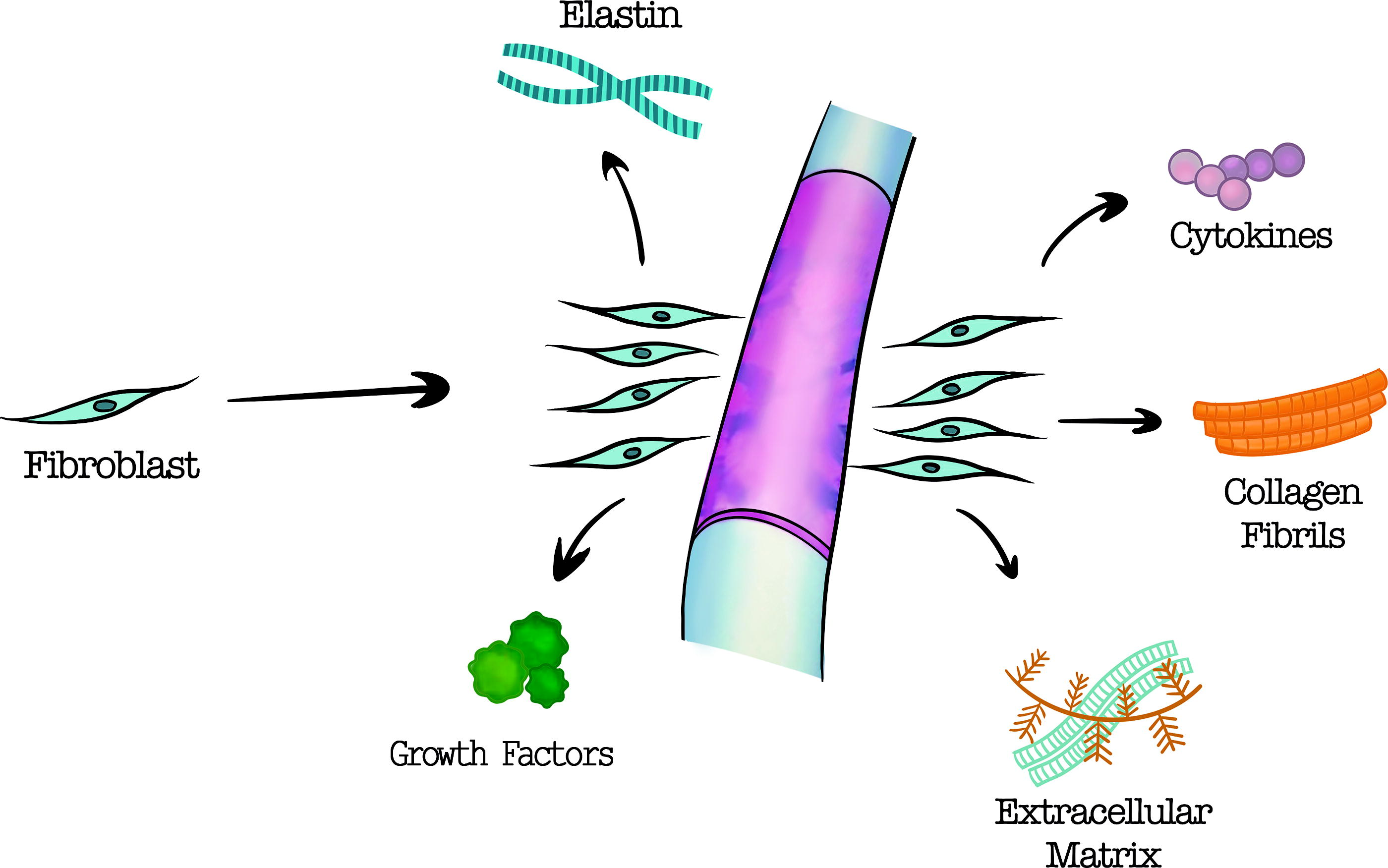
Fibroblasts migrate to the injury site, proliferate, and secrete extracellular matrix components. In addition, cytokines and growth factors secreted by fibroblasts further induce the adhesion formation. Physical barrier fabricated from biomaterials can effectively inhibit this process.
Types of Materials and Their Role in the Prevention of Adhesion
CEL, celecoxib; CS, chitosan; CSNFMS, core-sheath nanofibrous membrane; HA, hyaluronic acid; MSs, microspheres; PCL, poly(ε-caprolactone); PDA, 2,6-pyridinedicarboxaldehyde-polyethylenimine; PEG, poly(ethylene glycol); PELA, poly(ethylene glycol); PEU, poly(ether urethane); PLA, poly(lactic acid); PLLA, Poly(L-lactic acid); PNIPAM, poly N-isopropylacrylamide; PSBMA, poly(3-[2-(methacryloyloxy)ethyl](dimethyl-ammonio]-1-propanesulfonate); PU, poly urethane.
Natural materials against peritendinous adhesion
Hyaluronic acid (HA) is a nonspecific hydrophilic polysaccharide found in the synovial fluid of the human body that acts as a lubricant for tendon gliding and provides certain nutrients for it. HA not only has good viscoelasticity, moisture retention, and anti-inflammatory properties but also inhibits the proliferation and migration of fibroblasts. At present, it has become one of the most commonly used antiadhesion materials in clinical practice.23,24 Activation of the TGF-β1/Smad3 signaling pathway plays an important role in the process of peritendinous fibrosis. Therefore, blocking Smad3 and its downstream signaling pathway may attenuate the degree of peritendinous adhesion and improve tendon healing. 25 Based on the role of the above signaling pathways in tendon adhesion, Cai et al. 9 embedded matrix metalloproteinase-2 (MMP-2)-degradable gelatin-methacryloyl (GelMA) microspheres (MSs) encapsulated with Smad3-siRNA nanoparticles in HA hydrogel to construct a self-healing and deformable HA hydrogel. In previous studies, it has been found that the extrusion force between the gliding tendon and the peritendinous tissue is easy to cause the destruction of the implanted material, and the material debris can cause an inflammatory and immune response and aggravate the degree of peritendinous adhesion.26–29 Therefore, as a physical barrier, hydrogel needs to have excellent abilities for self-healing and deformability. In addition, GelMA MSs degraded responsively due to the increase of MMP-2 after tendon injury, thus achieving the on-demand release of siRNA nanoparticles and effectively inhibiting the proliferation of fibroblasts and the formation of peritendinous adhesion tissue, suggesting that this composite barrier can be used as an ideal material for preventing tendon adhesion.
Chitosan (CS) is a natural component of shells and other crustaceans. It has unique biological properties such as nontoxic, antibacterial, antioxidant, and hemostatic, so it is used in the field of medicine. In addition, CS has attracted extensive attention in the field of tissue regeneration because of its good mechanical properties, biocompatibility, and biodegradability. 30 In view of the defect of the rapid degradation rate of CS, Fakhraei et al. 10 fabricated poly(ε-caprolactone) (PCL)/CS composite nanofiber membrane by electrospinning. CS is hydrophilic, whereas PCL is hydrophobic. The combination of the two can appropriately improve the degradation rate of the nanofibrous membrane, which was beneficial to maintain the integrity of the membrane during the tendon healing process. In addition, scholars have conducted several studies on the phenomenon that CS can reduce the adhesion of fibroblasts. The study shows that it may be related to the positive charge on the surface of CS caused by amino protonation, but some scholars still hold different views on this phenomenon. Poly(N-isopropylacrylamide) (PNIPAM) is a famous temperature-responsive polymer, which exhibits reversible sol-to-gel phase transition behavior in water. When the temperature is lower than the lowest critical solution temperature (LCST), the hydrophobic interaction between the isopropyl groups was counteracted by the hydrogen bonds between the hydrophilic amide groups and water molecules. At this point, PNIPAM tends to dissolve in an aqueous solution, and the polymer chains exhibit flexible and expanded random coil-like conformations. As the temperature increases, some of the hydrogen bonds are destroyed and the hydrophobic interaction between the isopropyl groups enhances; the PNIPAM chains dehydrate and aggregate into a tightly packed globular-like conformation, transforming into a gel state.31–33 This phase transition ability will ensure that temperature-responsive hydrogel turns into gel after injection and forms a physical barrier around the injured tendon when the temperature is raised to physiological temperature. Moreover, the LCST of PNIPAM can be adjusted over a wide temperature range by adding more hydrophilic components or hydrophobic monomers through copolymerization based on thermodynamic principles. 34 In another study, injectable PNIPAM hydrogels grafted with CS and HA were demonstrated to effectively block the penetration of NIH 3T3 cells. In vivo, studies in rear-limb flexor tendon repair models revealed that the barrier PNIPAM hydrogels were more effective than currently available commercial barrier materials at reducing tendon adhesions. 11
Synthetic materials against peritendinous adhesion
PCL is widely used as a medical biomaterial and drug carrier because of its good biodegradability, biocompatibility, and low toxicity. PCL nanofibrous membrane fabricated by electrospinning exhibited the potential to prevent the formation of adhesions. With the average pore size of 0.87 μm, the membrane was able to obstruct the penetration of extrinsic fibroblasts, while allowing the exchange of nutrients and waste required for tendon healing. The researchers further grafted CS on the surface of the PCL nanofibrous membrane, which was demonstrated to reduce the adhesion of fibroblasts. The ability to prevent fibroblast penetration and attachment made the CS-grafted membrane a promising approach against peritendinous adhesions. 12 Chen et al. 13 embedded silver (Ag) nanoparticles in the HA/PCL nanofibrous membranes (HA/PCL+Ag NFMSs) to simulate the physiological functions of the fibrous and synovial layers in the tendon sheath. The NFMS has lubrication and antibacterial effects through a slow release of HA and rapid release of silver nanoparticles, respectively, and HA and silver nanoparticles play a synergistic role in reducing the attachment and proliferation of fibroblasts without significant cytotoxicity. Inspired by the commercial antiadhesion barrier membrane SurgiWrap®, Chen et al. 3 designed a new type of core-sheath nanofibrous membrane (CSNFMS) with poly(lactic acid) (PLA) instead of PCL in the previous sheath structure. This membrane enables to better control the release of lubricant HA from the core structure. In addition, the researchers also adjusted the release rate of HA and silver nanoparticles by changing the core-sheath ratio in order to better exert their synergistic effect and further optimize the antiadhesion performance of CSNFMS. However, the limitation of this study is that the risk factors of inflammatory response that may lead to tendon adhesions were ignored. Therefore, the addition of the NSAID drug ibuprofen (IBU) to NFMS may provide a better therapeutic outcome.
Polyurethane (PU) is a kind of compound synthesized on the basis of PCL, which consists of alternating soft and hard segments. The continuous matrix is formed by soft segments with a low glass transition temperature (Tg), making the material exhibit flexibility. Hard segments with high Tg often have a tendency to aggregate into domains by physical cross-linking. These domains mainly play a strengthening role in the continuous matrix with low Tg, improving the mechanical properties of the material. 35 The microphase separation structure makes it to have good flexibility and mechanical properties, so it has been used in tissue engineering. 35 Hsu et al. 14 synthesized a waterborne biodegradable polyurethane (WBPU) membrane, and the hydrophilicity of the material was improved by adding ionic groups to the chemical structure. Through the rabbit tendon injury model, it was proved that the antiadhesion effect of WBPU membrane group was superior to that of PCL membrane group and untreated control group. In recent years, PU has been increasingly used in the fabrication of antiadhesion barrier membranes.
Similarly, nanofibrous membranes prepared by poly(ethylene glycol) (PEG) also exhibited desirable antiadhesion function by preventing fibroblast migration and adhesion. 36 Previous studies have shown that PEG blocks with a molecular weight of approximately 20,000 g/mol are required in the structure of physical barrier membranes to maintain good mechanical and antiadhesion properties. 37 PEG with a molecular weight of less than 20,000 g/mol can be removed by urine, whereas PEG with a larger molecular weight is removed through urine at a quite slow rate. As the molecular weight of PEG increases, the main metabolic pathway shifts from the kidney to the liver. Its metabolites will gradually accumulate in the liver and kidney, causing liver and kidney damage. 38 In order to solve this problem, Zebiri et al. 17 modified PEG (molecular weight about 20,000 g/mol) into polyether urethane (PEU) by adding degradable urethane bonds. Based on this, a new PLA-PEU-PLA triblock copolymer antiadhesion membrane was designed. It was confirmed that the degradation products of this membrane could be metabolized smoothly by the kidney, avoiding the disadvantages of the tendency to cause liver and kidney damage. Meanwhile, due to the presence of urethane bonds and hexamethylene in the structure of PEU, the membrane exhibits excellent elasticity and ductility. In addition, the degradation rate is slow to adapt to the pathological process of peritendinous adhesion formation, which can effectively play the role of antiadhesion.
By introducing PEG into poly(L-lactic acid) (PLLA), a biodegradable polymer material PLLA-poly(ethylene glycol) (PELA) was synthesized. PELA is considered an ideal drug carrier because of its excellent performance in drug loading and delivery and its diverse degradation modes.39,40 Compared with PCL, PELA has better flexibility and hydrophilicity, making it a promising antiadhesion material. Li et al. 18 fabricated an electrospun bilayer biomimetic tendon sheath composed of an outer layer of celecoxib (CEL)-PELA fiber membrane and an inner layer of hyaluronic acid-PELA fiber membrane. The outer layer can act as a physical barrier and release CEL to inhibit the proliferation of NIH/3T3 fibroblasts and collagen expression by inhibiting the phosphorylation of ERK1/2 and Smad2/3, thereby reducing inflammation and adhesion to surrounding tissues. 41 In contrast, the inner layer releases hyaluronic acid to promote tendon gliding and healing.
Poly(lactic acid-co-glycolic acid) (PLGA) is a kind of degradable functional polymeric organic compound that is polymerized by lactic acid and glycolic acid. It has good biocompatibility and membrane-forming properties. In view of the fact that its degradation products lactic acid and glycolic acid are both products in the human metabolic pathways, PLGA has no significant toxicity to the human body when applied in the field of biomaterials. Although previous studies have found that PLGA membrane is even more effective than Seprafilm® in preventing abdominal adhesion in rats, the hydrophobicity has always limited the development and application of PLGA in the field of antiadhesion membrane. Li et al. 19 developed a PEG/PLGA nanofibrous membrane by electrospinning technology, which significantly improved the hydrophilicity of the barrier membrane. The effect of the composite membrane on preventing adhesion was confirmed in the rat cecum injury repair model. At the same time, the researchers found that with the increase of PEG concentration, the hydrophilicity of the membrane was better, but the best antiadhesion effect could be achieved only with moderate hydrophilicity. Therefore, the optimum concentration of PEG needs to be further studied. In recent years, as a suitable technique for surface modifications, cold atmospheric plasma (CAP) has attracted wide attention because of its great application potential in biomedicine, material processing, environmental protection, and other fields. ElKhatib et al. 42 found that the hydrophilicity of PLGA fibers can be improved without changing the structure of PLGA fibers by N2 plasma treatment. Studies have shown that the increase of oxygen-containing functional groups on the surface of PLGA fibers is the main reason for the improvement of hydrophilicity after CAP treatment. The accelerated and bombarded ions generated from a short distance cause the formation of highly active free radicals on the surface of PLGA, which then react with oxygen in the air. Specifically, after CAP treatment, the content of C-C/C-H bonds in PLGA fiber decreases, while the content of C-O and C=O bonds increases, which means that the content of oxygen and the ratio of O/C increase significantly.43,44 In a word, CAP treatment provides a new way to improve the property of PLGA.
Fang et al. 21 designed a salt-sensitive zwitterionic physical hydrogel, namely, poly 3-[2-(methacryloyloxy)ethyl](dimethyl)-ammonio]-1-propanesulfonate (PSBMA) polymer hydrogel. With the increase of the concentration of sodium chloride, the gel-solution transition occurs in PSBMA hydrogel, which makes PSBMA hydrogel dissociate completely under physiological conditions. In the animal experiment, it was found that the inflammatory reaction and the degree of adhesion in PSBMA hydrogel group were significantly better than those in the control group and HA hydrogel group. It is speculated that PSBMA hydrogel may effectively inhibit fibrosis and improve postoperative adhesion by activating TGF-β1/Smad7 and inhibiting TGF-β1/Smad3 signaling pathway. 45 Jiang et al. 22 synthesized a PSBMA hydrogel using micellar polymerization techniques, which improved the mechanical properties of the hydrogel. The advantage of this hydrogel lies in the deformation and dislocation of the micelles as dynamic cross-linking sites when subjected to external stresses, which make the hydrophobic chains entangled within the micelles slide and the stresses disperse thus preventing the hydrogel from fracture and therefore tougher. Of course, since ionic monomers destabilize the micelles, the micellar polymerization technique is only applicable to electrically neutral monomers and not to charged monomers.
Current studies have mainly focused on placing antiadhesion barriers, which mimic the natural structure of tendon sheath to block the infiltration and adhesion of fibroblasts. However, most of the antiadhesion barriers reported appear to be bioinert and lack the ability to regulate the fibroblast activities in the implanted areas.5,46,47 To this regard, incorporating the bioactive factors into the antiadhesion barriers might bring benefits to the prevention of peritendinous adhesions.
Li et al. designed a drug-loaded electrospun nanofiber membrane (ENM), which was composed of PLA and dicumarol conjugates. In addition to serving as a physical barrier against fibroblast penetration, the drug-loaded ENM could also reduce fibroblast proliferation and adhesion. 15 Xiang et al. reported a lubricated hydrogel patch with the ability to release gene nanoparticles to silence the key gene in the activation of fibroblasts. The outer layer of the patch was fabricated by PEG-based polyester hydrogel under electrospinning technology to form a lubricated and antiadhesion surface, which exhibited exceptional antiadhesion capacity. Nanoparticles encapsulating ERK2-siRNA were then incorporated into the PEG-based polyester hydrogel, which further targeted ERK2 gene and its downstream signaling pathway in fibroblasts. The synergistic effect of lubricated surface and gene silencing was proved to significantly block the formation of tendon adhesions in vivo. 20 In another research, Liu et al. 16 confirmed that ERK2-siRNA poly-L-lactic acid(PLLA)/hyaluronic acid membrane in the form of electrospun membrane mediated by 2,6-pyridinedicarboxaldehyde-polyethylenimine (PDA) can be used as an effective physical barrier to prevent peritendinous adhesion, which promotes the application and development of siRNA-related gene silencing in the field of prevention and treatment of peritendinous adhesion.
Macrophage
In addition to fibroblasts, macrophages also play a crucial role in the formation of tendon adhesion (Fig. 2). Based on their different roles in tendon repair, activated macrophages could be classified into two subtypes, the pro-inflammatory M1 macrophage and anti-inflammatory M2 macrophage. After being recruited to the injury site, the M1 macrophages could secrete pro-inflammatory cytokines which induce excessive inflammatory response and stimulate the proliferation and activation of fibroblasts, thereby contributing to the adhesion formation.4,48,49 The role of M2 macrophages in tendon healing is complex, as they support the anti-inflammatory response and facilitate tissue remodeling in the early stages, but their overactivity in the later stages may lead to an imbalance in the healing process, shifting the healing process from endogenous to exogenous repair, leading to scar tissue formation, and exacerbating tendon adhesions.50,51 Recent studies have identified a new subgroup of secreted phosphoprotein 1 (SPP1) positive macrophages that can exacerbate adhesion by influencing fibroblast proliferation and extracellular matrix deposition through the CD44 pathway. 52 In conclusion, the role of M2 macrophages in tendon healing is complex and requires further investigation.
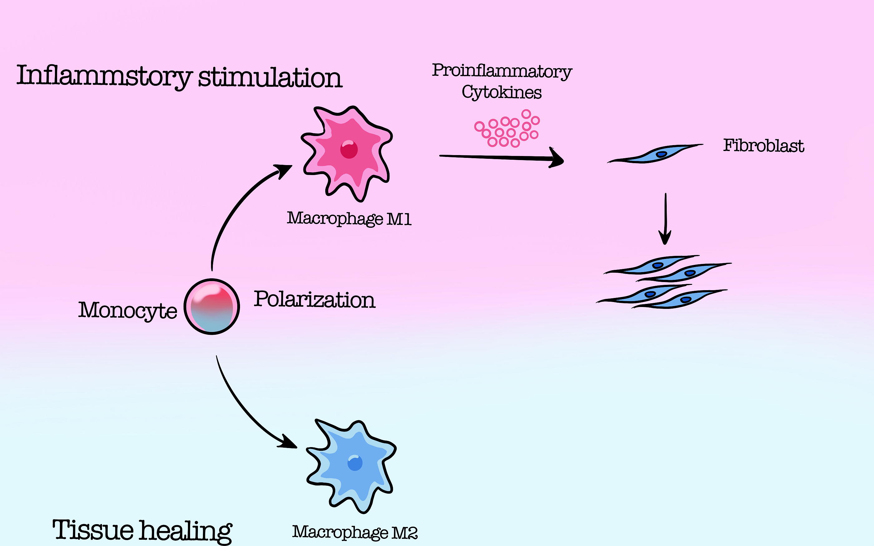
During the repair process after tendon injury, monocytes are activated into two subtypes, pro-inflammatory M1 macrophages and anti-inflammatory M2 macrophages. Upon recruitment to the injury site, M1 macrophages could secrete pro-inflammatory cytokines that induce excessive inflammatory response and stimulate proliferation and activation of fibroblasts, thereby promoting adhesion formation.
Furthermore, macrophages have been suggested to be responsible for the pathogenesis of foreign body reaction (FBR) after implantation. When the foreign biomaterials are transplanted into the body, macrophages recognize and degrade the material through releasing enzymes and reactive oxygen species (ROS). The activated macrophages release pro-inflammatory chemokines and cytokines, which recruit fibroblasts and other immune cells. Together, these cells aggregate around the implanted biomaterial and secrete a variety of bioactive factors, ultimately forming a dense fibrous capsule around the material.53–55 Multiple strategies have been reported to suppress the detrimental inflammation induced by macrophage, including drug-loaded membranes, scaffolds for gene silencing, and decellularized materials.9,56,57 (Table 2)
Classification of Materials and Their Functions in Preventing Adhesion
BDDE, 1,4-butanediol diglycidyl ether; CUR, curcumin; CEL, celecoxib; ECM, extracellular matrix; HA, hyaluronic acid; IBU, ibuprofen; NF-κB, nuclear factor-kappa B; MGF, mechano-growth factor; PCL, poly(ε-caprolactone); PEG, poly(ethylene glycol); PLA, poly(lactic acid); SF, silk fibroin; SSAD, skin secretion of Andrias davidianus.
Natural materials against peritendinous adhesion
In an experiment carried out by Wiig et al., 58 it was found that the prognosis of the injured finger could be significantly improved using sodium hyaluronate loaded with synthetic peptide PXL01 around the tendon in the repair of the flexor tendon after hand injury, but the specific mechanism had not been clarified. In this regard, Edsfeldt et al. 59 conducted related research. It showed that PXL01 in HA could promote the expression of proteoglycan 4 (PRG4/lubricin) after tendon repair, while decreasing the expression of inflammatory mediators such as IL-1β and IL-6, thus reducing the resistance to tendon gliding and inhibiting the formation of tendon adhesion. However, since HA itself also has certain anti-inflammatory effects, whether PXL01 and HA can synergistically inhibit inflammation still needs further research to confirm. In vivo study by Chen et al. 60 on rabbit flexor tendon rupture model showed that compared with Seprafilm® (a commercial antiadhesion film), the hyaluronic acid nanofibrous membrane (NFMS) loaded with IBU fabricated with Fe3+ and 1,4-butanediol diglycidyl ether can effectively reduce local inflammation and prevent tendon adhesion. It should be noted that when the IBU loading exceeds 30%, due to the high concentration of released IBU, it will be cytotoxic and detrimental to tendon healing.
The amnion is a semipermeable membrane that is differentiated from trophoblast cells. Its surface is smooth, without blood vessels, nerves, and lymphatic, and has a certain degree of elasticity. Compared with ordinary amnion, decellularized amniotic membrane has better histocompatibility, lower antigenicity, stronger permeability, and can be completely degraded in vivo, which makes it an ideal biomaterial. 61 Liu et al. 61 used the decellularized amniotic membrane as a mechanical barrier in Henry chicken tendon injury model. Compared with medical membrane group and control group, the inflammatory response at the injury site in the decellularized amniotic membrane group was mild and significantly reduced peritendinous adhesion, suggesting that amniotic membrane, as a natural biomaterial, can effectively inhibit exogenous healing, promote endogenous healing, and prevent tendon adhesion. In recent years, Sang et al. 62 further revealed the mechanism of decellularized amniotic membrane promoting tendon healing and preventing peritendinous adhesion, that is, promoting endogenous healing and inhibiting exogenous healing by releasing growth factors such as TGF-β1 and basic fibroblast growth factor (bFGF). Some scholars have found that freeze-dried amniotic membrane transplantation can promote the healing of flexor tendons in zone II and prevent adhesion. Despite the prevention and treatment of tendon adhesion being effective, it also exhibits the characteristics of poor mechanical properties, fast dissolution, easy slippage, and so on. 70 In order to overcome these defects of amniotic membrane materials, Liu et al. 63 used electrospinning technology to coat both surfaces of freeze-dried amniotic membrane with PCL nanofibers to form multilayer composite membranes to optimize amniotic membrane materials. The experimental results in the rabbit tendon injury model showed that the multilayer composite membrane structure mimicking the natural tendon sheath could effectively prevent the invasion of surrounding adhesive tissue and maintain good tendon gliding.
Silk fibroin (SF) is a kind of natural polymeric fibrous protein produced by silkworms, which has attracted wide attention from researchers because of its excellent biocompatibility, biodegradability, and mechanical properties. Electrospun SF scaffold was used as an antiadhesion barrier, which can promote the healing of injured Achilles tendons in rabbits and effectively reduce the adhesion of surrounding tissues. 71 Pagán et al. 71 further confirmed the promising application of silkworm gut fiber braid as a tissue engineering scaffold for tendon and ligament. Nadri et al. 64 fabricated an IBU/PEG/SF nanofiber membrane with a core-shell structure by coaxial electrospinning technique. The membrane was found to enable sustained release of IBU to the injury site, attenuate the post-traumatic inflammatory response, reduce local vasodilation in the injured area, and thus reduce the degree of tissue adhesion. Its excellent antiadhesion and anti-inflammatory properties have been confirmed in the rat abdominal injury model.
Local injury can lead to the release of ROS, which in turn induce oxidative stress at the site of tendon injury. Oxidative stress can cause inflammation response, increase vascular permeability, and lead to the release of inflammatory factors such as TNF-α, IL-1, and IL-6. Inflammation will then aggravate oxidative stress to form a vicious circle that promotes the formation of adhesive tissue.72,73 Curcumin (CUR), a natural compound found in turmeric, is derived from the rhizome of the plant turmeric and has great anti-inflammatory and antioxidant properties. 74 Zhang et al. 65 used poly(ethylene glycol dimethacrylate-co-1,2-ethanedithiol) [poly (EGMA-coEDT)] to design an oxidative stress-responsive electrospun membrane loaded with CUR/CEL. The sulfur-containing polymer in the electrospun membrane reacts with ROS and is oxidized to sulfoxide or sulfone, which significantly enhances the hydrophilicity and realizes the responsive release of drugs under oxidative stress; when the inflammatory reaction is obvious, CUR and CEL release more rapidly and play a synergistic role in inhibiting cell proliferation and collagen synthesis, thus achieving long-term effective antiadhesion effect.
Dang et al. 66 reported a self-healing and deformable bilayer skin secretion of Andrias davidianus (SSAD)-derived hydrogel. Compared with other materials used for tendon injury repair, SSAD-derived hydrogel can promote the migration of tendon stem cells because they contain insulin-like growth factor, stromal cell-derived factor, and glycine which are beneficial to tendon healing. In addition, SSAD-derived hydrogel also has antioxidant and antibacterial properties, which help to reduce peritendinous adhesion. The use of self-healing and deformable hydrogels is expected to be a reliable solution to avoid accidents such as slippage and rupture of antiadhesion barrier membrane in the process of tendon sliding.
Synthetic materials against peritendinous adhesion
PLA is a commonly used antiadhesion material because of its excellent mechanical properties, biocompatibility, and biodegradability. Biomaterials have been widely used in the field of tissue engineering. However, when biomaterials such as PLA are implanted into the human body, they will activate the body’s immune system, trigger foreign body reaction and aseptic inflammation, promote the proliferation of fibrous tissue, and then lead to the formation of postoperative adhesion tissue. Studies have found that M1-type macrophages and the inflammatory microenvironment they mediate play an important role in the above process, which provides a potential target for intervention and treatment of FBR and tissue adhesion. 75
Nuclear factor-kappa B (NF-κB) is a transcription factor involved in the regulation of cell signaling and inflammatory response. NF-κB phosphorylation plays a crucial role in promoting the classical activation of macrophages (M1 phenotype). Inhibition of NF-κB-mediated M1 polarization of macrophages is expected to improve implant-induced FBR. 67 Wang et al. 67 designed a PLA membrane loaded with JSH-23 (selective NF-κB inhibitor) (JSH-23/PLA-M) by electrospinning technology. It has been proved that JSH-23/PLA-M could accurately and effectively inhibit NF-κB phosphorylation and PLA-M-related M1 polarization, exhibiting superior anti-inflammatory and antiadhesion effects. Lu et al. 56 also found that inhibition of NF-κB phosphorylation could downregulate the expression of type III collagen and ultimately reduce the formation of peritendinous adhesion. At the same time, it is also able to upregulate the expression of type I collagen in the tendon and promote healing of the injured tendon, which is consistent with the previous findings. All of these results suggest that NF-κB related signaling pathway is closely related to peritendinous adhesion, and inhibiting NF-κB can effectively prevent the formation of fibrosis.67,68
In order to mitigate FBR, Song et al. explored the application of mechano-growth factor (MGF) to modify the surface of the PCL fibrous scaffold. The MGF-modified scaffold was demonstrated to induce the M2 phenotype polarization in vivo. The surface modification of the scaffold significantly decreased the thickness of fibrotic tissue formed around the scaffold in vivo. 49 Another study also focused on the surface modification of biomaterials to direct macrophage polarization, in which the scholars functionalized the PCL/SF fibrous scaffold with decellularized ECM components secreted by mesenchymal stem cells. The ECM-functionalized scaffold could induce macrophage polarization toward M2 phenotype in vitro and significantly reduce in vivo FBR triggered by pro-inflammatory macrophages. 69
Discussion
In recent years, scholars have used various natural and synthetic materials to fabricate antiadhesion barriers in the form of hydrogels, nanoparticles, and nanofibrous membranes. These advancements have overcome the limitations of previously reported antiadhesion barriers in terms of biodegradability, biocompatibility, and mechanical properties, which were insufficient to meet practical needs. In addition, by incorporating drugs, stem cells, and growth factors into the implant materials, the antiadhesion barriers not only simply serve as the physical barrier but also assume an essential role in regulating cell behaviors, improving the regenerative microenvironment, and promoting tendon healing.
Although the development of antiadhesion barriers has made remarkable achievements, the inappropriate degradation rate, uncontrolled release of loaded drugs, and the toxic and side effects caused by implanted materials and their degradation products remain problems that are thorny issues that pose difficulties for scholars to resolve. In light of these challenges, the author presents the following prospects for the optimization and development of the antiadhesion barrier membrane in the future: (1) To optimize the physical and chemical properties of the materials by physical or chemical methods to enhance their suitability for tendon injury healing; (2) To explore the mechanism of tendon healing and peritendinous adhesion formation, and give full play to the loading function of the implanted materials. (3) On the basis of fully understanding the physical and chemical properties and pharmacological effects of drugs, the “regulation switch” for the targeted release of drugs is designed to limit the release area of drugs or other substances in order to improve the utilization rate and reduce unnecessary side effects.
Footnotes
Authors’ Contributions
P.Z. and J.H.: Drafted the manuscript; X.L., Y.L. and S.P.: Edited and revised the manuscript; S.L.: Approved final version of article.
Disclosure Statement
No competing financial interests exist.
Funding Information
This work was supported by the National Natural Science Foundation of China (No. 81902234, 82172408, and 81772314); Principle Investigator Innovation Team of Both Shanghai Sixth People’s Hospital and Shanghai Institute of Nutrition and Health, Shanghai Jiao Tong University Medical College “Two-hundred Talent” Program (No. 20191829); The Second Three-Year Action Plan for Promoting Clinical Skills and Clinical Innovation in Municipal Hospitals of Shanghai Shenkang (No. SHDC2020CR4032); Shanghai Engineering Research Center for Orthopedic Material Innovation and Tissue Regeneration (No. 20DZ2254100); National Natural Science Foundation of China (No. 82302740); and Shanghai Pujiang Program (No. 23PJ1410200). Original Exploration project (22ZR1480300) and Outstanding Academic Leaders (Youth) project (21XD1422900) of Shanghai Science and Technology Innovation Action Plan.