




Abstract
Developing an in vitro model of gingival connective tissue that mimics the original structure and composition of gingiva for clinical grafting is relevant for personalized treatment of missing gingiva. Using tissue engineering techniques allows bypassing limitations encountered with existing solutions to increase oral soft tissue volume. This review aims to systematically analyze the different currently existing cellularized materials and technologies used to engineer gingival substitutes for in vivo applications. The Preferred Reporting Items for Systematic Reviews and Meta-Analyses guidelines were followed. An electronic search on PubMed, Scopus, Web of Science, and Cochrane Library databases was conducted to identify suitable studies. In vivo studies about gingival substitutes and grafts containing oral cells compared with a control to investigate the graft remodeling were included. Risk of bias in the included studies was assessed using the Systematic Review Center for Laboratory animal Experimentation (SYRCLE) 10-item checklist. Out of 631 screened studies, 19 were included. Animal models were mostly rodents, and the most used implantation was subcutaneous. According to the SYRCLE tool, low-to-unclear risk of bias was prevalent. Studies checked vascularization and extracellular remodeling up to 60 days after implantation of the cellularized biomaterial. Cells used were mostly fibroblasts and stem cells from oral origin. Grafts presenting vascularization potential after implantation were produced by tissue engineering technologies including cell seeding or embedding for 14, cell sheets for 2, microsphere for 1, and extrusion 3D bioprinting for 2. Components used to build the scaffold containing the cells are all naturally derived and are mainly fibrin, gelatin, collagen, agarose, alginate, fibroin, guar gum, hyaluronic acid, and decellularized extracellular matrix. The most recurring crosslinking method was using chemicals. All studies except one reported vascularization of the graft after implantation, and some detailed extracellular matrix remodeling. Current solutions are not efficient enough. By assessing the relevant studies on the subject, this systematic review showed that a diversity of cellularized biomaterials substituting gingival connective tissue enables vascularization and extracellular remodeling. Taking the results of this review into account could help improve current bio-inks used in 3D bioprinting for in vivo applications compensating for gingival loss.
Impact Statement
This systematic review focused on existing cellularized materials used for large viable gingival connective tissue substitutes grafted in animals and sought to identify those promoting tissue integration. It is newly shown here that vascularization and extracellular matrix remodeling of the graft after implantation can be achieved using a variety of cell types, tissue engineering technologies, and naturally derived components. However, human studies are needed before implementing these tissue-engineered constructs routinely in clinics. Moreover, focusing on gingival tissue engineering is a step forward in the complex research of multilayered constructs for periodontal surgery.
Introduction
Periodontal defects, impairing the specialized tissues that both surround and support the teeth to maintain them in the jawbones, are a public health issue. 1 The most prevalent periodontal defect is gingival recession, affecting more than two-thirds of the population worldwide, according to a recent meta-analysis. 2 Gingival recession is described as the apical shift of the gingival margin past the cemento-enamel junction of a tooth, 3 which irreversibly results in exposure of the root surface to the oral environment. Moreover, untreated gingival recession tends to worsen despite good patient motivation. 4 Gingival recession has a negative impact on patients’ oral health-related quality of life, 5 and requires surgical treatments.
The gold standard for increasing soft tissues around teeth and implants is the subepithelial connective tissue graft (SCTG), which is an autogenous graft. 6 However, autogenous grafts present several limitations: increased morbidity, requirement of a large second surgical site in a usually painful area of the oral cavity, and sometimes cannot be performed due to donor site insufficiency. 7 Therefore, allografts and xenografts were introduced as alternative solutions. Currently commercialized soft tissue substitutes help avoid a second surgical site and display interesting results. Still, their use is not as clinically efficient as the SCTG.8,9 Besides, these biomaterials can be expensive, require rigorous procedures, and xenografts involve the use of animals. To go further, it was reported that the stability of the gingival margin is not maintained over time using acellular dermal matrices and collagen matrices from porcine origin compared to autologous connective tissue graft procedures. 10 These limitations encouraged alternatives from tissue engineering, which combine autologous cells and a substitute matrix mimicking gingival connective tissue to increase the thickness of the missing gingiva. Large viable constructs can be engineered using conventional fabrication techniques (e.g., electrospinning) or using additive manufacturing (e.g., 3D bioprinting) . 11
The hypothesis of this study was that cellularized biomaterials mimicking gingival connective tissue properties may exhibit the best grafting characteristics in vivo, in terms of mechanical properties, vascularization, and extracellular matrix (ECM) remodeling. This systematic review aimed to analyze the current English-language literature and compare the compositions and outcomes of tissue engineered cellularized gingival grafts in vivo. As human trials are not yet widespread, this review specifically focused on animal experiments analyzing gingiva regeneration before any potential human application.
Methods
Protocol and registrations
This systematic review was conducted following the Preferred Reporting Items for Systematic Reviews and Meta-Analyses (PRISMA) 12 guidelines. The protocol was registered on the Prospective Register of Systematic Reviews (PROSPERO) database (registration number CRD42023391576) and is accessible at: https://www.crd.york.ac.uk/prospero/display_record.php?ID=CRD42023391576.
Review question
Using the Population, Intervention, Control, and Outcomes format, 13 this review addresses the following question: “To compensate for gingival loss, what are the different cellularized materials used to engineer gingival substitutes for in vivo application?”.
Search strategy
The University of Bordeaux library network allowed the conduct of an electronic search on four databases: PubMed, Scopus, Web of Science, and Cochrane Library. Medical subject headings terms were combined with keywords and Boolean operators to search databases from November 2022 to December 2023. The search was restricted to full-texts available in English, with abstract. No publication date limit was applied. The following search query was used: ((gingiva* OR “oral mucosa” OR “oral connective tissue”) AND (graft OR construct OR substitute OR “tissue scaffolds” OR biomaterials OR “biocompatible materials” OR “tissue engineering” OR biofabrication) AND (cellularized OR cell* OR fibroblast)) AND “in vivo”.
Inclusion and exclusion criteria
Studies were included if they met the following criteria: in vivo studies about engineering a gingival substitute, a graft containing oral cells and/or used to regenerate the connective tissue of an oral defect, grafted animals compared with a control (no graft or graft without cells), and remodeling of the graft (vascularization and/or ECM). In vitro models, ex vivo studies, and in silico studies were excluded. Studies without control and single-case studies were not included, as well as studies about commercial products without addition, acellularized grafts, and substitutes containing only keratinocytes.
Study selection
Two independent reviewers (CD and AN) screened titles and abstracts to identify studies meeting the inclusion criteria. Discussion with two others (RS and CM) resolved any disagreement. Full texts of selected abstracts were then assessed for data extraction.
Data extraction
Relevant data were extracted from available included studies using tables made according to the following parameters: study description (year, author, experimental groups, control groups, number of animals per group), animal model (species, sex, age when the graft was implanted, time of follow-up), and intervention of interest (matrix, cellular parameters with type of cells/cell numbers/added factors, type of scaffold, tissue engineering technology, in vitro incubation before implantation). The primary outcome was the study of graft remodeling after implantation: vascularization (histological markers, number of vessels, hemoglobin concentration), volume maintenance in time and mechanical characteristics. Secondary outcomes focused on the synthesis of new matrix macromolecules, enzymatic presence, cell proliferation, cell diffusion, and inflammation.
Quality assessment of studies and analysis of the data
Methodological quality of each study was assessed using the Systematic Review Center for Laboratory animal Experimentation (SYRCLE) 10-item checklist. 14 Formatting was done with the robvis visualization tool. 15 Discussion with other reviewers resolved any disagreement. Data analysis was performed in a narrative way because the information obtained did not enable meta-analysis.
Results
Study selection and characteristics
The PRISMA flow diagram detailing selection process is presented in Figure 1. Our primary search yielded 631 articles after manual removal of duplicates. Titles and abstract screenings resulted in 58 studies sought for retrieval (kappa inter-observer agreement between the two reviewers was 0.51). One article selected was retracted from publication and consequently excluded. After full text reading, 39 articles out of the 58 assessed for eligibility were excluded for not meeting the inclusion criteria (kappa inter-observer agreement between the two reviewers was 1). In the end, 19 studies were included in this systematic review: 14 developed oral mucosa and gingiva substitutes,16–29 whereas 5 developed soft tissue substitutes not specific to gingiva but were included because they used cells extracted from gingiva.30–34 Regarding institutions, five studies were from Spain and three from China, whereas the others were from diverse countries across America, Asia, and Europe. Four16,18,20,22 out the six Spanish studies and two27,28 out of the four Chinese studies were from the same team, and thus went further in their work. Most of the included studies were published between 2015 and 2023, whereas the other five were issued between 2007 and 2011.

PRISMA flow diagram of the online selection process. PRISMA, preferred reporting items for systematic reviews and meta-analyses.
Risk of bias assessment
Risk-of-bias assessment ranked from low to high according to the SYRCLE checklist is presented in Figures 2 and 3. Overall, bias associated with baseline characteristics and attrition bias were the lowest (risk of bias domains 2 and 8, respectively). Two studies21,26 mentioned following the Animal Research Reporting In Vivo Experiments (ARRIVE) guidelines. 35 Nonetheless, most studies lacked details regarding randomization and blinding (risk of bias domains 1, 3, 4, 5, and 6). Corresponding risks of bias were judged “unclear,” as recapitulated in Figure 3. Two studies did not mention the number of animals studied and consequently showed the highest risk of bias.18,27
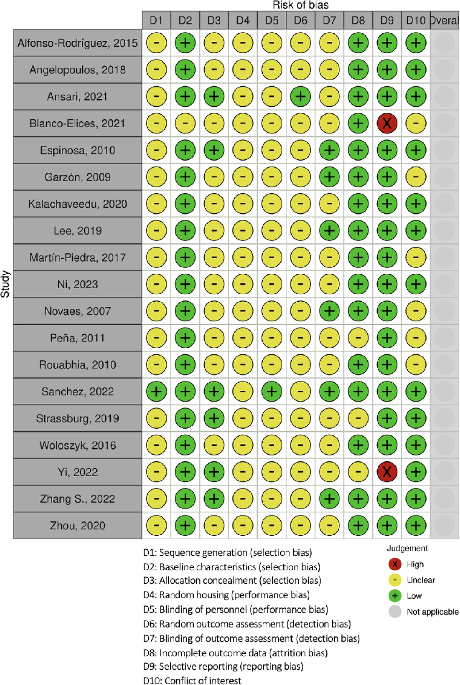
Traffic-light plot of quality assessment for each included study using SYRCLE checklist and robvis tool. SYRCLE, systematic review center for laboratory animal experimentation.

Summary plot of quality assessment for the included studies using SYRCLE checklist and robvis tool. SYRCLE, systematic review center for laboratory animal experimentation.
Biomaterials characterization
Main compounds
Biomaterials used in the included studies are detailed in Table 1. Briefly, the recurring main compounds were fibrin (42%),16,18,20–22,24,27,33 gelatin (26%),27,28,30,32,33 collagen (26%),17,19,25,26,29 agarose (21%),16,18,20,22 alginate (16%),27,28,30 fibroin (5%), 34 guar gum (5%), 31 hyaluronic acid (HA) (5%), 32 and decellularized ECM (11%).23,29 The decellularized ECM originated from human dermis 23 and rabbit artery branches. 29 Crosslinking was mostly done chemically (91% of the crosslinking methods reported). One study used ultra-violet radiation to do so for the methacrylated gelatin used. 30
Composition of the Construct and Tissue Engineering Technology Used for the Included Studies
Since the tissue of interest for this review is the connective tissue, the number of epithelial cells was not extracted.
A.b., Aristolochia bracteolata; ACVM, acellular vascular matrix; ADSC, adipose tissue stem cell; A.i., Acalypha indica; BMSC, bone marrow stem cell; dADSC, differentiated adipose tissue stem cell; dBMSC, differentiated bone marrow stem cell; dDPSC, differentiated dental pulp stem cell; DF, dermal fibroblast; DPSC, dental pulp stem cell; GEC, gingival epithelial cell; GF, gingival fibroblast; GK, gingival keratinocyte ; GMSC, gingival mesenchymal stem cell; HLC-I, human-like collagen type I; HUVEC, human umbilical vein endothelial cell; HWJSC, human umbilical cord Wharton’s Jelly stem cell; L.i., Lawsonia inermis; ndADSC, nondifferentiated adipose tissue stem cell; ndBMSC, nondifferentiated bone marrow stem cell; ndDPSC, nondifferentiated dental pulp stem cell; OEC, oral epithelial cell; OMF, oral mucosa fibroblast; OMK, oral mucosa keratinocyte; SDF, skin dermal fibroblast; SEK, skin epithelial keratinocyte; T.p., Thespesia populnea; VEC-like cell, vascular endothelial-like cells; VEGF, vascular endothelial growth factor.
Additional factors
Furthermore, several studies included additional factors. The study testing microspheres crosslinked with HA and polylysine aimed to enhance soft tissue regeneration. 32 A couple of studies using fibrin-based scaffolds added tranexamic acid, a clinically approved antifibrinolytic, to prevent gel fibrinolysis. 36 Calcium chloride was used in several studies to induce polymerization of the construct. One study added the arginine-glycine-aspartic acid tripeptide to compensate for the very low mechanical strength of gelatin and increase the overall mechanical properties of the hydrogel. 30 Another one added vascular endothelial growth factor (VEGF) to stimulate angiogenesis. 17 One study even investigated the benefits of adding traditional Indian medicinal plant extracts to stimulate wound healing: Acalypha indica, Aristolochia bracteolata, Lawsonia inermis, and Thespesia populnea. 31
Mechanical characteristics
Six out of the 19 included studies reported measurement of mechanical values (modulus of elasticity, compression modulus, tensile stress) on their engineered construct before implantation.27–31,33 Only Zhou et al. measured those values using a cellularized construct. 29 Results varied between 3.2 kPa and 38,780 kPa. However, techniques used were different, and thus results reported differed, as detailed in Table 2.
Characteristics of the Construct and its Behavior After Implantation for the Included Studies
ECM, extracellular matrix; IL-10, interleukin-10; TNF-alpha, tumor necrosis factor-alpha; VEGF, vascular endothelial growth factor; vWF, von Willebrand factor.
Tissue engineering technologies
Table 1 gathers tissue engineering technologies used in the selected studies. The most recurring technologies were cell embedding (used in 63% of the included studies)16–18,20–22,24,27,28,30,32,33 or cell seeding (used in 68%)16,19–26,29,31,33,34 on membranes (47%),19,20,23,25,26,29,31,33,34 hydrogels (37%),16–18,22,27,28,30 and cell-sheets (11%).21,24 In most cases, fibroblasts were embedded in the construct while epithelial cells were seeded. However, Ni et al. developed crosslinked polylysine-HA microspheres that were coated with a noncrosslinked HA gel and laden with cells. Those microspheres were produced using a water-in-oil emulsion with a phosphate buffer, 4-(4,6-Dimethoxy-1,3,5-triazin-2-yl)-4-methylmorpholinium chloride (DMTMM), liquid paraffin, and the Span-80 emulsifier. 32 Two studies used electrospinning, a method that involves generating ultrathin fibers using electrostatic force and is the most adapted method to large-scale preparation, and obtained a matrix which was then seeded.31,33 Two studies used 3D extrusion bioprinting, consisting in pushing an ink through a narrow nozzle to produce thin fibers able to be added layer by layer, with cells embedded in their bio-ink to obtain an hydrogel.27,28 Six studies described an in vitro incubation before implantation. Among them, four studies left the construct in air-liquid condition to promote epithelization for 1 to 3 weeks,16,20,22,25 whereas two reported a 7-day incubation before implantation to let the construct mature.19,34
Biological aspect
Five studies used autologous animal cells,19,21–23,26 whereas 14 used human cells, with one study conducted on chorioallantoic membranes (CAMs) and 13 in mice. Included studies worked with cells that can be distinguished between stem cells and other types. The most reported types of cells are gingival fibroblasts23,25,27–29,33,34 and oral mucosa fibroblasts (OMF).16,18–22,24 The others tested mesenchymal stem cells (MSCs) from gingiva (GMSCs),17,26,30–32 dental pulp (DPSCs),17,18,34 bone marrow (BMSCs),18,28 and adipose tissue (ADSCs).18,22 Three studies tested the angiogenic potential of more specific cells: human umbilical cord Wharton’s Jelly stem cells (HWJSCs), human umbilical vein endothelial cells (HUVECs), and vascular endothelial-like cells (VEC-like cells).16,18,29 To investigate the epithelialization process of the graft, seven studies added epithelial cells to their biomaterials, more precisely keratinocytes for six studies16,20–22,24,25,33 and HWJSC for one. 16 Beside coculture with epithelial cells, Blanco-Elices et al. cocultured OMF with three types of MSCs either differentiated or not for the experimental groups and used OMF with HUVECs as a positive control, 18 whereas Zhou et al. cocultured gingival fibroblasts with VEC-like cells. 29 The type and number of cells added to the engineered scaffold are detailed in Table 1.
In vivo experiments
Characteristics of the animal studies
Experimental models for each included study are detailed in Table 3. Most studies compared experimental groups with negative controls, whereas one-third used both negative and positive controls. Moreover, 21% of the included studies reported several experimental groups to compare various cell types introduced in their construct.17,18,33,34 The total number of animals used ranged from 6 to 50. Most studies were carried out on rodents (12 on mice,16–18,20,24,25,27–30,32,33 two on rats21,31), whereas two were conducted on rabbits,19,22 two on dogs,23,26 and one on chicken embryos. 34 Furthermore, similarities were noted between the mice species used: 92% did not produce T lymphocytes, and so presented an immunodeficiency of cell-mediated immunity, and 83% were hairless. Those characteristics imply the incapacity to reject xenografts. Procedures performed were mostly dorsal subcutaneous implantation (13 studies out of 19), whereas five studies focused on oral implantation,19,21–23,26 and one was a CAM assay. 34 No oral implantation was performed on mice, but it was done on bigger animals (rat, rabbit, dog) for easier manipulation. Ni et al. performed subcutaneous implantation of their cellularized microspheres loading them in a gelatin sponge. 32 Cellularized biomaterials were mostly bioengineered by the teams reporting the studies, whereas four biomaterials were commercial options with cells embedded inside or seeded onto.17,23,25,26 Remodeling of the graft was mainly investigated using histological analysis. All included studies presented biocompatible constructs.
Description of the Intervention of Interest for the Included Studies
AACT, autologous artificial oral connective tissue; ACVM, acellular vascular matrix; ADMA, acellular dermal matrix allograft; AG, alginate/gelatin; AGH, ag + nano-hydroxyapatite; BMSC, bone marrow stem cell; CAF, coronally advanced flap; CAOME, complete autologous oral mucosa equivalent; CMX, xenogeneic collagen matrix; dADSC, differentiated adipose tissue stem cell; dBMSC, differentiated bone marrow stem cell; dDPSC, differentiated dental pulp stem cell; DPSC, dental pulp stem cell; GelMA, gelatin methacrylate; GeL, gelatin; GF, gingival fibroblast; GK, gingival keratinocyte; GMSC, gingival mesenchymal stem cell; HDPSC, human dental pulp stem cell; HFOM, human oral mucosa fibroblast; HGF, human gingival fibroblast; HLC-I, human-like collagen type I; HOM, human oral mucosa stroma; HUVEC, human umbilical vein endothelial cell; iPAG, injectable platelet-rich fibrin + AG; ndADSC, nondifferentiated adipose tissue stem cell; ndBMSC, nondifferentiated bone marrow stem cell; ndDPSC. Nondifferentiated dental pulp stem cell; pl-HAM, polylysine-hyaluronic acid microsphere; R120, percentage of the ratio of plants extracts used (20% of 4:4:1:1); SDF, skin dermal fibroblast; SEK, skin epithelial keratinocyte; VEC-like cell, vascular endothelial-like cells.
Follow-up time points
Follow-up time points per included study are listed in Table 2. Among the 39 time points reported, the most recurrent time points were at 7 days for 8 of them, 14 days for 8, and 28 days for 6. Follow-up was mostly done short-term, with experiments ending after 1 week up to a month for 58% of the mentioned studies.16–18,20,21,24,29–31,33,34 Moreover, the furthest time point was at 12 weeks post-implantation in the work by Martín-Piedra et al. 22 Inflammation was mostly investigated by lengthy follow-up, between 2 and 8 weeks. Late time points are necessary to assess remodeling and to check the biodegradability of the implanted construct.
Vascularization remodeling
Remodeling specificities regarding vascularization are detailed in Table 2. All the included studies showed vascularization after implantation except one. 33 Five confirmed vascularization inside the grafted construct using immunohistochemistry to check the presence of common vascularization network markers: VEGF, clusters of differentiation 31 and 34 (CD31 and CD34), von Willebrand factor (vWF).18,25,27,30,32 Six studies investigated the number of vessels after implantation.17–19,23,27,28 Blanco-Elices et al. counted per mm2 the number of vessels positive for the labeling of VEGF, CD31, CD34, and vWF. To go deeper, they even reported that about half of the vessels were from human origin and the others were from the host mice. 18 One study looked at the hemoglobin concentration inside the newly formed vessels and showed an increase for the two experimental groups with DPSCs and GMSCs similar to the positive control with HUVECs (G1 and G2: 500 μg/mL; C+: 600 μg/mL; C−: 100 μg/mL). 17
ECM remodeling
Because the clinical objective is the long-term increase of gingiva thickness, the volume maintenance over time is crucial to evaluate. Two studies explored this point in a qualitative manner on histology slides: Peña et al. stated a progressive increase in the thickness of the chorion during the 3-weeks post-implantation on immunostaining for vimentin, 24 and Ni et al. showed an increase of subcutaneous soft tissue volume at both 3- and 6-weeks groups with microspheres without/with GMSCs on hematoxylin and eosin (HE) staining. 32 To go in depth, Lee et al. quantified the mucosa thickness on Masson’s trichrome staining and reported a thicker mucosa with the cellularized construct after 21 days compared to the control. 21 Half of selected studies looked at the synthesis of new matrix macromolecules. As detailed in Table 2, these studies mostly focused on histological aspect and the synthesis of new collagen fibers synthetized by the host. None focused on detecting the presence of new ECM enzymes by the host, such as the matrix metalloproteases or the tissue inhibitors of matrix metalloproteases.
Grafted cells behavior
Behaviors after implantation of the cells within the engineered scaffold are presented in Table 2. Four studies explored cell proliferation rate:17,18,24,29 Angelopoulos et al. quantified human MSCs inside the graft using human leukocyte antigen-A immunostaining, Blanco-Elices et al. detailed the number of positive endothelial cells per mm2 for VEGF/CD31/CD34/von vWF, Peña et al. used vimentin to detect fibroblasts’ growth, and Zhou et al. showed histological sections stained with HE to evaluate the total number of cells in the scaffold complex. They all reported an increase in the number of cells studied in the experimental groups compared to the controls. Ni et al. studied the effect of their microspheres on endogenous stem cells recruitment by immunofluorescence with stems cells surface markers, and they showed positively co-stained CD44/CD90/C77 cells around them at weeks 3 and 6. 32 Furthermore, two papers focused on cell infiltration.17,33 On one hand, Angelopoulos et al. showed red blood cells in some tubular structures 12 days after implantation and the scaffold’s invasion by human cells in both groups with MSCs. 17 On the other hand, Strassburg et al. checked for immune cells implicated in the initial start of tissue repair. They found that mouse leukocytes positive for CD11b in the electrospun gelatin from the host infiltrated the grafted construct, more in the construct with skin cells than in the one with gingival cells. 33 Nine studies investigated inflammation after grafting their cellularized engineered construct, as presented in Table 2. Many studies used animals with a reduced immune system, as developed in part 3.7.1. Characteristics of the animal studies.
Discussion
This systematic review aimed to report the state of the art on cellularized biomaterial substituting gingiva tested in vivo and promoting remodeling. To our knowledge, this review is the first focusing on gingival tissue engineering after implantation using cellularized constructs. Our hypothesis could not be confirmed, as most of the approaches achieved vascularization and ECM remodeling of the graft after implantation, without necessarily detailing the mechanical properties. Moreover, most of them used cells and molecules that have been described in the gingival connective tissue.
Besides one retracted article, the search query used for this systematic review did not select human studies. A complementary search allowed us to find ongoing clinical trials using gingival cells loaded in a scaffold for alveolar bone defect treatment for periodontitis without any published outcome (NCT03137979 and NCT03638154) and one with promising published results (Abdal-Wahab et al., 2020, 10.1111/jre.12728). It was not included in the review because this review concentrates on soft tissue regeneration. Cellularized biomaterials tested in humans for maxillofacial application often focus on bone tissue (and not on connective tissue) in the context of periodontitis. However, one ongoing clinical trial is comparing the regenerative and differentiation potential of gingival MSCs from several harvesting sites for future dental tissue regeneration applications (NCT03570333). The use of cellularized biomaterials to mimic and increase gingival connective tissue seems to be a research field that still needs time and effort before human application.
Cell sources were diverse across the included studies. When autologous cells were used, experiments were conducted on big animals (rat, rabbit, dog). Four studies compared cells to find the best vascular remodeling after cellularized engineered scaffold implantation in vivo.17,18,33,34 Indeed, two models did not find a significant difference in vascularization remodeling after implantation for their gelatin/fibrin construct with human fibroblasts from skin derma and gingiva 33 or for their silk fibroin construct with human DPSCs and gingival fibroblasts. 34 The article by Blanco-Elices et al. suggests that predifferentiated MSCs from dental pulp and bone marrow have a vascularization potential similar to HUVECs and better than those that are not predifferentiated and predifferentiated MSCs from adipose tissue when added in coculture with gingival fibroblasts in their construct. 18 To go further, GMSCs and DPSCs compared in vivo showed a similar number of vessels and hemoglobin concentration, but in vitro GMSCs showed better proliferation and migration potentials and a higher ability to form angiogenic tubules compared to DPSCs. 17 Besides being the cells from the native tissue aimed to be constructed, using gingival cells for tissue engineering offers several advantages. As they are routinely removed and considered biological waste during dental procedures, gingiva is easily accessible and gingival cells culture is well-known. Regarding cell quantities, most studies reported cell numbers in a range between 105 and 106, but they usually did not specify the total volume of the finished construct. Indeed, cells seem to represent 5% of the connective tissue for healthy human gingiva. 37 Thus, comparing the number of cells inside the total volume of the construct could have been interesting to see how well the native cell concentration was reproduced.
The matrices of all included studies were based on naturally derived components, mainly proteins and polysaccharides, to engineer their constructs. The rationale for using natural polymers is to guarantee several requirements of a successful gingival graft, such as biocompatibility, biodegradability, hydrophilicity for soft tissues, and functional similarity to native ECM. The frequent use of fibrin can be explained by its rapid degradation, its minimal foreign body response, and its use as a sealant for wound healing. One of the studies used Matrigel®, a commercial product mostly composed of laminin and collagen IV, which are derived from a murine tumor called Engelbreth–Holm–Swarm. 38 However, Matrigel® can present variation in composition even within the same batch, and consequently in its biochemical properties. 39
Technical specifications needed for a cellularized construct to be implanted in vivo include cell viability, biocompatibility, and mechanical properties similar to the native tissue it aims to reproduce. All constructs analyzed in this systematic review were cytocompatible and were tolerated in vivo. Concerning the mechanical properties, comparing the constructs was not possible due to the heterogeneity of measurements. Some studies calculated alternatively the “modulus of elasticity,” the “tensile modulus,” or the “compression modulus.” Simply put, all these terminologies describe how a material can easily stretch and deform. 40 Only one study tested the material stiffness, which is a material’s capability to resist elastic deformation. Thus, these measurements are complementary but not similar. Studies measuring mechanical values did not use the same device or settings and therefore could not be compared. Moreover, one major issue was the lack of consensus concerning reference values of human gingival connective tissue mechanical properties. 41 Indeed, one-third of the studies tried to replicate the original mechanical properties, but they did not use the same parameters.
Concerning all the tissue engineering techniques encountered in this review, 3D bioprinting offers advantages when cells are needed. Indeed, constructs can be customized to fit the defect in clinical application. The defect geometry can be digitalized with optical impression (intraoral scanner) or imaging segmentation (computed tomography-scan, cone beam computed tomography, magnetic resonance imaging). Thanks to automatization, the construct production is reproducible and fast. Another advantage of bioprinting is the differential cell distribution within layers of the cellularized construct. Each bio-ink can exhibit a specific cell type, and cell density can be controlled in the printed construct.
Most of the constructs included in this review needed additional chemical factors for crosslinking. Because they could modify the properties of the construct, new technologies present promising potential. For instance, the use of nucleotide lipids can give auto-assembly properties to a gel and thus produce crosslinker-free scaffolds. One example in the literature tested their nucleotide lipid-based hydrogel with gingival fibroblasts to show cytocompatibility. 42
Vascularization was mostly explored by histological analysis, and few studies quantified the number of vessels and the hemoglobin concentration after implantation. However, it was surprising that none explored the branching length per field, the number of junctions per field, or the number of meshes per field. Those characteristics are typically explored to assess vascularization remodeling.
Various limitations were met during this systematic review. A meta-analysis of the studies was not relevant due to the restricted number of studies and the heterogeneity in materials and methods. Another limitation was the low number of studies focusing exclusively on connective tissue, leading to the inclusion of studies on bone-gingiva constructs. Research on gingival connective tissue engineering can expand to these types of multilayered constructs for periodontology surgery. In these cases, the extraction of results concerning strictly the connective tissues was not always possible. An additional limitation was the risk-of-bias judgement using the SYRCLE checklist. Most items were judged unclear due to a lack of clear statements in the included studies, even for research teams declaring adherence to the ARRIVE checklist. Thus, it is possible that some studies do not report all technical details important for compiling results. As each approach combined multiple protocol specificities, correlations between one composition parameter and one specific outcome were difficult to isolate. Researchers working on cellularized biomaterials used as gingiva substitutes should be more rigorous in their reporting of animal studies and should more closely follow the ARRIVE guidelines. In addition, stiffness of the biomaterial is usually a limitation for handling and was not systematically reported. Mechanical characterization should be more complete to better report the chemicophysical properties of the material used. For now, research on cellularized biomaterials used as gingiva substitutes aims to show proof of concept. In doing so, there is usually no study of the biomolecular aspect. Indeed, articles report only graft integration, vessel formation, and sometimes ECM remodeling.
Nonetheless, gingival cells as a cell source in sufficient numbers, natural components with a solution to add stiffness to better replicate the native gingival connective tissue, and 3D bioprinting as a tissue engineering technique all seem like interesting parameters to combine to obtain a graft that may be used for human application one day. Further studies seem necessary to validate which cellularized biomaterial is the most suitable to human application to compensate for gingival loss. In the literature, several hydrogels not yet tested in vivo showed potential for increasing gingival thickness. For instance, Oliveira et al. developed an extrudable hydrogel composed of methacrylated-collagen and methacrylated-HA, 43 an interesting lead to use gingival cells and go a step further toward human application. Other promising candidates should arrive in a couple of years in the literature, offering new perspectives on the subject.
Conclusion
This systematic review showed the multiplicity of cellularized biomaterial used to engineer large viable connective tissue constructs tested in vivo. Various cell types from several cell sources could be used to do so. Few studies integrated coculture in their constructs to promote vascularization. Vascularization and ECM remodeling of the graft after implantation occurred in most of the included studies. Remodeling after implantation could be achieved using several tissue engineering technologies to engineer constructs made of naturally derived components. These results will enable us to set up experiments based on all the parameters, the diversity of which shows that there is a certain amount of freedom.
Footnotes
Authors’ Contributions
C.D.: Investigation, Writing—Original Draft. R.S.: Writing—Reviewing and Editing. C.M.: Writing—Reviewing and Editing. A.N.: Methodology, Writing—Reviewing and Editing.
Disclosure Statement
No competing financial interests exist.
Funding Information
This systematic review is part of the PhD project of Camille Déchelette. This PhD is granted by the PERIOPRINT Project (ANR-22-CE52-0004-01), funded by the French National Research Agency (