




Abstract
Human vocal folds (VFs), a pair of small, soft tissues in the larynx, have a layered mucosal structure with unique mechanical strength to support high-level tissue deformation by phonation. Severe pathological changes to VF have causes including surgery, trauma, age-related atrophy, and radiation, and lead to partial or complete communication loss and difficulty in breathing and swallowing. VF glottal insufficiency requires injectable VF biomaterials such as hyaluronan, calcium hydroxyapatite, and autologous fat to augment VF functions. Although these biomaterials provide an effective short-term solution, significant variations in patient response and requirements of repeat reinjection remain notable challenges in clinical practice. Tissue engineering strategies have been actively explored in the search of an injectable biomaterial that possesses the capacity to match native tissue’s material properties while promoting permanent tissue regeneration. This review aims to assess the current status of biomaterial development in VF tissue engineering. The focus will be on examining state-of-the-art techniques including modification with bioactive molecules, cell encapsulation, composite materials, and in situ crosslinking with click chemistry. We will discuss potential opportunities that can further leverage these engineering techniques in the advancement of VF injectable biomaterials.
Impact Statement
Injectable vocal fold (VF) biomaterials augment tissue function through minimally invasive procedures, yet there remains a need for long-term VF reparation. This article reviews cutting-edge research in VF biomaterial development to propose safe and effective tissue engineering strategies for improving regenerative outcomes. Special focus is paid to methods to enhance bioactivity and achieve tissue-mimicking mechanical properties, longer in situ stability, and inherent biomaterial bioactivity.
Introduction
Human vocal fold (VFs) have distinct layers of submucosal connective tissue sandwiched between the laryngeal epithelium and the thyroarytenoid muscle and anchored by the arytenoid cartilage (Fig. 1A). 1 During phonation, the VF stretch longitudinally between 5 and 15 mm, while the superficial lamina propria (LP) oscillates with up to 0.5 mm amplitude.1,2 These three layers are the superficial, intermediate, and deep LP (Fig. 1B). Advanced imaging studies using multiphoton microscopy and optical coherence tomography found a continuous transition between LP layers, further elucidating the intricate organization of VF extracellular matrix (ECM) and microvasculature, which requires tissue-specific regenerative solutions.3–7

VF Anatomy and Injection Laryngoplasty.
The ECM structure plays an important role in supporting phonation.2,8,9 Collagens are the primary constituents of VF ECM, contributing to 40–50% of total protein. 10 Collagen I fibers form the overarching structural network, regulate cell distribution, and recruit cells for wound healing.10,11 Collagen III and elastin bundles contribute to VF resilience, tensile strength, and viscoelasticity. Hyaluronan is an abundant glycosaminoglycan that helps regulate wound healing and dampens mechanical stress from high-frequency phonatory oscillation.11,12
VF atrophy, sulcus, scarring, and unilateral paralysis/paresis are candidates for injection laryngoplasty, where a biomaterial is injected to augment VF function and closure.13–16 Age-associated VF atrophy is characterized by tissue bowing and reduced viscoelasticity that may impact LP or muscle.17–21 Sulcus vocalis is related to fibrotic alterations characterized by groove formation and epithelium invading the superficial LP. 22 VF scarring is generally caused by surgical interventions, radiation, or trauma, where healthy LP is replaced by excessive fibrous collagen deposition.12,17 VF paralysis/paresis is unilateral or bilateral VF immobilization caused by injury to the recurrent laryngeal nerve from cervical or thoracic surgical intervention, tumor formation, trauma, radiation, and chronic inflammation.23–26 Bilateral VF paralysis may require reinnervation or airway bypass surgeries. However, unilateral VF paralysis and paresis account for up to 75% of injection laryngoplasty treatments.27–29
Injection laryngoplasty is performed in operating rooms or doctor’s offices (Fig. 1C).30–32 In-office injection laryngoplasty offers relatively low risk and cost as well as immediate testing of glottal function. The materials are injected into the lateral aspect of the thyroarytenoid muscle, or the paraglottic space.32–35 Material placement is critical to treatment outcomes. Superficial injection causes improper biomaterial localization, tissue stiffening, and impeded vibration.36–38 While there is interest in developing regenerative biomaterials and delivery techniques for the superficial LP, intramucosal injections are limited to free-drug formulations such as steroids and growth factors.36,39–41
In this article, the primary goal is to assess the successes and limitations of current VF injectable biomaterials and review the state-of-the-art of VF biomaterials under development. We also evaluate advanced tissue engineering techniques that can help stimulate regeneration, mimic mechanical properties, and control material degradation toward permanent VF augmentation.
Overview of Existing VF Biomaterials Used in Clinics
The first injectable VF biomaterial, Teflon, caused severe granuloma for 5–10 years following injection and removal caused by tissue morbidity. 42 Also, collagen (Zyplast®) and gelatin-based (Gelfoam®) injectables were used for VF augmentation for nearly 20 years but have been discontinued due to short-term efficacy and chance of allergic reaction to bovine collagen.43,44 Atelocollagen, a similar bovine collagen material, continues to be used in Japan. 45 Cymetra™ is a discontinued micronized decellularized dermal matrix sourced from human cadavers that lasted 6 weeks to 6 months and reduced the risk of allergic reaction, decreased foreign body response, and improved voice outcomes compared to bovine collagen.46,47
These examples emphasize the need for long-term safety evaluations and follow-up. 42 At present, common biomaterials for injection laryngoplasty include hyaluronan, calcium hydroxyapatite with carboxymethylcellulose, autologous fat, and silk hyaluronan (Table 1). 48
Common VF Injectable Biomaterials
VF, vocal fold; FDA, United States Food and drug administration.
Restylane® and Juvederm® are produced from hyaluronan microparticles and reduce excess collagen I deposition as well as augment the glottal wave and voice quality.49,50 These hyaluronan-based products are approved as dermal fillers but are used off-label for voice applications. 48 Treatment with HA-based products may improve phonation quality for at least 6 months. 51
Calcium hydroxylapatite microparticles are a bioceramic injectable filler suspended in a carboxymethylcellulose gel.47,51 Calcium hydroxyapatite, marketed as Radiesse® and Prolaryn Plus®, is FDA approved for injection laryngoplasty. The residence time of calcium hydroxylapatite is relatively long, up to 24 months.47,51 However, calcium hydroxylapatite injection outcomes may be variable, and cause deposits in the superficial LP and deleterious foreign body response.52,53 VF stiffening, impaired mucosal wave, edema, and hypervascularity were also reported with calcium hydroxylapatite.48,52–54
A significant limitation of the aforementioned injectables is a lack of capacity to remodel tissue.13,55,56 In contrast, autologous fat and silk hyaluronan show potential to stimulate tissue deposition for long-term efficacy.
Autologous fat grafts contain stem and endothelial cells that contribute to tissue remodeling and vascularization.55–57 About 30–40% of excess volume is commonly used to offset resorption, but optimal volume is difficult to predict as resorption varies from 20% to 60%.58–60 Overinjection of autologous fat can alter VF shape and mechanics, cause dysphonia, and require surgery to remove the excess.57–59 Autologous fat may also induce acute and chronic inflammation, necrosis, and fibrosis.55–57
Silk microparticles in a crosslinked hyaluronan carrier are new FDA-approved injection laryngoplasty biomaterials.33,61,62 The silk microparticles incorporate bioactive properties to promote cellular infiltration and tissue remodeling.33,62–65 Silk hyaluronan improves control over degradation, wherein the hyaluronan degrades more rapidly while silk microparticles are retained for up to 18 months. 63 While the remodeling effects are promising, about 32% of patients in a large clinical study required additional injection laryngoplasty procedures after a median of 106 days.33,62 Serious complications including edema, dyspnea, and difficulty swallowing were reported with low incidence.62,64
There remains an unfilled need for a minimally invasive, injectable VF biomaterial that effectively restores tissue structure and functions to reduce the requirement for repeated treatments. Tissue engineering strategies are required for injectable VF biomaterials with more permanent efficacy. In the following section, we review options for biomaterial composition, enhancing of bioactivity with biomaterial functionalization and encapsulation of growth factors and cells, and control of mechanical and degradation properties through composite materials and crosslinking.
Choosing Polymers for VF Injectables
Injectable VF biomaterials are fabricated from polymers derived from natural sources or synthesized in laboratories, each of which carry advantages and limitations (Table 2). Selecting polymers for VF injectables relies on desired properties, including bioactivity, cell response, structure, mechanics, and biodegradability.
Advantages and Disadvantages of VF Biomaterials Under Investigation
ECM, extracellular matrix; dECM, decellularized extracellular matrix; VF, vocal fold.
Natural materials
Natural polymers are selected for biocompatibility, bioactivity, porous ECM-like structure, or immunomodulatory capacity.66,67 On the other hand, natural polymers have high variability, and may lack mechanical tunability or degrade before achieving permanent effect unless crosslinked or in composite.66,68 In addition to those in clinical VF biomaterials, other natural polymers including alginate, chitosan, gelatin, and decellularized extracellular matrix (dECM) are under active investigation for favorable regenerative, mechanical, and degradation properties.
Collagen and gelatin are used in VF biomaterials for their capacity to promote cell adhesion, migration, and proliferation.66,69–71 However, collagen-based hydrogels exhibit rapid degradation and poor mechanical and structural stability.66,69 Despite the discontinuation of existing collagen and gelatin VF injectables, crosslinked and composite collagen and gelatin formulations are under investigation.68,72,73
Hyaluronan and silk-based biomaterials are under development to improve treatment duration and remodeling capacity.74–76 Hyaluronan provides a damping function within the VF, regulates cell function, and modulates inflammation with reduced immunogenicity compared to collagens.44,77 Silk fibroin has well-established biocompatibility due to its use in sutures with highly tunable mechanical and structural properties.69,70 However, biomedical grade silk fibroin is expensive, depends on nonstandardized hand processing and environmental conditions, and is sold in unstable aqueous solution. 78 Production variability leads to differences in bioactivity, degradation, and mechanical properties. 78
Sodium alginate is a biocompatible, antibacterial polysaccharide derived from brown seaweed that degrades into glucose-like monosaccharides that can be safely metabolized.66,69,79,80 Alginate lacks innate cell adhesion sites, but its bioactivity and mechanical properties are highly tunable through variable molecular weight, chemical modification, and crosslinking. 80 In VF injectables, alginate is under investigation for cell and growth factor delivery, and as a composite to provide improved mechanics and durability to regenerative biomaterials.74,76,79,81
Chitosan is a biocompatible, antimicrobial polysaccharide derived from the exoskeletons of arthopods with low immunogenicity. 66 In VF applications, chitosan has been shown to reduce fibroblast proliferation and excess collagen deposition. 82 However, chitosan has a high swelling ratio, lacks elasticity, and is unstable at neutral pH, limitations that may be circumvented by crosslinking and use in composites.71,79,83,84
dECM is produced by removing cells from tissue. Its composition of ECM proteins, glycosaminoglycans, and other regenerative molecules helps stimulate innate wound healing processes.41,85,86 VF-specific dECM has high batch to batch variability and poor scalability, but commercial sources from larger organs such as small intestinal submucosa (SIS) show regenerative potential in the VF.87,88 Both VF and SIS dECMs have been shown to downregulate fibrotic genes, reduce excess collagen I deposition, and stimulate LP restoration, though these hydrogels possessed weak mechanical properties.86–90 dECM also has potential to stimulate other VF-relevant processes including recruitment of endogenous stem cells, angiogenesis, and formation of tissue-specific structures.41,91–93
Synthetic materials
Synthetic polymers have standardized fabrication processes and high tunability. However, synthetic polymers do not possess innate bioactivity and may integrate poorly with surrounding tissue or produce toxic byproducts. 66 Also, polymer synthesis may involve toxic materials, and require extensive purification.94,95 Synthetic polymers under investigation for VF applications include polyethylene glycol (PEG), poly ε-caprolactone (PCL), and polyurethane (PU).79,96–98
PEG is a nonimmunogenic, amphiphilic polymer comprised of ethylene oxide monomers that degrades slowly through oxidative degradation.99–101 PEG has extensive chemical modification capacity for modulating bioactivity, degradation, and mechanical properties.96,97,99 However, some patients exhibit pseudoallergy to PEG, and PEG derivatives used at high concentrations or functionalized with molecules like acrylates may cause cytotoxicity.100,101
PCL possesses favorable viscoelasticity at physiological temperature and is biodegradable over 2–3 years, though its hydrophobicity limits cell adhesion and proliferation.102,103 The mechanical properties of PCL are high compared to soft tissues, but may be tuned by using composites. 103 In rabbits with VF paralysis, PCL microspheres in a Pluronic F127 carrier augmented VF function for 12 months.98,102
PU has been used as a composite material for injectable VF biomaterials. 104 PU are flexible and durable block copolymers that can be synthesized with hydrolytic or enzymatic degradability depending on linkages between its constituent monomers. 105 PU degradation products and impurities retained after synthesis may be cytotoxic, though safer methodologies are under development.105–107
Enhancing Bioactivity of Injectable VF Biomaterials
To better match material degradation with the wound healing rate, the bioactivity of injectable VF biomaterials may be enhanced by functionalization with bioactive motifs and encapsulating growth factors or cells within a bioactive material.
Conjugation with bioactive motifs
Conjugating polymers with biomolecular motifs can increase bioactivity.96,98,108 Conjugation is performed via crosslinking the functional groups of the bioactive molecule and the polymer.109,110 For example, arginylglycylaspartic acid (RGD), a peptide found in fibronectin and laminin, is a commonly used to enable cellular adhesion to polymers without innate adhesion sites.108–111 Heparin, a glycosaminoglycan, is another common conjugation choice that is used to attract and sequester growth factors released by surrounding cells or control the release rate of encapsulated growth factors.96,98,112,113
Injectable microparticles for VF augmentation were produced from PEG functionalized with maleimide and thiol groups that crosslinked by thiol-Michael addition. 108 The PEG-maleimide backbone was modified with thiol-functionalized RGD. After injection to rabbit thyroarytenoid muscle in annealing polymer solution, exposure to visible light produced a microporous annealed particle (MAP) hydrogel. 114 RGD-conjugated PEG and the MAP structure provided a favorable environment for cellular adhesion, infiltration, and vascularization.108,114
The Heparin and RGD-conjugated MAP hydrogel is the first to demonstrate tissue remodeling with long-term VF augmentation in an animal model. 96 The RGD-conjugated MAP hydrogel was further conjugated with thiolated heparin to sequester growth factors and improve tissue integration (Fig. 2A). 96 MAP hydrogels and a Restylane® control were injected intramuscularly to a rabbit VF paralysis model. After 14 months, all six rabbits treated with MAP maintained augmentation. The biomaterial was replaced by collagen-rich neo-tissue with distinctive morphology compared to surrounding muscle.

Regenerative Effects of Enhancing the Bioactivity of VF Injectable Biomaterials.
Encapsulation of growth factors
Growth factors are signaling cytokines secreted by cells that stimulate cellular activity, inflammation, and tissue remodeling. 115 Basic fibroblast growth factor (bFGF) and hepatocyte growth factor (HGF) are used clinically to treat VF scarring due to fibroblast modulation and antifibrotic activity.39,115 Free growth factors have several limitations including instability, rapid inactivation, and poor targeting resulting in safety concerns including tumorigenesis.116,117 Encapsulating growth factors in biomaterials for intramuscular injection may slow release and improve target site retention.74,98
Encapsulation in a VF injectable has been performed by calcium chloride crosslinking, trapping the HGF within the “egg-box” network of a hyaluronan-alginate hydrogel. 74 In vitro, HGF release was sustained over 3 weeks as the porous “egg-box” facilitated slow diffusion.74,118 When the hydrogel was injected into injured VF LP, vibrational and viscoelastic properties improved compared to controls treated with HGF alone.
Growth factors may also be bound to injectable microspheres with growth factor binding sites.98,119 Gelatin microspheres with bFGF have reduced scarring when injected to injured rabbit and canine VF LP.119,120 Heparin-immobilized PCL microspheres with bound bFGF and HGF in a Pluronic F127 carrier showed ECM deposition and muscular regeneration 1 month after injection to a VF paralysis rabbit model, a regenerative effect that could not be achieved with PCL alone. 98
Cell delivery
Codelivery of cells with a biomaterial may provide a synergistic effect on VF regeneration. In VF animal models, stem cells have been shown to remodel fibrotic tissue and restore mechanical function.121–123 In a nonrandomized clinical study, patients received injections of autologous mesenchymal stem cells (MSCs) to the LP and muscle following surgical resection of scarring. 122 Up to 75% of patients (n = 16) showed improved VF function. Encapsulating cells in a biomaterial improves cellular viability and retention in the target tissue and provides the cells with bioactive and mechanical cues to enhance VF augmentation.122,123
Encapsulation of stem cells in hyaluronan has improved retention in VF LP or muscle and reduced fibrosis in rat and rabbit models.121,124 However, a hyaluronan-MSC group performed comparably to MSCs alone in a preliminary clinical study. 122 Encapsulation in a biomaterial with remodeling capacity or longer-term stability improve outcomes.
Delivery of urine-derived stem cells (USCs) in a SIS dECM hydrogel was recently explored in a heat-injury rabbit VF LP model (Fig. 2B). 86 SIS dECM-only, USC-only, and SIS-USC groups all demonstrated lower inflammatory marker expression (CD86, TNF-α) and reduced collagen I/collagen III ratio compared to LP without treatment after 2 and 8 weeks, respectively. The SIS dECM-only and USC-only groups induced comparable responses, while the SIS-USC group induced an enhance synergistic effect on VF regeneration.
Modulating the Mechanical, Physical, and Degradation Properties of VF Biomaterials
Mimicking in vivo VF mechanical properties and degradation at a rate that matches regeneration are necessary for biodegradable materials to permanently augment VF function. Alongside biomolecular cues, the mechanical (stiffness, viscoelasticity) and physical (porosity, swelling) properties of a biomaterial help direct cells to generate neo-ECM characteristic of VF. 125 VF mimicry is complicated by its layered structure and varied thickness.3,126 VF exhibit compressive moduli of 3.9–5.4 kPa on the medial LP surface, 2.7–3.2 kPa on the superior LP surface, and 1.3–2.7 kPa on the thyroarytenoid muscle. 126 A range representative of the muscle or LP is typically targeted in biomaterial design.
Degradation properties are also critical, as bioactive materials cannot achieve permanent VF augmentation if they degrade before tissue remodeling is complete. 66 Despite the bioactivity and VF-mimicking storage modulus of SilkVoice®, its stability varies, and reinjection are still frequently required.62,63 The use of composite materials and chemical crosslinking can tune biomaterial properties to achieve VF mimicry and controlled degradation.
Composite VF biomaterials
Natural, ECM-derived polymers with innate bioactivity may be combined with more stable polymers and the composition ratio and crosslinking method tuned to produce an injectable composite that better mimics human VF than either alone (Table 3).72,81
Advantages and Disadvantages of Conventional Crosslinkers
For instance, thiolated hyaluronan was crosslinked with PEG-diacrylate (PEGDA) to produce a hydrogel with tunable viscoelasticity for VF injection, and supplemented with collagen I collagen III to achieve a native-like structure. 72 Inclusion of collagens was necessary to retain encapsulated fibroblasts and increased the storage modulus from 0.16–0.21 kPa to 0.65–1 kPa within the 0.3–1.2 k range of adult VF LP.72,127 HA-PEGDA prevented the fibroblast-mediated contraction associated with collagen-only hydrogels. The inclusion of natural materials and a synthetic polymer improved tissue-mimicking and bioactivity.
Advantages and Disadvantages of Click Crosslinking Methods
HVFF, Human Vocal Fold Fibroblast; IEDDAC, inverse electron demand Diels–Alder cycloaddition; MSC, mesenchymal stem cell; MAP, microporous annealed particle; PCL, poly ε-caprolactone; PEG, polyethylene glycol; RGD, arginylglycylaspartic acid; SPAAC, strain promoted azide-alkyne cycloaddition; TCO, transcyclooctene.
The impact of material composition on mechanical properties is illustrated by the varied properties of calcium-crosslinked alginate microspheres in composite with poly-L-lysine or chitosan (Fig. 3A). 79 The VF Young’s modulus by nanoindentation ranges between 0.5 and 6 kPa, matching the Alginate-only (∼3 kPa) and Alginate-poly-L-lysine (∼2 kPa) microspheres but not alginate-chitosan (∼12 kPa).79,128,129 Low swelling ratios are favorable for stability and biological response predictability. Alginate-poly-L-lysine microspheres showed the lowest ratio at 9% after 48 h, while alginate-only microspheres swelled by 34%. Alginate-chitosan microspheres swelled by over 80%, contraindicating use due to risk of airway obstruction.
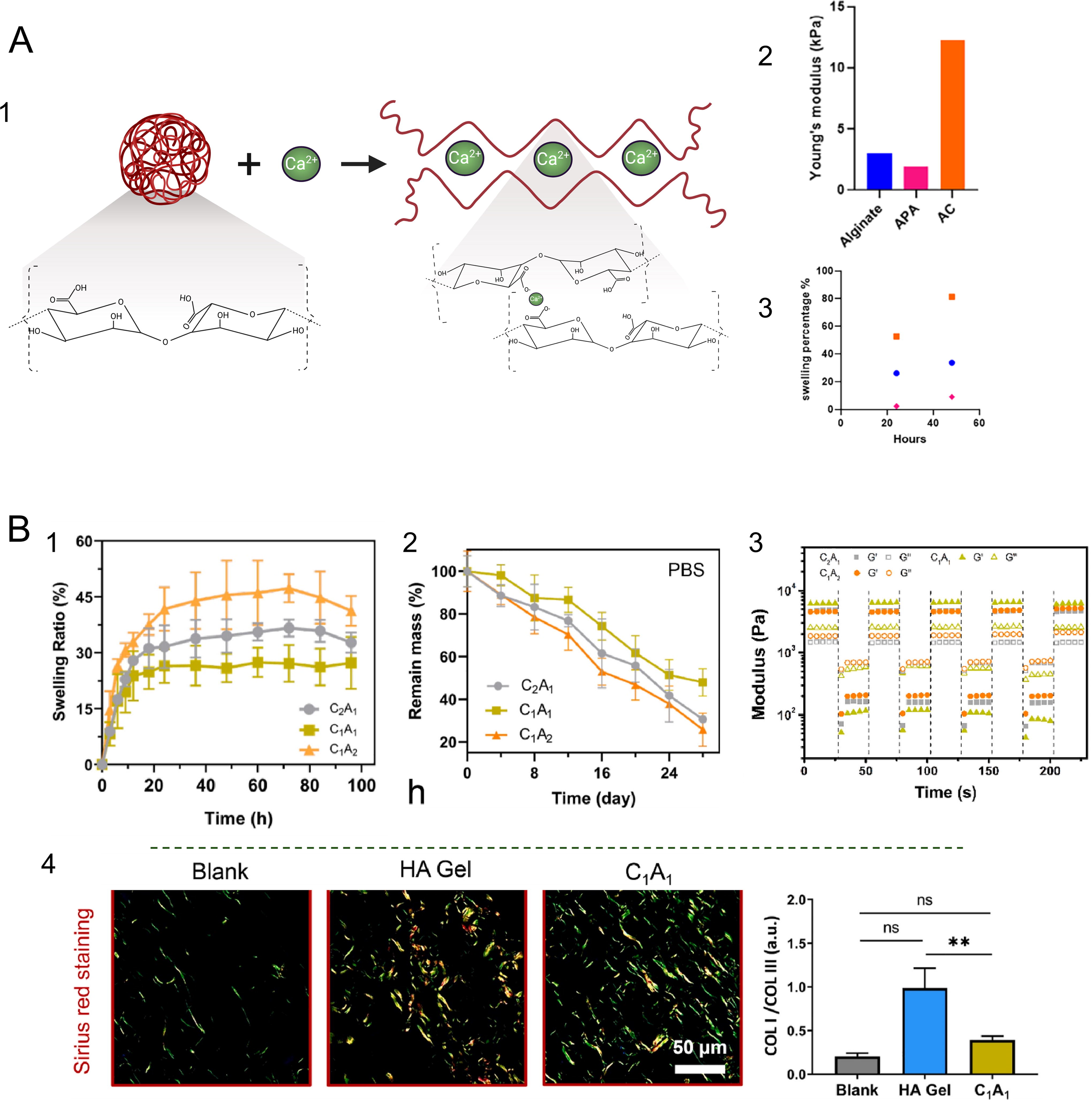
Alginate-chitosan hydrogels exhibited more favorable swelling characteristics when crosslinked with Schiff base and borate ester bonding and visible light methacrylate polymerization (Fig. 3B). 130 Tuning the alginate-chitosan ratio revealed that a 1:1 ratio was most stable with a swelling ratio of ∼26% after 48 h compared to 1:2 (∼35%) and 2:1 (∼45%) ratios. The 1:1 alginate-chitosan hydrogel retained the greatest mass by at least 1.5 times after a 28-day hydrolytic degradation test. Intramuscular injection of the 1:1 hydrogel in a VF paralysis rabbit model better maintained volume, exhibited a smaller glottal gap, and stimulated neo-ECM production after 12 weeks compared to a hyaluronan control. 130 In sum, composite biomaterials provide vast opportunities for tuning biomaterial properties to produce a synergistic effect on regeneration.
Conventional crosslinking of VF biomaterials
Crosslinking method fundamentally impacts network structure, mechanical properties, and degradation rate of a biomaterial. Desirable properties for crosslinkers in VF injectables include reaction specificity, nontoxic byproducts, and capacity for safe in situ crosslinking.
Chemical and ionic crosslinking are used to fabricate VF injectables, such as 1,4-butanediol diclycidyl ether (BDDE) in Juvederm®, Restylane®, and SilkVoice®. 131 BDDE slows enzymatic degradation by obstructing hyaluronidase cleavage sites. Although BDDE is toxic, it is removed before patients are exposed. 131 However, BDDE-crosslinked hyaluronan degrades without repairing tissue, necessitating reinjection.
Dialdehydes such as glutaraldehyde and glyoxal react with amine groups and are used to strengthen mechanical properties and provide resistance to thermal and enzymatic degradation in collagen, gelatin, and glycol-chitosan VF injectables.83,119,132 However, dialdehydes are cytotoxic at low concentrations and the reaction is difficult to control with multiple possible reaction products.43,133–136
Carbodiimide crosslinking with 1-Ethyl-3-(3-dimethylaminopropyl) carbodiimide (EDC) and N-hydroxysuccinimide (NHS) couples amine and carboxylic acid groups and is used to improve mechanical stability. EDC-NHS forms a favorable network structure for cellular integration but crosslinks DNA, proteins, and cell surface molecules and produces a toxic isourea byproduct.135–137
Genipin crosslinking is less cytotoxic and reacts specifically with primary amines, making it useful for controlling crosslinking density.138,139 In VF applications, genipin-crosslinked gelatin hydrogels exhibited low swelling ratios, slow enzymatic degradation, and improved mechanical strength compared to carbodiimide-crosslinked hydrogels. 68 However, cytotoxicity and hinderance of cell infiltration in genipin-crosslinked biomaterials may increase at physiological temperature, and there is a shortage of clinical safety evaluation.138–141
Dialdehyde, carbodiimide, and genipin crosslinkers may be safe for microparticles that can be sufficiently purified prior to injection but are not recommended for in situ gelation to prevent cytotoxicity and crosslinking of ECM proteins in the native tissue. 136
Ionic crosslinking with calcium chloride, used to fabricate alginate and chitosan biomaterials, is rapid and biocompatible.74,79,142 However, limitations include rapid, uncontrollable crosslinking causing heterogenous bond distribution and difficulty of injection and ion leaching resulting in loss of structural stability. 143
The side reactions, cytotoxicity, and heterogeneity inherent to conventional crosslinkers led to the development of bioorthogonal click chemistry, which may be more suitable for in situ gelling VF injectable applications. 144
Bioorthogonal click chemistry in VF biomaterials
Bioorthogonal click chemistry describes selective reactions that link molecules at high yield, do not require purification, and proceed safely in vivo.144–148 Bioorthogonal click chemistries for in situ gelling VF injectables include Thiol-X, strain promoted azide-alkyne cycloaddition (SPAAC), and inverse electron demand Diels–Alder cycloaddition (IEDDAC) (Table 4).144,148
The MAP and HA-PEGDA hydrogels discussed previously were fabricated by the thiol-Michael thiol-X reaction.96,144,148,149 The thiol-Michael reaction occurs through a thiol attacking an electron-deficient alkene, catalyzed by a base, primary amine, or phosphine. 149 A limitation is that catalysts may cause side reactions including disulfide bond formation, acrylate dimerization, and Aza-Michael addition.149,151
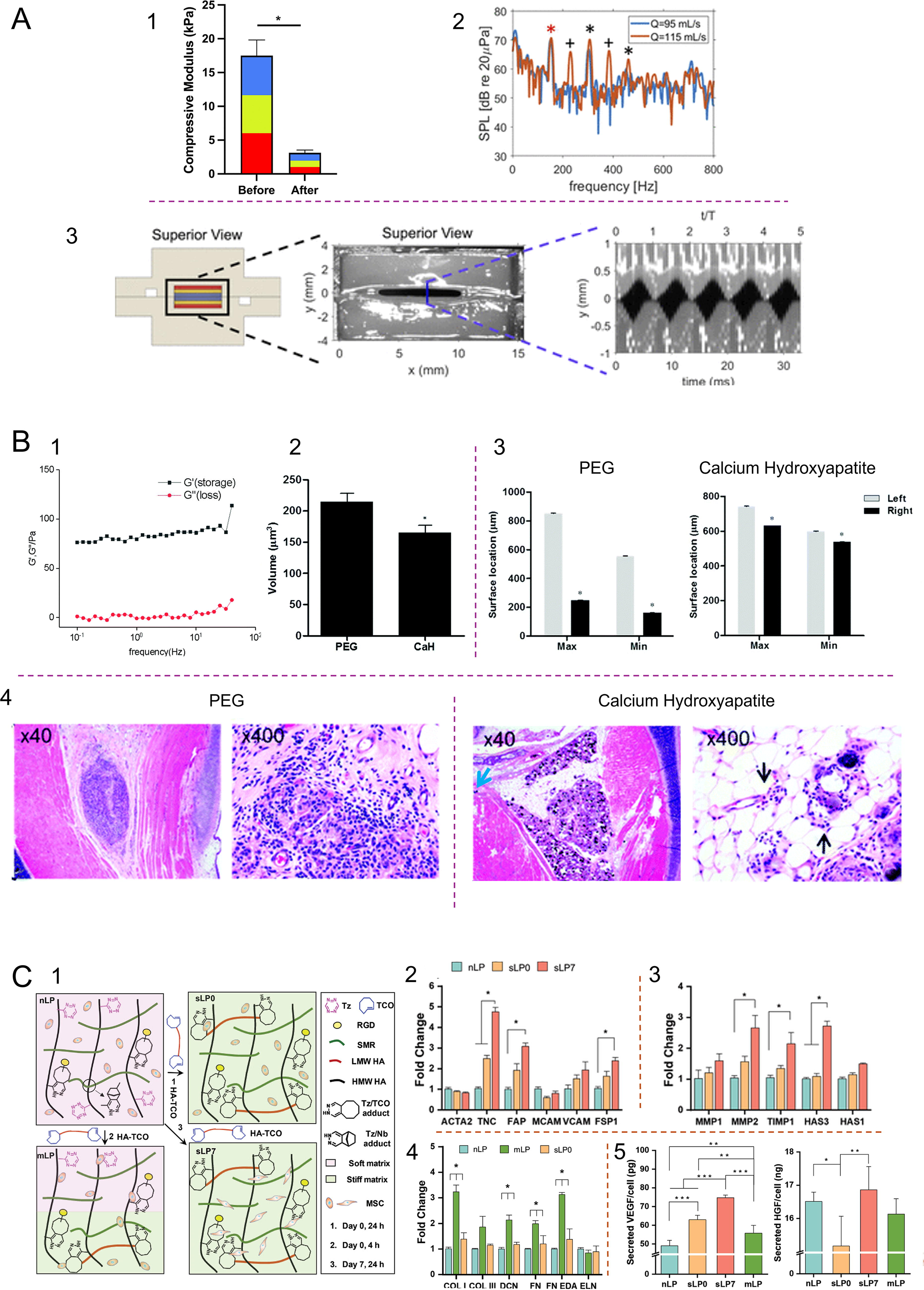
The light-catalyzed, radical-mediated thiol-ene reaction is a thiol-X without side reactions that proceeds between a thiol and an electron-rich alkene. 152 It has not yet been used to fabricate a VF injectable, but has been used to model the VF LP by reacting hyaluronan-norbornene of increasing concentrations (1–2.5%) with a UV-activated dithiol to produce three layers with differing stiffness. (Fig. 4A). 153 A bench test showed that the scaffold could oscillate within the normal human speaking range (about 150 Hz). 153 To prevent UV-induced tissue damage, VF injectables may be produced using visible light-catalyzed thiol-ene chemistry. 152
Click Chemistry in VF Tissue Engineering
SPAACs involve the reaction between strained ring cyclooctynes and aliphatic azides to form a triazole bond. 154 SPAACs do not require an external catalyst, but disadvantages include difficulty of cyclooctyne synthesis and low reaction yield. 154 A VF hydrogel was produced from by SPAAC from four-arm PEG functionalized with azide and dibenzocyclooctyne (Fig. 4B). 97 The storage modulus of the PEG hydrogel were low compared to native VF, less than 100 Pa. Nevertheless, the click PEG hydrogel showed increased stability compared to Radiesse® in a VF paralysis rabbit model after 16 weeks, albeit minimal cellular infiltration. Additionally, the PEG hydrogel remained within the thyroarytenoid muscle, while Radiesse® migrated to surrounding tissue and caused foreign body giant cell accumulation and vasculitis. 97
IEDDACs are rapid reactions between dienes and alkenes or alkynes that forms a six-membered ring, do not require catalysts or react with moieties in tissue, and can occur at room and physiological temperatures.154–156 IEDDACs have not yet been used for VF injectables, but have been used to model fibroblast differentiation during LP maturation.157,158
MSCs were first encapsulated in soft hydrogels formed by reaction between hyaluronan-tetrazine and a norbornene-functionalized MMP-degradable peptide, then stiffened by secondary reaction with hyaluronan-transcyclooctene after7 days (Fig. 4C). Compared to hydrogels without secondary crosslinking, MSCs within the delayed-stiffening hydrogels upregulated expression of fibroblast (Fibroblast Activation Protein, Ferroptosis Suppressor Protein 1) and neo-ECM (collagen I, collagen III, fibronectin) markers matrix metalloproteinases, hyaluronan synthase 3, and regenerative growth factors (VEGF, HGF). To achieve delayed stiffening in an injectable biomaterial, a slow secondary crosslinker could be included in a rapidly gelling hydrogel. Overall, click chemistry shows great potential in VF tissue engineering for producing bioactive, injectable composites with tunable mechanical, physical, and degradation properties.
Discussion and Future Prospects
Injection laryngoplasty provides a minimally invasive, relatively low-cost procedure. However, there remains a need for a reconstructive injectable biomaterial capable of permanently augmenting VF function. To achieve this goal, a balance is required between biodegradability and sufficient biomaterial longevity to stimulate VF repair. Neither biomaterials requiring reinjection nor nondegradable materials are ideal for long-term VF augmentation.
Standard injection laryngoplasty may be capable of permanently restoring VF function if a regenerative biomaterial is replaced with neo-ECM as it degrades, but cannot treat fibrotic conditions in the superficial LP. 96 Surgical techniques to preventing vibratory disruption in intramucosal biomaterial injection are under investigation alongside biomaterials with scar-remodeling potential.39,82 Ultrafast laser surgery has been proposed to aid with localization by subsurface ablation, i.e., the precise removal of tissue to form a void within the LP.36,159 When subsurface ablation was performed in a preliminary canine study (n = 1), biomaterial localization was observed within the void indicating the surgical techniques may aid in retention at the target region by creating a defect for the injectable biomaterial to fill. 159
Intramucosal injection with autologous fat has been tested clinically with variable outcomes. 60 Researchers have begun to explore the possibility of using dECM hydrogels to remodel scars in the LP, though these studies have not yet reached the clinical phase.41,86 In future development of intramucosal biomaterials, it is critical to mimic the native mechanical and physical properties of the LP, as mechanical mismatch is one of the reasons superficial injection of existing VF biomaterials disrupts vibratory function.
The need for minimally invasive permanent VF restoration may be fulfilled by injectable VF biomaterials with improved stability that stimulates reconstruction. Promising results have been achieved in vitro and in animal models for VF-mimicking composites of mechanically tunable polymers and regenerative ECM-based proteins or polysaccharides. Bioorthogonal click chemistry can enable improved safety for in situ gelling crosslinked biomaterials and incorporate features such as delayed stiffening to stimulate cellular behavior toward wound healing. These advanced tissue engineering techniques will greatly improve the efficacy of minimally invasive intramucosal and injection laryngoplasty treatments.
Footnotes
Authors’ Contributions
M.B.: Conceptualization (lead), writing—original draft (lead). H.O.: Writing—review and editing (supporting). M.Y.: Writing—Review and Editing (supporting). M.T.: supervision (supporting), writing—review and editing (supporting). N.Y.K. L.-J.: Supervision (lead), conceptualization (supporting), writing—review and editing (lead).
Author Disclosure Statement
The authors declare that they have no known competing financial interests or personal relationships that could have appeared to influence the work reported in this article.
Funding Information
This review was supported by the National Sciences and Engineering Research Council of Canada (RGPIN-2018–03843, RGPIN-2024-04235, and