
Abstract
Electric fields (EFs) offer a powerful tool for manipulating cells and modulating their behavior, holding significant promise for regenerative medicine and cell biology. We provide a comprehensive overview of the effects of different types of EF on eukaryotic cells with the special focus on physical mechanisms and signaling pathways involved. Direct current EF induces electrophoresis and electroosmosis, influencing cell migration, proliferation, and differentiation. Alternating current EF, through dielectric polarization and dielectrophoresis, enables cell manipulation, trapping, and sorting. Pulsed EF, particularly high-intensity, short-duration pulses, induces reversible and irreversible electroporation, facilitating drug and gene delivery. The review covers some technological aspects of EF generation, emphasizing the importance of experimental setups, and integration with microfluidic platforms for high-throughput analysis and precise manipulations. Furthermore, the synergistic potential of combining EFs with optical tweezers is highlighted, enabling fine-tuned control of cell positioning, intercellular interactions, and measurement of biophysical properties. Finally, the review addresses limitations of EF application, such as field heterogeneity and potential side effects, and outlines the directions for future studies, including developing the minimally invasive delivery methods.
Impact Statement
The application of electric fields (EFs) for cell manipulation and modulation of cellular functions and behavior is a promising task in regenerative medicine and cell biology. This article provides systemized and structured information about EF parameters, induced effects in cells in vitro, and involved signaling pathways and introduces novice biomedical engineers to research in this field.
Introduction
Electric fields (EFs) make an integral part of the environment, play a significant role in various biological processes, and find their application in biology and medicine. 1 EFs are ubiquitous, from global atmospheric phenomena to localized signals that regulate the development and activity of living organisms. 2 At the micro level, cells generate EFs as a result of differences in ion concentrations on either side of the cell membrane and currents flowing through membrane transport proteins. These endogenous EFs can regulate the processes of differentiation, proliferation and migration, control apoptosis, and cell morphology.3–5
Endogenous EFs arise in organisms as a result of the spatial organization of ionic currents created during wound healing, tissue formation, embryo development, and nervous system functioning.6–10 At the same time, cells are exposed to exogenous EFs generated by both natural and artificial sources. Artificial EFs are used in medicine and biology for diagnosis, treatment, and manipulation of cells. 1
Electrical stimulation is actively used in medicine to treat various diseases. The effects of EFs on synaptic plasticity, learning, and memory ability have been proven.11–14 Deep brain stimulation is a recognized treatment for Parkinson’s disease. 15 The effects of transcranial direct current stimulation (tDCS) are mentioned in the context of treating depression, schizophrenia, and other neurological and psychiatric disorders. 16 The perspectives of transcranial alternating current stimulation (tACS) are scrutinized for the treatment of dementia, attention deficit hyperactivity disorder, and obsessive-compulsive disorder. 17 Temporary interference EFs are a promising area of application in medicine, which allow effect on deep-lying structures of the brain with minimal effect on superficial layers. 18
EFs can be effectively used to manipulate cells in culture: to direct cell migration (galvanotaxis)19–22 and to create cellular patterns23,24 and bioengineering constructs with defined architectonics. 25 EFs are also utilized for cell reorientation in adhesive cultures, 26 cell sorting, 27 and electroporation—formation of pores in the cell membrane, 28 which opens up a wide range of possibilities for tissue engineering, cell therapy, and development of new biomedical technologies.
Understanding the effects of exogenous EFs on cells is an urgent and dynamically developing area of scientific research. Interpreting the mechanisms of cells–EF interaction will extend opportunities for the development of new methods of diagnostics, treatment, and manipulation of cells.
In this review, we analyze the types of EFs applied in medicine and biology and their effects on cells. The effect of EF on migration, differentiation, proliferation, functional activity, and cell death is scrutinized in this article. The fundamental mechanisms of cell–EF interaction are observed, and prospects for practical application are discussed.
Technological Aspects of EF Application in Cell Biology
As already mentioned, EFs play a crucial role in the functioning of cells and tissues, influencing processes at the molecular, cellular, and tissue levels. Cells generate EFs ranging from a few mV/mm to ∼100 mV/mm, which are essential for life support, substances transport, signal transduction, and regulation of numerous other processes.1,29 In addition, cells can perceive and respond to external EFs, both natural and artificial.
Experimental setups used to generate EFs have different configurations that depend on the objectives of the experiment, the type of cell culture to be stimulated, and the mode of stimulation (Fig. 1). EF stimulation can be carried out through direct coupling—electrodes are in direct contact with tissue, cell culture, or medium (the use of electrode substrate can also be mentioned here 30 ) and through capacitive coupling—there is a dielectric layer between the electrodes and the stimulation object. 31
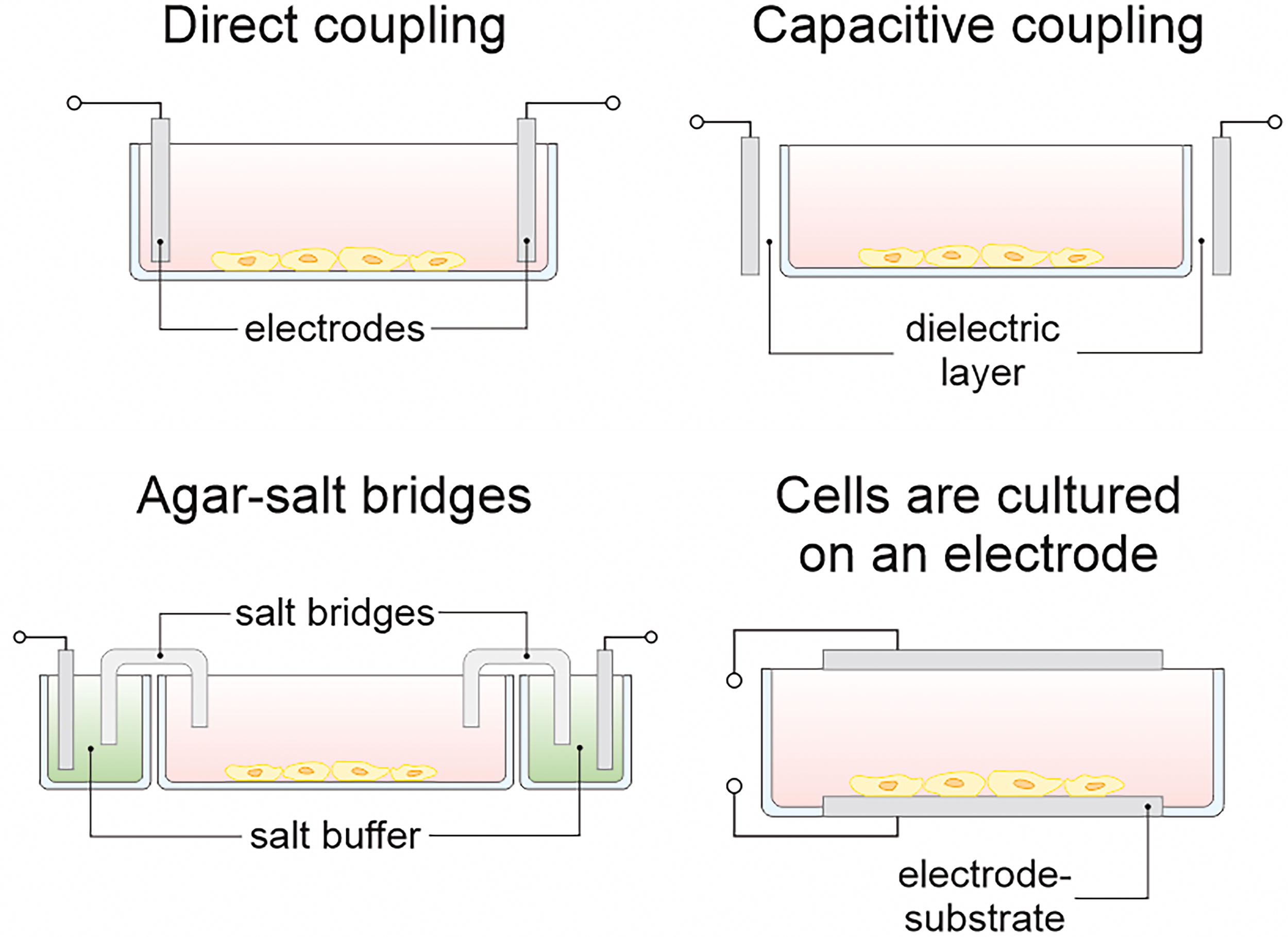
Major configurations of experimental setups for studying electric field-induced effects on cells in vitro: Direct coupling is characterized by immersion of electrodes directly into cell medium; capacitive coupling provides transmission of electric signal through dielectric layer, application of agar-salt bridges prevents products of electrochemical reactions from getting into cell medium; incorporation of electrode substrate into the stimulation system allows cultivation of cells on electrically conductive substrate.
The selection of materials is an important step in the design of the EF stimulation system, which determines the possibility of maintaining the viability of cell cultures during the experiment. The ideal electrode material should possess several key properties: high electrical conductivity, biocompatibility to minimize adverse cellular reactions, chemical inertness to prevent corrosion and degradation in the culture medium, and suitability for microfabrication to create electrodes with desired geometries. Noble metals such as gold and platinum are commonly used as electrode materials due to their excellent electrical conductivity, inherent corrosion resistance, and biocompatibility.32–34 Agar-salt bridges are included in stimulation systems to prevent the entry of electrochemical reaction products emitted at the electrode–electrolyte boundary interface into the cell culture.35–37 The use of conductive polymers as a substrate or for incorporation into three-dimensional (3D) constructs, such as hydrogel structures, offers great opportunities for tissue engineering. 35 An important feature of such chemical components is the ability to control their stiffness and biocompatibility. The use of conductive synthetic or natural polymers in tissue engineering constructs improves adhesion, stimulates proliferation and differentiation, and maintains high cell survival. 38 Electrically conductive hydrogels (CHs) with high conductivity, special mechanical properties, and biocompatibility have great potential in peripheral nerve regeneration.39,40 Polypyrrole, poly(3,4-ethylenedioxythiophene), and polyaniline are CHs that have been investigated in the context of restoring innervation in rats in vivo. 41 In addition to physically replacing part of the peripheral nervous system, CHs are able to affect the proliferation and differentiation of neighboring cells. Conductive biomaterials that autonomously generate electrical signals in response to external stimuli such as optical radiation, magnetic field, or mechanical stress have been widely studied in recent years. 42
The effect of EFs on cells depends on its characteristics such as strength, frequency, waveform, and duration of exposure. Increasing technical capabilities and the development of new designs and systems for electrical stimulation will result in the modeling of EFs of various configurations and the evaluation of its effects on cells and tissues both in vitro and in vivo. The diversity of experimental setups will expand the range of applications of EFs and take biological and medical research to a next level. In the following, we review the effects of the three main types of EFs—direct current EF (DC EF), alternating current EF (AC EF), and pulsed EF (PEF)—on cells and tissues and discuss their mechanisms and potential applications in biology and medicine.
Direct current EF
DC EF is characterized by a constant EF strength vector. In a biological context, the effect of DC EF on cells and tissues triggers various cascades of biophysical and biochemical modifications. These alterations are based on fundamental physical principles governing the interaction of charged particles and EFs. One such principle is electrophoresis. Charged molecules such as ions, proteins, and other biomolecules tend to move toward the electrode with the opposite sign of charge affected by the DC EF. This can lead to redistribution of charged components within the cell and in the intercellular space, which affects metabolic processes, activation or inhibition of signaling pathways, and changes in the functional activity of the cell.43–45
DC EF also induces polarization of the cell membrane and intracellular structures. 46 The cell membrane, which is a dielectric, undergoes polarization under the action of an external EF. This leads to a membrane potential shift and can affect the activity of ion channels and membrane transporters. Another important mechanism is electroosmosis. Electroosmosis is the movement of liquid induced by an applied EF across a porous material, charged surface, or membrane. In porous tissues, electroosmosis occurs due to the electrical double layer (EDL) that forms at charged interfaces (such as cell membranes). The EDL consists of a fixed charge layer on the surface and a diffuse layer of mobile counter-ions. The applied EF acts on these mobile ions, and their movement, along with their associated water molecules, generates a bulk fluid flow. This mechanism is capable of inducing solute transport in tissues and cell migration and enabling the delivery of therapeutic agents.47–49
Various experimental setups are used to investigate the effect of a DC EF on cells in vitro. An experimental setup, engineered by Ende et al., 50 examined the electrotaxis of T cells and was designed using two silver chloride electrodes immersed in PBS solution reservoirs connected to the cell culture chamber by agar-salt bridges. The electrotaxis of T cells was discovered to depend on cell type, substrate coating, and EF strength. Srirussamee et al. 51 employed a system with platinum electrodes embedded in the lid of a 6-well plate that generated an EF directly in the culture medium. DC EF affected the expression of immediate early genes such as c-FOS and c-JUN and the metabolic activity of mesenchymal stem cells (MSCs). Cho et al. 52 developed a setup integrated with a platform to measure cellular traction on a hydrogel substrate. In this setup, the EF was generated using silver chloride electrodes located in reservoirs of nutrient medium connected to the cell culture chamber by agar-salt bridges. The EF stimulated a directional migration and reorientation of intercellular stress. A microfluidic device made of polymethyl methacrylate with platinum electrodes was used by Li et al. 53 to precisely control the EF parameters. The electrotaxis of lung cancer cells was shown to be heterogeneous and cell type dependent.
In spite of the differences in design, all of the reviewed setups are united by a common operating principle: generation of a DC EF between two electrodes and the effect on cells cultured in a special chamber. An important feature of these systems is the necessity of controlling the uniformity and strength of the EF to ensure reproducibility of experiments.
The generation of an EF in cell cultures by direct immersion of electrodes in the medium is accompanied by electrochemical processes at the electrodes. These reactions trigger the production of reactive oxygen species (ROS), which modulate transgene expression through the activation of the kelch-like ECH-associated protein 1/NF-E2–related factor 2 (KEAP1/NRF2) signaling pathway. 54 This effect was used to develop DC-actuated regulation technology (DART) programming of gene expression by direct current stimulation. 55 The results demonstrated the possibility to maintain normoglycemia in type I diabetic mice using a portable DART-based device. In general, electrogenetics is based on the stimulation of gene expression and protein synthesis through the transfer of free electrons sourced from conducting materials and molecular proteins. 56 Thus, the application of wearable devices for electrogenetic programming and other purposes could be a major breakthrough in cell and gene therapies. For instance, a wireless-powered electrical bandage contact lens was presented for corneal wound healing by DC EF stimulating the migration and proliferation of corneal epithelial cells. 57
The application of DC EF in biology and medicine is particularly useful in the study of phenomena characterized by directional effects. Cell motility consisting of the cell migration and reorientation can be induced by a DC EF. 52 Galvanotaxis has been widely explored in the context of wound healing, embryonic development, and tissue regeneration.10,58 Directed cell migration induced by DC EF can be used to create tissue-engineered constructs with a defined architecture, as well as for targeted cell delivery to the area of injury. 59 DC EF promoted keratinocyte and fibroblast migration, 27 stimulated angiogenesis, 60 cytoskeleton reorganization, and collagen fiber alignment. 61 DC EF can also affect the proliferation and differentiation of various cell types. 62 For instance, DC EF promoted fibroblast proliferation, 63 stimulated the differentiation of neuronal progenitor cells (NPCs) into neurons, 20 and enhanced osteogenic differentiation of MSCs. 51 Endogenous EFs in the wound play an important role in healing, and application of external DC EF can enhance these endogenous fields and accelerate healing processes. 64
DC EF is a promising tool for manipulating cells and tissues and developing new methods of treatment. However, it is necessary to further study the mechanisms of DC EF effects on cells and tissues as well as to optimize the stimulation parameters to achieve maximum efficiency and safety.
Alternating current EF
AC EF is characterized by cyclic alterations in the value and direction of the field strength vector. The effect of AC EF on cells and tissues differs from the effect of DC EF and is determined by a number of specific physical mechanisms. The interaction of AC EF with biological objects is based on the phenomena of dielectric polarization and dielectrophoresis (DEP). Cells and tissues as dielectrics are polarized under the effect of an EF. Dipoles of water molecules and other polarizable molecules of the cell are oriented according to the field direction in an alternating field, cyclically changing their orientation depending on the frequency of the field. This leads to the conversion of some of the EF energy into heat. 65 This heating, particularly in nonuniform AC EFs, can lead to AC electrothermal (ACET) flow.66–68 ACET flow arises when a nonhomogeneous AC EF is applied to a conductive fluid, such as a biological buffer. Joule heating, caused by the passage of electric current through the conductive fluid, generates temperature gradients. These temperature gradients create local variations in the electrical conductivity and permittivity of the fluid. The nonhomogeneous EF then interacts with these induced gradients in conductivity and permittivity, resulting in a net force on the fluid and generating localized fluid motion, often in the form of vortices.
DEP is a phenomenon in which a dielectric particle, such as a cell, is subjected to a force generated by a nonuniform EF.69–71 These forces depend on the dielectric properties of the cell and the environment, as well as the frequency and gradient of the EF. DEP can cause cells to be attracted or repelled from a region of high field strength, opening up possibilities for manipulating cells—trapping, sorting, and moving them. For instance, DEP can be used to create well-structured layered tissue constructs composed of different cell types 72 or to trap, sort, and manipulate cells in microfluidic devices.23,69,73
AC EFs are particularly well-suited for separation of cells due to the frequency-dependent nature of DEP. By selecting the frequency of the applied AC field, it is possible to exploit differences in the dielectric properties of different cell types to induce selective movement, enabling label-free, continuous sorting based on intrinsic cellular characteristics.74,75 Various microfluidic device designs leverage this principle. For instance, Zhao et al.
76
used microfluidic chips with asymmetrical orifices and embedded electrodes to create an EF gradient, separating live and dead yeast cells and measuring their lateral migration at different frequencies ranging from 1 kHz to 103 MHz. Similarly, Punjiya et al.
77
employed a two-electrode geometry (a half-ring trap and a ground electrode) to generate a negative DEP trap, applying a frequency of 7 kHz. Some designs combine DEP with other forces. Sun et al.
68
combined DEP with ACET flow, using a bipolar electrode for focusing and switching particles. Oshiro et al.
78
integrated hydrodynamic filtration and DEP, where filtration by fluid flow presorts cells by size, performs buffer exchange and DEP, with slanted electrodes, and performs the final separation. Varmazyari et al.
79
investigated different 3D electrode configurations (triangular, rectangular, and semicircular) for separating cancer cells (MDA-MB-231) and white blood cells. Semicircular electrodes performed best, minimizing the required EF strength to
Beyond cell separation, AC EFs also enable precise manipulation of individual cells. Lv et al. 80 combined hydrodynamic trapping with negative DEP for selective release of cells, such as human leukemia K562 cells, using individually addressable microelectrodes. Zheng et al. 81 demonstrated two-dimensional cell movement of HeLa and human breast cancer MCF-7 cells (Michigan Cancer Foundation-7) with a quadrupole-electrode unit by modulating AC signal phases and amplitudes at a frequency of 100 kHz. Chen et al. 82 used a wireless bipolar electrode array for capturing circulating melanoma cells, optimizing the frequency to 50 kHz after characterizing the dielectric properties of melanoma cells and peripheral blood mononuclear cells. Although these DEP-based techniques offer a high degree of control over individual cells, it is crucial to remember that exposure to EFs, particularly the nonuniform fields inherent to DEP, carries the risk of inducing cell stress or damage.
Various experimental systems have been used to generate an AC EF and explore its effect on cells. An experimental setup with eight gold electrodes arranged in a circle on a glass substrate was utilized by Abadijoo et al. 32 to generate AC EF. The field frequency was 100 kHz and the voltage amplitude was 3 V. The results demonstrated that AC EF was able to inhibit the clonal expansion of activated lymphocytes in vitro. Kunti et al. 23 developed a setup consisting of two parallel electrodes separated by an insulating layer with a hole. This design allowed to create a nonuniform EF and consequently a temperature gradient, which led to the concentration and pattern formation of bacterial and yeast cells. Hernández-Bule et al. 83 designed a setup with two steel electrodes to generate AC EF with a frequency of 448 kHz and a current density of 50 µA/mm2. Increased proliferation of human MSCs in AC EF was demonstrated. Draz et al. 66 demonstrated enhanced immunoassays using a novel ACET electrode design featuring tilted electrodes. This design generated in-plane microfluidic mixing vortices with a single-phase AC signal typically at 1 MHz, resulting in improved fluid replenishment. This approach achieved a 6-time enhancement in HER2 cancer biomarker staining and a 75% reduction in assay time compared with traditional methods. Not only classical setups involving multiple electrodes can be used to create an EF but also physical phenomena such as piezoelectric or triboelectric effects. The combination of the piezoelectric effect with tissue engineering technologies is applied in accelerating wound healing.84,85 Ultrasound stimulation initiated voltage generation on a piezoelectric membrane in rats in vivo and promoted fibroblast migration and proliferation. 86 In the context of osteogenesis, the use of piezoelectric membrane scaffolds provided the electrical potential necessary for wound healing, corresponding to physiological parameters, and demonstrated antimicrobial and immunomodulatory functions. 87
The applications of AC EF in biology and medicine are extremely diverse. Tumor Treating Fields (TTFields) are low-voltage AC EFs with amplitudes of 1–4 V/cm and frequencies of 100–300 kHz. 88 TTFields are capable of affecting cancer cell division by disrupting mitotic spindle formation and leading to cell death. 88 TTFields are already used in clinical practice to treat glioblastoma. AC EF is utilized to stimulate neural tissue by affecting neuronal excitability and synaptic plasticity.11,89,90 AC EF can also be used to manipulate cells in vitro: to sort, trap, move, and form cellular patterns.23,69 The application of AC EFs in biology and medicine is constantly expanding as the understanding of the mechanisms of their effects on cells and tissues improves.
Despite the design differences, the operating principle of these systems is based on the effect of AC EF on cells cultured in vitro. To achieve the desired effect (e.g., DEP and modulation of cellular functions), it is necessary to precisely control the field parameters, such as frequency, amplitude, and waveform, as well as the duration of exposure.
Pulsed EF
The application of PEF is based on the effect of short pulses of an electrical signal on the stimulation object. Additional variable parameters of the PEF are the shape and width of the pulse. Rectangular pulses are usually used for cell stimulation but other signal profiles can also be applied.91–94
PEF of high intensity and short duration can result in electroporation.88,95 Electroporation is subdivided into reversible (RE) and irreversible (IRE) depending on the effects caused. The parameters of the applied EF, such as intensity, duration, and number of pulses, determine the type of electroporation induced. RE initiates temporary membrane permeabilization followed by restoration of cellular homeostasis and is used as electrochemotherapy (ECT), gene electrotransfer (GET), and calcium electroporation (Ca-EP). An important step in the delivery of DNA molecules by PEFs is to overcome several cellular barriers: the interstitial barrier, which is the space of extracellular matrix and intercellular junctions, the cell membrane, the cytoplasmic barrier, and the nuclear envelope. 96 ECT, GET, and Ca-EP use low-frequency 1–10 Hz rectangular pulses of about 100 µs duration and intensity amplitude up to 1000 V/cm. Stimulation with 6–8 pulses of this intensity is able to initiate RE permeabilization of the cell membrane and delivery of therapeutic agents and genes into the cytoplasm.97,98 Electroporation allows the delivery of various molecules such as DNA, RNA, and drugs into the cell, which is widely used in biotechnology and medicine.99,100
Various systems and experimental setups are used to generate PEFs. Hernández et al. 34 demonstrated a setup consisting of eight pairs of gold electrodes coated with platinum for stimulating embryoid bodies (EBs). EBs were exposed to biphasic current pulses of 1 Hz frequency, 1 ms duration, and field strength of 65 or 200 mV/mm. The results showed that short-term electrical stimulation promoted cardiac differentiation of human induced pluripotent stem cells (hiPSCs). An electrical cell stimulation and recording apparatus system was presented by Abasi et al. 101 that provided cell culture stimulation on a membrane insert, generating a homogeneous EF by a pair of planar titanium electrodes and recording alterations in transendothelial electrical resistance (TEER) using electrical impedance spectroscopy (EIS). This system has been successfully tested on human umbilical vein endothelial cells (HUVECs) and proved the efficacy of PEF stimulation to activate proliferation and accelerate cell monolayer formation. 102 Triboelectric stimulation has potential for application in regenerative medicine, such as muscle stimulation and wound healing.84,103 Bone marrow mesenchymal stromal cells were exposed to pulsed current using a triboelectric nanogenerator, which induced cell rejuvenation by maintaining pluripotency and proliferative potential. 104 Triboelectric stimulation suppressed the expression of senescence-associated genes such as p53, p21, and p16 and reduced β-galactosidase levels and senescence-associated secretory phenotypes.
Overall, the area of development of new constructions is continuously evolving and actively providing original technical solutions for EF studies in biology and medicine. Figure 2 presents variants of real experimental setups designed for in vitro cell stimulation and accounting for different methods of electrode integration.
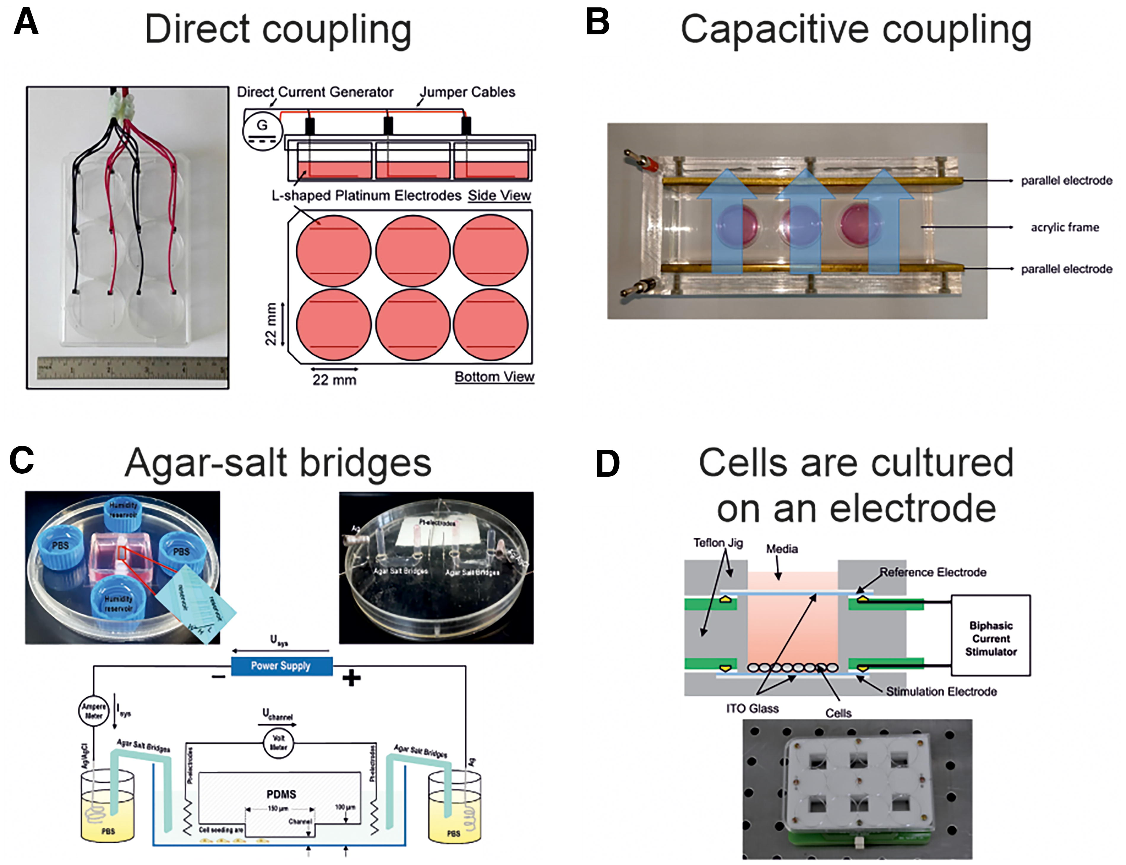
Experimental setups with different types of electrode integration for cell stimulation in vitro. (
The choice of the method of cell stimulation by EF and the development of the installation largely depend on the application and the goals of the experiment, as they have a number of advantages and limitations, briefly characterized in Table 1. The use of CHs, portable stimulation technologies, tribo- and piezoelectric effects in tissue engineering, and cell therapy will provide new approaches and methods for the treatment of diseases.
Advantages and Limitations, as Well as Possible Application Scenarios, of Different Types of Electric Field Generation Systems
The table contains the main types of experimental setups and the limitations and advantages associated with their use, which can be compared to select a suitable one for a particular purpose. ROS, reactive oxygen species.
EFs as Sensory Systems for Assessing Cellular Status and State
Assessment of mechanical and electrical properties of cells is a practical tool to monitor and analyze its status, phenotype, and viability. Current methods for measuring cell viability, such as trypan blue, propidium iodide and tetramethylrhodamine, and methyl ester staining, are intended for irreversible, destructive analysis of cell culture. 112 Stain-free single-cell methods for high-throughput analysis are able to circumvent the mentioned limitation and become a promising approach to accurately identify the cellular state. For instance, DEP phenomenon was utilized to detect staurosporine-induced apoptosis in suspension and adherent cultures of Jurkat and HeLa cancer cells with consideration of apoptotic cells. 116 The efficacy of paclitaxel and vinblastine treatment of carcinoma cells was determined by induced mitosis arrest at the G2/M phase, which resulted in deviation of amplitude and phase characteristics of cell impedance compared with untreated ones. 119
A range of electrical and mechanical cell parameters such as cytosol conductivity, membrane capacitance, Young’s modulus, cell radius, and fluidity can be measured by EIS and impedance flow cytometry (IFC) in combination with impedance-deformability approaches.113,120,121 Electric cell-substrate impedance spectroscopy is used to analyze the impedance of a cell layer adhered to an electrode substrate. EIS is based on the measurement of impedance of a static cell in a wide range of frequencies, which provides comprehensive information about its state, but the throughput of this method is low. The principle of IFC is the registration of impedance at several frequencies (often at 2 or 4), which significantly increases the throughput, but often reduces the amount of information about the electrical properties of the cell. To measure mechanical properties, the impedance-deformability approach is often used, which is based on the deformation of a cell in a constriction channel.
Approaches based on the application of the electrical impedance method allow high-precision analysis of different mechanical and electrical properties of cells and its classification in a heterogeneous suspension. Microfluidic technologies that combine flow analysis capabilities and integration with electrode systems are widely used for this task. 122
Multimodal electrical–mechanical properties were used for phenotyping, cytoskeleton, and cell stiffness assessment. 121 Impedance-deformability approach and impedance recording at frequencies of 0.25, 0.45, 0.75, and 1.2 MHz were applied to measure mechanical and electrical properties of HepG2, MCF-7, and MDA-MB-468 cancer cell lines. This comprehensive approach significantly increased the accuracy of cell classification in the microfluidic system. Not only impedance value serves as a characteristic for cell state assessment but also the opacity spectrum as the ratio of magnitude of high-frequency impedance to magnitude of low-frequency impedance. To expand the possibilities of impedance spectrometry, the parameter “complex opacity” was proposed, which takes into account the phase of the received signal and, as a consequence, the complex impedance. 112 Increasing the throughput of the microfluidic system can be achieved by varying the applied aspiration pressure and trapping cells in parallel by multiple hydrodynamic traps. 123
The rotation spectrum of a cell is also used to determine its electrical properties. Rotation in 3D space and orientation of cells in the preferred direction is necessary to perform targeted drug delivery and to study fundamental molecular signaling processes. 124 A platform with four bipolar electrodes was proposed to rotate cells three dimensionally in an external rotating EF, which allowed noncontact, noninvasive manipulation of cells. 115 IFC was used to sort and desalt cell suspension as a preparation step for mass spectrometry. 118 Such approaches aim to improve the performance of sample preparation processes for subsequent cell diagnostic testing.
To improve the sensitivity of the IFC method, constriction channels have been incorporated. The cell when passing through the constriction fills the entire channel space and closes the electrical circuit, thus reducing the contribution of fluid to impedance. However, in microfluidic devices with mechanical channel constriction, there is a high probability of cell deformation, membrane rupture, or blockage of the flow channel. Recently, a new approach of constricting the fluid flow with the cell sample using perpendicular flow of insulated sheath fluids has been implemented. 125 In this report, the classification accuracy obtained with K562, Jurkat, and HL-60 cell lines reached 99.8%. This method is expected to increase the throughput of impedance flow approaches in microfluidic systems up to 1000 cells/s. IFC was also applied for analyzing cell viability in 3D cultures. It was determined that the impedance opacity value characterizes the change in cell membrane permeability and, as a consequence, cell viability. 106
The complex application of EIS and IFC supports the manipulation, sorting, and classification of cells in microfluidic systems. A platform for parallel single-frequency analysis of a cell in dynamic flow and spectral analysis of a static trapped cell was used to determine the interchangeability and mutual amplification of EIS and IFC.118 Thus, the complementarity of these methods has been demonstrated, the combined use of which increases the efficiency of determining the electrical properties of the cell.
Obtaining hybrid cells with highly efficient methods in microfluidic systems has been made possible by the introduction of EF techniques. The combination of DC EF and AC EF for two-cell electrospinning and release, respectively, was used to obtain melanoma hybridoma with high survival in a single microfluidic system. 107
An important question in the design of microfluidic devices is the selection of the architecture of the integrated electrode systems. Coplanar and parallel electrodes are widely used but lead to distorted results due to nonuniform EF distribution. “Sandwich” devices can be a solution to this problem and increase the efficiency of impedance approaches in cellular technologies. 126
Microfluidic systems with integrated TEER measurement are gaining popularity. 127 TEER recording with EIS provides a wide range of cell layer impedance and, as a consequence, enhanced information about the cellular state. A system of parallel electrodes on polycarbonate substrates has been used to assess proliferation and differentiation of mucociliary human airway epithelium during long-term cultivation. 111 Planar electrode systems, including flexible thin-film electrodes, can also be integrated into microfluidic platforms.128,129 The advantage of such systems is scalability to electrode arrays used for impedance recording of multiple cell layers/barriers simultaneously. 130 Moving electrode systems are a promising technological solution for spatial TEER measurement, allowing the analysis of different regions of the cell barrier. 131
Schematic images and photographs of real microfluidic devices are shown in Figure 3. The ability to change the architecture of microfluidic channels and electrode systems makes such devices applicable to a multitude of applications, including sorting, rotation, fusion, detection, and property measurement.
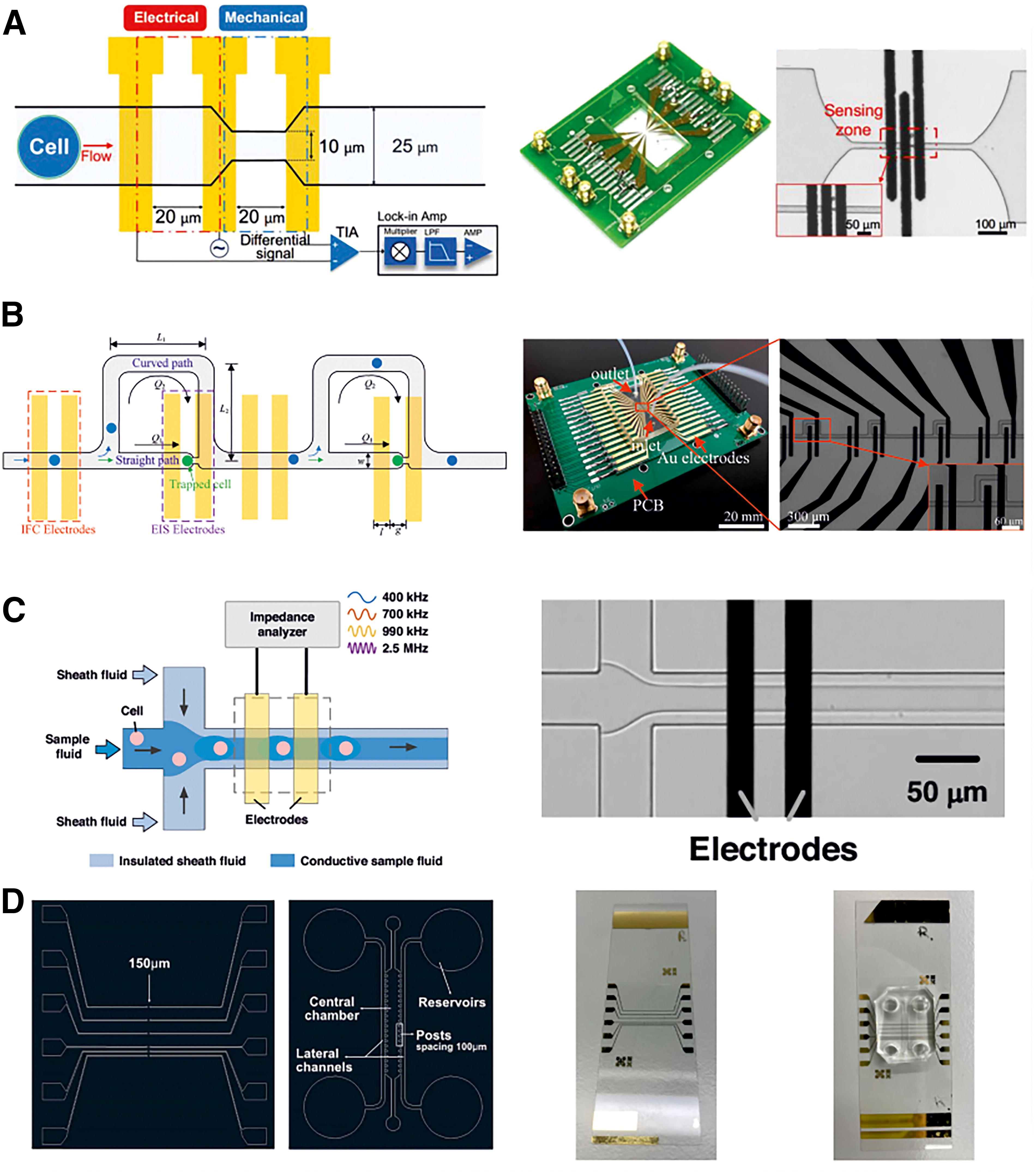
Microfluidic device designs used to measure mechanical and electrical properties of cells and analyze cellular state and status.
The described microfluidic platforms with integrated electrode systems have broad prospects of use for diagnostic purposes: for the analysis of peripheral blood cells such as erythrocytes, leukocytes, neutrophils,118,125,132,133 and as a sensor to assess the efficacy of drugs acting at tumor cells.116,117
EFs as Modulators of Cell Proliferation, Differentiation, Migration, and Cell Death
Specific examples of the application of EFs, along with their corresponding field characteristics and affected cell types, are summarized in Table 2.
Electric Field-Induced Effects on Different Cell Types
Biological effects such as migration, proliferation, differentiation, functional activity, and cell death induced by electric field with different stimulation parameters in eukaryotic cells are presented.
Cell migration, proliferation, and differentiation in vitro
Directed cell migration, or galvanotaxis, is the movement of cells in response to DC EF exposure. This phenomenon has been widely investigated in the context of wound healing, 59 embryonic development, 153 and tissue regeneration.136,154 DC EF promoted the migration of keratinocytes27,52,135 and fibroblasts19,27 into the wound area, facilitated the formation of cellular patterns, and directed tissue growth, 154 stimulated angiogenesis, 60 and alignment of collagen fibers. 61 This is especially important for creating tissues with anisotropic structure, such as nerves, muscles, and vessels.102,139 For instance, Lang et al. 26 demonstrated that DC EF can be used to align Schwann cells and fibroblasts, creating oriented cell monolayers that can serve as a basis for nerve regeneration. Zhao et al. 60 revealed that application of DC EF to endothelial cells stimulates their production of vascular endothelial growth factor, a key growth factor.
The direction of cell migration (to the cathode or anode) in DC EF depends on the cell type and stimulation parameters. Li et al. 53 identified that lung cancer cells demonstrated different directions of galvanotaxis depending on the cell line. Differences in migration directions of certain cell types also enable noninvasive sorting in cocultures. Leal et al. 27 showed that directional migration of keratinocytes and fibroblasts initiated by DC EF enabled the separation of a coculture of cells into two monocultures. The mechanisms underlying galvanotaxis are not fully investigated but are thought to be related to the asymmetrical distribution of ion channels and receptors on the cell membrane, as well as cytoskeleton rearrangement and lamellipodia formation. 19
The effect of EFs on cell proliferation is also being actively studied. DC EF can both stimulate and inhibit proliferation of different cell types depending on the stimulation parameters and cell type. Thus, Urabe et al. 63 reported that PES increased the proliferation of human skin fibroblasts. Electrical stimulation at 448 kHz promoted the proliferation of MSCs. 83 However, researchers observed an inhibitory effect of DC EF on proliferation of HeLa and U937 leukemia cells.140,155
The application of EFs to stimulate proliferation may be a useful method in mass expansion of cell cultures, especially in combination with process of cultivation in bioreactors. 156 Electrical stimulation systems can be integrated into the bioreactor design and provide a constant controlled effect on cells.
The mechanisms underlying the effects of EFs on cell proliferation are probably related to cell cycle disturbances and 136 changes in the expression of genes encoding growth factors.134,137 For instance, Hernández-Bule et al. 83 reported that electrical stimulation led to activation of proliferating cell nuclear antigen and mitogen-activated protein kinase/ extracellular signa-lregulated kinase 1/2 (MAPK/ERK1/2) signaling pathway, which play an important role in cell cycle regulation.
EFs can also affect the differentiation of cells, guiding their development toward a specific cell phenotype. The use of EFs for directed differentiation of stem cells will overcome one of the key problems of tissue engineering—lack of donor organs and tissues. Several studies have shown that electrical stimulation can initiate controlled differentiation of stem cells. 145 Ariza et al. 20 demonstrated that DC EF stimulated the differentiation of NPCs into neurons, and Srirussamee et al. 51 found that electrical stimulation enhanced osteogenic differentiation of MSCs. The mechanisms underlying this phenomenon may include activation of specific signaling pathways, such as the PI3K/Akt/GSK−3β/β−catenin signaling pathway, 144 alteration of the activity of the osteogenic marker, alkaline phosphatase, 137 and changes in the epigenetic landscape of the cell.157,158 EFs contribute to the creation of a favorable microenvironment for tissue growth and development. For instance, Weizel et al. 159 presented a novel design of an electrical stimulation device that can simultaneously affect multiple scaffolds with cartilage cells, promoting their viability and differentiation. In another research, Hernández et al. 34 used electrical stimulation to promote cardiac differentiation of hiPSCs, accompanied by increase in cardiac gene expression and the percentage of beating EBs. This result highlights the potential of electrical stimulation to target stem cell differentiation and create functional tissues.
Overall, EFs are a promising tool for regulating cell migration, proliferation, and differentiation. Understanding the mechanisms of EFs effect on these cellular processes opens a wide range of opportunities for the development of new approaches to disease treatment, tissue regeneration, and engineering.
Functional activity of cells
EFs have a significant impact not only on basic cellular processes such as migration, proliferation, and differentiation but also on the functional activity of various cell types. They are capable of modulating cell signaling, ion transport, metabolism, gene expression, and other processes underlying cellular activity.
One example of the effect of EFs on the functional cell activity is the modulation of synaptic plasticity in nervous system. Transcranial DCS can modify neuronal excitability and enhance long-term potential in vivo,14,89 underlying mechanisms of memory encoding and consolidation. For example, Yu et al. 14 demonstrated that anodal tDCS increased brain-derived neurotrophic factor levels in the hippocampus, which may explain its positive effects on synaptic plasticity and cognitive function.
AC EF can also affect the functional activity of neurons. tACS can synchronize neuronal activity in specific frequency range and modulate cognitive functions in vivo. 160 Particularly, tACS is able to modulate gamma brain rhythms that are associated with attention, working memory, and other cognitive functions. 11 tACS affected synaptic plasticity in brain tissue ex vivo, triggering changes in synapse morphology. This confirms the ability of tACS to modulate synaptic transmission and affect learning and memory processes.
In addition to affecting neural tissue, EF can modulate the functional activity of other cell types as well. DC EF regulated the rate and direction of migration of NPCs through calcium-dependent mechanisms by affecting actin polymerization and cell outgrowth in vitro. 108 This research demonstrated that DC EF not only navigates cell movement but also affects cell intrinsic mechanisms responsible for migration.
Low-voltage PEFs (LV-PEFs) are able to induce morphological changes in cells without reducing viability and electroporation effects. In contrast to high-voltage PEFs (HV-PEFs), whose biological effects are accompanied by thermal damage, LV-PEF reduces heating effects by decreasing the amplitude of stimulation simultaneously with increasing the number of applied pulses. PEFs caused remodeling of the cytoskeleton of HUVECs in vitro due to redistribution and accumulation of actin microfilaments at the periphery of the cytoplasm and promoted localization of the tight junction proteins zonula occludens-1 (ZO-1) and VE-cadherin around the nucleus, and consequently disruption of gap junctions and partial destruction of the endothelial monolayer.138,141
PEFs are interesting not only for the delivery of plasmids containing specific genes but also as a way to control the expression of genes introduced into the cell. LV-PEF of eight pulses of 100 µs duration was used to stimulate mammalian cells transfected with the inducible expression vector pNF-kB-secreted embryonic alkaline phosphatase (SEAP) in vitro. 161 Human cervical carcinoma cells Hep-2c demonstrated an active increase in SEAP protein expression upon stimulation with 0.165 kV/cm, but membrane permeability remained unchanged. This result indicates the possibility of applying microsecond EFs to control gene expression.
In the field of oncology, EFs can lead to disruption of mitotic division, causing arrest of cell cycle and apoptosis. For example, TTFields inhibited the growth of various cancer cell lines in vitro and in vivo by disrupting mitotic spindle formation and causing cell destruction during cell division. 149 TTFields are already used in clinical practice to treat glioblastoma and show perspectives for the treatment of other types of cancer.147,162,163
In general, EFs have multifaceted effects on the functional activity of different cell types. They can modulate synaptic plasticity, cell signaling, ion transport, metabolism, and other processes underlying cellular activity. Understanding the mechanisms of EFs effect on the functional activity of cells opens up wide prospects for the development of new approaches to the treatment of various diseases, tissue regeneration, and the creation of new biotechnological constructs.
EF-induced cell death in vitro
EFs, in addition to their ability to modulate cellular functions, can also induce cell death by acting through a variety of mechanisms. This effect of EFs has found application in cancer therapy and has also been used to selectively kill undesirable cells in other medical applications.
One of the main mechanisms for the induction of cell death by EFs is electroporation. IRE disrupts the integrity of the cell membrane, leading to irrevocable changes in internal processes and cell death. The ability to initiate cell destruction defines IRE as a promising method in the treatment of cancers that are difficult to treat surgically. 142 IRE effect can be initiated by increasing the EF strength up to 3000 V/cm and/or pulse duration up to millisecond range. However, while keeping the same stimulation parameters used for RE but the number of pulses is significantly increased, high-voltage IRE effects can also be observed. Electroporation initiated apoptosis by activating the caspase cascade and other signaling pathways. 164 Cancer cell death during HV-PEF ablation is promoted not only by thermal effects and membrane pore formation but also by changes in pH throughout the stimulated field, increased synthesis of ROS, and a rapid decrease in cellular adenosine triphosphate (ATP) levels. 142 The loss of ATP in cancer cells by IRE initiates an energy crisis leading to the interruption of caspase signaling of apoptosis and is able to redirect the cell death process along the necrosis pathway. 150 This effect may enhance the efficacy of treatment of tumors with mutant apoptosis genes by PEFs. The main changes caused by the effect of EF on the cell and mediating cell death are presented below (Fig. 4).
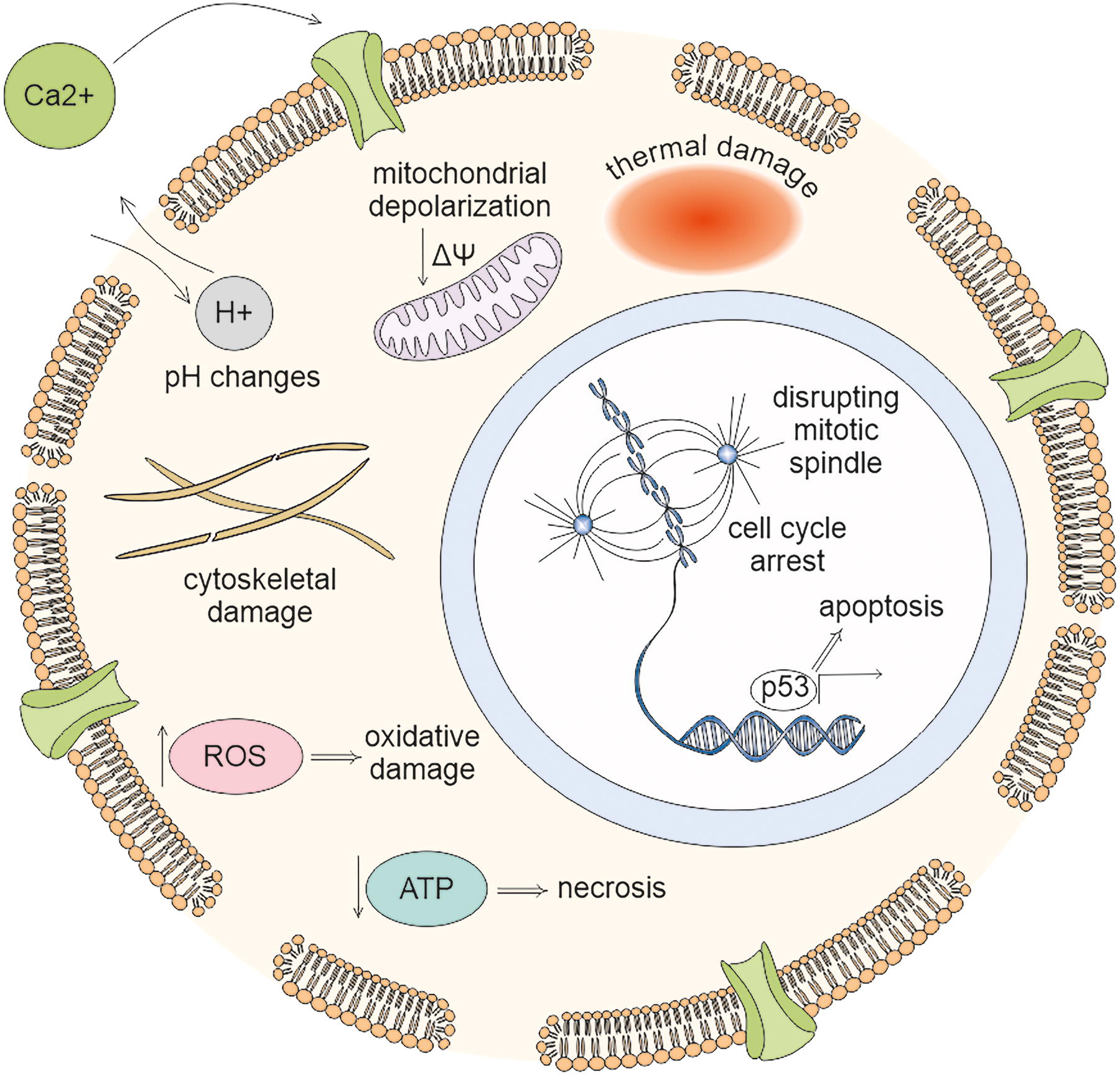
Electric field-induced processes leading to cell death: Electric fields promote membrane pore formation, activate the influx of calcium ions into the cell via VGCCs, modulate mitochondrial membrane potential, initiate changes in pH, ROS, and ATP levels, induce thermal and cytoskeletal damage, and disrupting mitotic spindle. ROS, reactive oxygen species.
The formation of pores in the cell membrane during electroporation can occur due to the reorientation of phospholipids as a result of the penetration of water molecules into the lipid bilayer. 165 PEFs are also able to initiate lipid peroxidation and, through electro conformational changes, modulate the activity of membrane proteins, in particular voltage-gated ion channels (VGICs). Using molecular dynamics modeling, it has been determined that PEFs promote pore formation in voltage-sensor domains (VSDs) of VGICs, which in turn can be accompanied by the dissociation of VSDs from the channel. 166 Such conformational changes are thought to result in VGIC dysfunction and a decrease in their sensitivity to alterations in membrane potential.
Nanosecond PEFs (nsPEFs) have been widely investigated for electroporation. Decreased pulse duration provides a reduction in undesirable effects such as electrochemical reactions accompanied by the release of by-products at the electrodes, whereas the delivery efficiency of therapeutic molecules is comparable to conventional microsecond pulse exposure methods. 98 nsPEFs induce the formation of micropores in the cell membrane, thereby activating VGICs and the flow of calcium ions into the cytosol. In this way, various cascade reactions are triggered, leading to the processes of proliferation, differentiation, and cell death. 167 nsPEFs caused the destruction of the cell cytoskeleton 168 and mitochondrial depolarization. 169
In cancer therapy, the use of TTFields to selectively kill cancer cells has been widely studied. TTFields are able to disrupt cancer cell division by affecting microtubules and other components of the mitotic spindle. 88 This leads to the arrest of cell cycle and induction of apoptosis in cancer cells, whereas healthy cells remain less damaged. In addition to electroporation and TTFields effects, EFs can induce cell death in other ways. EFs can also activate intracellular signaling pathways leading to apoptosis, such as the p53 signaling pathway.151,170,171 EFs can regulate the level of surface phosphatidylserine in cancer cells through a calcium-dependent pathway, thereby labeling them for subsequent detection. 172
The selectivity of the effect of EFs on cells can be achieved through different stimulation parameters such as frequency, intensity, and duration of pulses, as well as through differences in the electrical properties of different cell types.173,174 For example, cancer cells often have higher membrane conductivity compared with healthy cells, making them more susceptible to electroporation. 175
In general, the ability of EFs to induce cell death opens wide prospects for the development of new approaches to the treatment of cancer and other diseases, as well as for the development of new methods of cell engineering and biotechnology. However, it is necessary to further study the mechanisms of cell death induction under the action of EFs and optimize the stimulation parameters to increase the selectivity and efficiency of treatment.
Mechanisms and Signaling Pathways Mediating the Effects of EFs on Cells
EFs affect cells, involving various molecular mechanisms in the cellular response. The activity of signaling pathways can be controlled by exogenous stimuli and also by EFs, regulating the processes of differentiation, proliferation, migration, cell death, and metabolism. The main signaling pathways controlling cellular response to EF stimulation and involved physical phenomena and structural cellular alterations are reviewed below (Fig. 5).
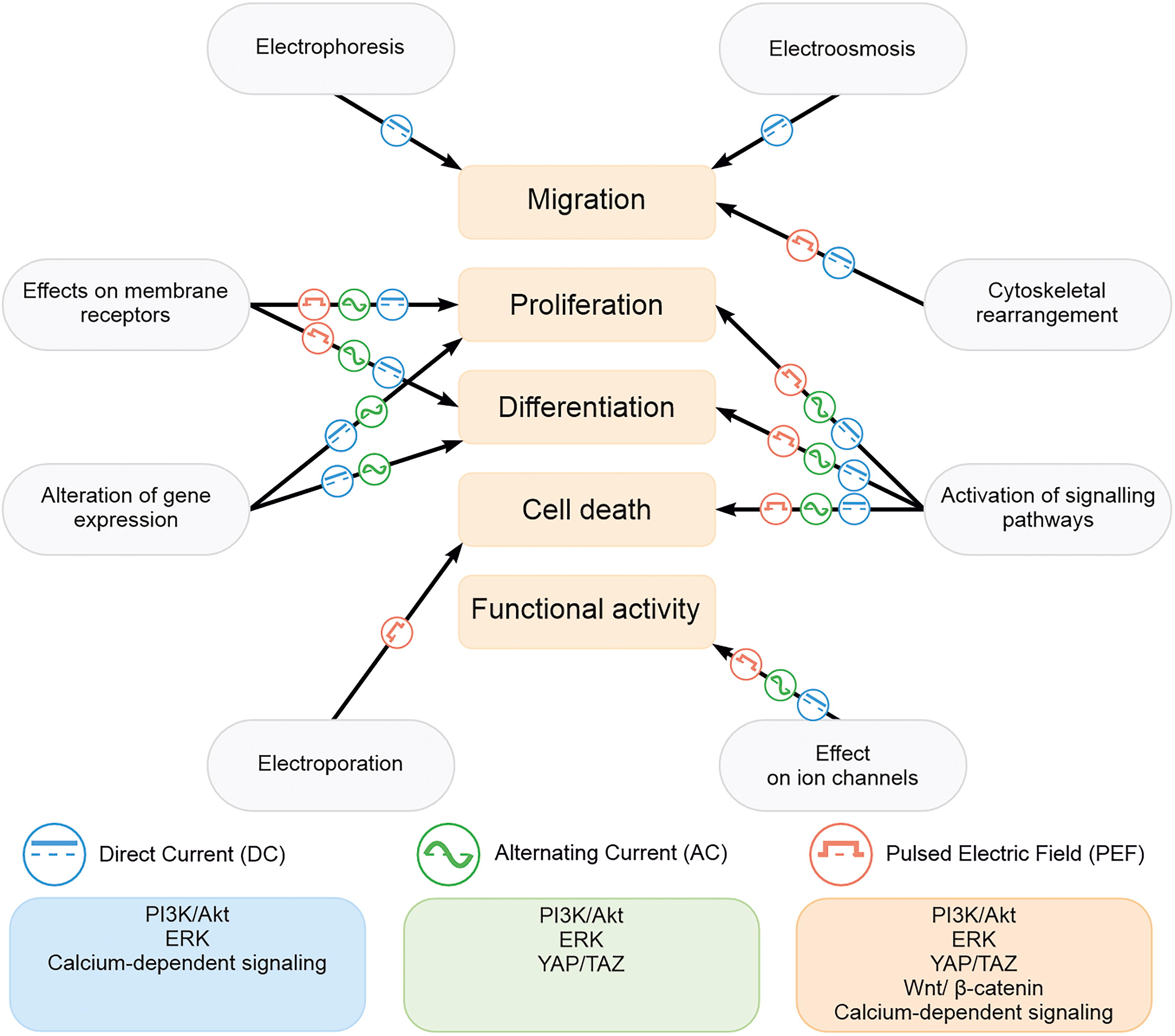
Biological effects and molecular signaling pathways involved in the cellular response affected with EF: Signaling pathways mediate cellular response to DC EF (blue rectangle), AC EF (green rectangle), and PEF (orange rectangle). Arrows relate physical phenomena and structural cellular alterations (labeled in gray ellipses) that cause biological effects through electric fields. AC EF, alternating current electric field; DC EF, direct current electric field; PEF, pulsed electric field.
EF induces calcium signaling pathways in the cell. Calcium in flux into cells via voltage-gated calcium channels can be induced by EF stimulation. 176 Ca2+-bound calmodulin is able to bind to specific calmodulin kinases that control osteoblast differentiation through cAMP response element-binding protein/cAMP response element (CREB/CRE) activation. 177 Depending on calcium dynamics and calmodulin binding, PI3K activates the mammalian target of rapamycin (mTOR) signaling pathway that also accompanies bone formation processes. 177 nsPESs are able to regulate calcium signaling and stimulate the expression of calcium-dependent genes c-FOS, c-JUN, EGR1, NURR-1, and β3-TUBULIN. 178
An increase in intracellular calcium level can activate a combination of two other signaling pathways Wnt/Ca2+ and Wnt/β-catenin, accompanied by translocation of β-catenin into the cell nucleus. 179 Through the Wnt/β-catenin pathway, nsPEFs are able to regulate chondrocyte differentiation. 180 Wnt/β-catenin is involved in embryogenesis, cell growth, and regeneration, but aberrant activity is often associated with tumorigenesis, so suppression of increased activity of the pathway may become a target for cancer therapy. 181 nsPEFs reduced migration and invasion of human pancreatic carcinoma PANC-1 cells through inhibition of the Wnt/β-catenin pathway, which indicates the feasibility of applying submicrosecond-range EFs to reduce metastasis. 152 Currently, there is no safe and fully proven molecular inhibitor of the Wnt/β-catenin pathway for use in anticancer therapy.182,183 Thus, the application of EFs, especially pulsed fields, to regulate Wnt/β-catenin dynamics could be a useful method in cancer treatment.
YAP (yes-associated protein) and TAZ (transcriptional coactivator with PDZ-binding motif) are the main effectors of the Hippo signaling pathway controlling the processes of cell proliferation, differentiation, apoptosis, tissue homeostasis, and the proper development of organs.184,185 Nuclear localization is considered a feature of YAP/TAZ transcriptional activity. Although the YAP/TAZ complex is mainly considered as a mechanotransducer 186 regulating the cellular response to mechanical stimuli, however, recent studies indicate its role in generating the cellular response to EFs. Stimulation of HUVECs with a low-frequency PEF initiated nuclear translocation of the YAP/TAZ complex, observed after stimulation and accompanied by proliferation. 102 YAP/TAZ also controls osteogenic differentiation by inducing increased expression of Runx2 and other bone markers.109,187 Increased expression and localization of YAP in the nucleus were observed in human dermal fibroblasts (HDFs) exposed to nsPEFs. 187 PEFs regulate the polarization of macrophages differentiated from human leukemia cell line THP-1, increase the ratio of anti-inflammatory macrophages M2 to proinflammatory macrophages M1, and stimulate YAP/TAZ expression, which is suggested to be a way to control the inflammatory response and play a role in wound healing. 143
Both YAP/TAZ and EFs affect self-renewal and stem cell fate and control various processes in cancer cells.104,148,188,189 However, to date, the contribution of the YAP/TAZ signaling pathway to EF-induced stem cell rejuvenation and oncogenic changes remains unexplored. Also, the participation of YAP in the processes of galvanotaxis and orientation has not been determined. The combination of exposure of cells to EFs and mechanical stimuli, such as shear stress and mechanical alteration of cell morphology, may be a useful tool to study the activity of YAP/TAZ and its involvement in cellular response formation.
The ERK signaling pathway plays an important role in physiological development of the organism as well as in various pathological conditions and controls multiple cellular processes including migration, proliferation, differentiation, survival, and metabolism. 190 The signaling cascades comprising ERK can be controlled by EFs and become a target for the regulation of cell development. MAPK-ERK1/2 activation accompanies cellular galvanotaxis processes in a constant EF45,134,191–193 and in low-frequency PEFs 194 and mediates osteogenic differentiation of MSCs. 195 LV-PEF demonstrated a neuroprotective effect on human neuroblastoma cells line SH-SY5Y exposed to toxic H2O2 and β-amyloid via ERK activation. 105
ERK dynamics can be controlled by exogenous stimuli, such as through epidermal growth factor. 31 EF stimulation may also be applicable to regulate the activity of the ERK signaling pathway. 196 A distinguishing feature of EFs application is the possibility of noninvasive stimulation by using capacitively coupled experimental setups that prevent the products of electrochemical reactions from entering cell cultures and toxic effects of ROS and pH changes on cells. Such a system was used in work, 93 where it was found that the activation or inhibition of ERK depends on the shape of the exogenous signal and the duration of external pulses. The results detected a correlated change in the activity of Ras-GTP, which is the membrane-binding proteins of the ERK pathway, indicating a possible susceptibility of Ras-GTP to EF. 93 Thus, modulation of MAPK-ERK1/2 activity by EFs may become a way to regulate cellular activity.
Another important signaling pathway activated by EF is the PI3K/Akt pathway. 31 EF stimulation initiates activation of endothelial nitric oxide synthase and enhanced synthesis of NO through increased PI3K expression and Akt phosphorylation, which are necessary for stimulation of angiogenesis. 197 The PI3K/Akt signaling pathway controls galvanotaxis,191,192,198 and it has an inhibitory effect on autophagy.199,200 Enhanced activation of the PI3K/Akt signaling pathway may indicate carcinogenic processes in the body; therefore, developing therapeutic strategies to treat cancer through PI3K/Akt inhibitors that increase sensitivity to radio- and chemotherapy has recently been actively explored.201,202 However, the application of PI3K/Akt inhibitors and chemotherapeutic agents imposes certain limitations, consisting in the formation of resistance to inhibitors and chemotherapeutic agents. 203 EFs can independently inhibit PI3K/Akt activity in glioblastoma cells and stimulate autophagy. 204 In combination with other PI3K/Akt inhibitors, EFs enhance antitumor effects.205,206 Administration of PI3K/Akt inhibitors increases the sensitivity of cancer cells to TTFields. 207
Thus, modulation of cell activity by signaling pathways involved in the formation of cellular response to EF stimulation has great potential for controlling cell activity and viability. To identify the effective ranges of physical parameters of EFs, it is necessary to further study their effects on different cell types. The use of EFs as activators or inhibitors of signaling pathways may become a promising method in the treatment of many diseases.
Limitations and Novel Opportunities in the Application of EFs in Cell Biology
Despite promising results, the application of EFs in biology and medicine faces a number of limitations that must be taken into account when developing new methods and devices. One of the main limitations is the inhomogeneity of the field distribution in biological tissues due to differences in the electrical conductivity of tissues and cells. This can lead to nonuniform effects of the field on cells and tissues, reducing the efficiency and selectivity of stimulation. For example, in a study 20 that investigated the effect of DC EF on rat hippocampal NPCs, DC EF was shown to stimulate the differentiation of NPCs into neurons. However, the effect was heterogeneous due to the heterogeneous field distribution in the culture. The authors note that new methods for creating more homogeneous EFs in biological tissues need to be developed to improve the efficiency and homogeneity of stimulation.
Another limitation is the side effects that electrical stimulation can cause. These can include tissue heating, electrolysis, free radical formation, and cell membrane damage.51,69 In the article, 51 it was found that electrical stimulation of human MSCs resulted in the generation of hydrogen peroxide which is toxic for mammalian cells. To minimize side effects, it is necessary to carefully select stimulation parameters, taking into account the chemical composition of the culture medium, and to develop new stimulation methods that would reduce undesirable effects. Finally, the efficacy and safety of electrical stimulation depend on a variety of parameters such as frequency, voltage, pulse duration, waveform, and stimulation mode. Optimizing these parameters to achieve the desired effect can be a challenging task requiring many experiments with different cell and tissue types.
To expand the potential applications of EFs in biology and medicine, the combined use of EFs with other technologies is being actively explored. These can be microfluidic technologies, magnetic fields, ultrasound, optical methods, and other approaches.146,208–211 One of the most promising areas is the combined use of EFs with optical tweezers. 212 Optical tweezers are a tool that allows manipulation of microscopic objects, such as cells, using focused laser beams. The principle of operation is that the laser beam, as it passes through a dielectric object, refracts and changes its pulse. This change in momentum creates a force acting on the object and allows it to be held and moved in 3D space. Optical tweezers provide high precision and noninvasive manipulation, making them an ideal complement to electrical stimulation.
This approach is particularly useful for studying such phenomenon as electrokinetic interaction of cells arising due to their ability to polarize in an external EF. Under the action of an EF, the charges inside the cell redistribute to form an induced dipole moment

The schematic representation of an experimental setup for measuring cell–cell interaction forces in an electric field using optical tweezers: Optical tweezers (red beams), focused through an immersion lens, capture and position cells (green spheres) in a cuvette containing nutrient medium. A system of eight electrodes (gray) creates an electric field E, inducing a dipole–dipole interaction
Optical tweezers allow individual cells to be precisely positioned in specific regions of the EF,214,215 opening up opportunities to investigate the effects of EF on individual cells with high reproducibility. Moreover, optical tweezers can be used to measure forces acting on microscopic objects, including cells. For example, it is possible to measure the adhesion force of cells to a surface,110,216,217 intercellular adhesion, 218 or viscoelastic properties of cells.219–222 This allows us to quantify the force with which EF affects cells and gain a deeper understanding of the biophysical mechanisms.
The combined use of optical tweezers and DEP offers the potential to generate complex cellular patterns with high precision. Optical tweezers are used to grasp and move cells into desired positions, whereas DEP ensures that they are fixed and form a defined structure. For example, it is possible to create layered 3D tissue-engineered structures composed of different cell types.223–225 Optical tweezers also allow for controlled proximity and distance of cells from each other. In combination with EF, this creates a unique opportunity to study cell signaling and intercellular interactions in the presence of an EF. Optical tweezers grasp the cell, holding it in the focus of the laser beam. The gripping force depends on the power of the laser, the refractive index difference between the cell and the environment, and the size of the cell. Using optical tweezers, individual cells as well as small groups of cells (up to a few dozen cells) can be captured and manipulated.226,227 Such a combination is not only a powerful tool for solving applied problems but also a unique platform for fundamental research, in particular for studying collective and pairwise cell dynamics. This area is still understudied, and the use of the described combination of methods opens new perspectives.
Numerical models 228 can be used to analyze experimental data obtained with EF and optical tweezers. These models allow describing cell movement and interactions taking into account various factors such as adhesion, cytoskeleton mechanics, and signaling interactions.229,230 The development of such models is an important area of future research that will provide a deeper understanding of the principles of collective cell behavior. Digital models, verified and complemented by experimental data obtained with EF and optical tweezers, may become an indispensable tool to accelerate in vitro testing. This could lead to more efficient drug screening methods and the development of new approaches to disease treatment.
The combination of EFs and optical tweezers represents a powerful and versatile toolkit that opens new horizons in biology and medicine. This technology not only provides a high level of control over cells, allowing their position, orientation, and interaction to be manipulated with high precision, but also creates a unique platform for basic research. The combination of these techniques opens up the possibility for in-depth studies of the biophysical properties of cells, cell signaling mechanisms, and principles of collective cell behavior, which are key to understanding the development of multicellular organisms and the onset and progression of diseases. In the future, the synergy of EFs and optical tweezers may lead to revolutionary breakthroughs in medicine, opening up new possibilities for diagnosing and treating diseases. The high precision of manipulation provided by optical tweezers, combined with the ability to target cellular processes using EF, opens up new perspectives for creating highly effective methods of cell therapy and genetic engineering. Moreover, this technology has great potential for the development of tissue engineering, where precise control of cells and their interactions is key to creating functional tissues and organs.
Conclusion
The response of cells and tissues to EF stimulation leads to significant changes in cells’ functional activity and metabolic status, e.g., migration, proliferation, differentiation, and programmed cell death. Currently, there is a big progress in understanding the diverse physical mechanisms underlying these effects, including electrophoresis, electroosmosis, dielectric polarization, and DEP, as well as the key signaling pathways affected. It is well known that the response depends on EF parameters (intensity, frequency, pulse shape, and duration of exposure) and cell type which is important for the development of effective and safe methods of EF application. Deciphering the specific characteristics and outcomes of DC EF, AC EF, and PEF action is required for their applications in regenerative medicine, tissue engineering, cell biology, oncology, etc. The choice of EF type and stimulation parameters is determined by a specific task, e.g., modulation of cell migration or selective destruction of cells.
There are various technological issues that should also be taken into the consideration, including setup configurations, electrode materials, and integration with microfluidic systems, which, in particular, allow high-throughput analysis of cells, measurement of their electrical and mechanical properties, as well as precise manipulation. The synergistic use of EF and optical tweezers allows manipulating with cells and analyzing the cell-to-cell communication or collective cell behavior.
Despite significant progress, EF applications are limited by field heterogeneity and evident side effects. Promising areas of research include overcoming these limitations, studying the mechanisms of EF action, optimizing stimulation, and developing minimally invasive target drug delivery systems. Machine learning-based simulations combined with experimental data would advance the understanding of EF action and accelerate the development of new diagnostic and therapeutic approaches.
Footnotes
Authors’ Contributions
Conceptualization, D.A.B. and S.A.K.; writing—original draft preparation, D.A.B. and D.D.V.; critical revision and editing of the article, S.A.K., A.B.S., and S.O.Y.; visualization, D.A.B. and D.D.V.; project administration and funding acquisition, A.B.S. and S.O.Y.
Author Disclosure Statement
The authors report no conflicts of interest.
Funding Information
This work was supported by a grant from the Ministry of Science and Higher Education of the Russian Federation for major scientific projects in priority areas of scientific and technological development (project no.: 075-15-2024-638).