
Abstract
Organoid engineering is a rapidly expanding field that involves developing miniaturized, three-dimensional (3D) structures to mimic the architecture and function of real organs. It provides a powerful platform to investigate organ development, disease modeling, and personalized medicine. Recent advances in cell printing technology, also known as bioprinting, feature high-throughput potential, precise control, and enhanced reproducibility, enabling the deposition of living cells to generate complex, 3D biological structures. Cell printing with bioinks composed of cells and supportive biomaterials has been utilized to generate in vitro tissues and organs with intricate architectures and functionalities to investigate normal tissue morphogenesis and disease progression. The integration of cell printing technology and organoid engineering holds tremendous potential in biomedical research. Here, we summarize recent advances in cell printing technology in developing different organoid models, creating patient-specific tissue grafts, and utilizing these models and grafts in drug testing, as well as studying disease progression. Some of these bioprinted organoids have been utilized in clinical trials, highlighting the potential of cell printing technology in future applications in tissue and organ transplantation, as well as precision medicine.
Impact Statement
This article summarizes recent advances in integrating cell printing technology with three-dimensional tissue culture to develop organoid models. It discusses the advantages and limitations of three bioprinting technologies used in cell and organoid printing. The review also highlights the significant potential of cell printing technology in organoid model development and its applications in biomedical research and drug screening.
Introduction
Tissue structure and function rely on the appropriate interaction between cells and their microenvironment. 1 Compared with traditional two-dimensional (2D) tissue culture, organoid models offer a more biomimetic approach by recapitulating in vivo-like cell–cell and cell–extracellular matrix (ECM) interactions. The three-dimensional (3D) tissue structures of organoids in ECM gel or biocompatible material mimic the in vivo architecture and functionality of organs, which provides a revolutionary platform for studying tissue/organ development, disease progression, and drug responses in biomedical research. 2 Increasing evidence shows that organoid models significantly enhance the discovery of new treatments and deepen our understanding of diseases such as cancer, neurological disorders, and infectious diseases.3,4
Organoid models provide powerful tools for the advancement of biomedical research. However, conventional organoid construction is primarily based on spherical structure, which cannot fully capture the complexity and diversity of the in vivo microenvironment. Additionally, the phenotypic and functional variability of organoids in 3D culture poses challenges for result quantification and data analysis. These limitations lead to significant biases in evaluation and prediction, underscoring the practical importance and urgency of improving the traditional organoid models.
The reconstruction of complex organoids in tissue culture involves the combination of different cell types and ECM and the formation of subtissue structures. 5 As an engineering solution, cell bioprinting effectively connects cells, materials, and structures to facilitate these complex interactions. Over the past decade, there has been a growing effort to utilize this technique for organoid modeling (Fig. 1), including the fabrication of vascular networks 6 for the study of dysfunctional blood vessels, the engineering of bone scaffolds for tissue regeneration, 7 and the development of artificial intelligence models of organoids 8 for preclinical drug screening. 3D bioprinting techniques include inkjet, extrusion, and laser printing.9-11 Inkjet-based bioprinting technology offers a notable advantage in high-precision cell manipulation, making it particularly valuable for high-throughput cell sorting and arranging cells in ordered structures. Extrusion bioprinting techniques have expanded into specialized fields such as solid-phase displacement and digital light-curing printing, demonstrating enhanced performance in consistent structural formation and microstructure construction. Laser-based bioprinting allows for single-cell control with high precision, and its adaptability to different materials lies between that of inkjet and extrusion methods.
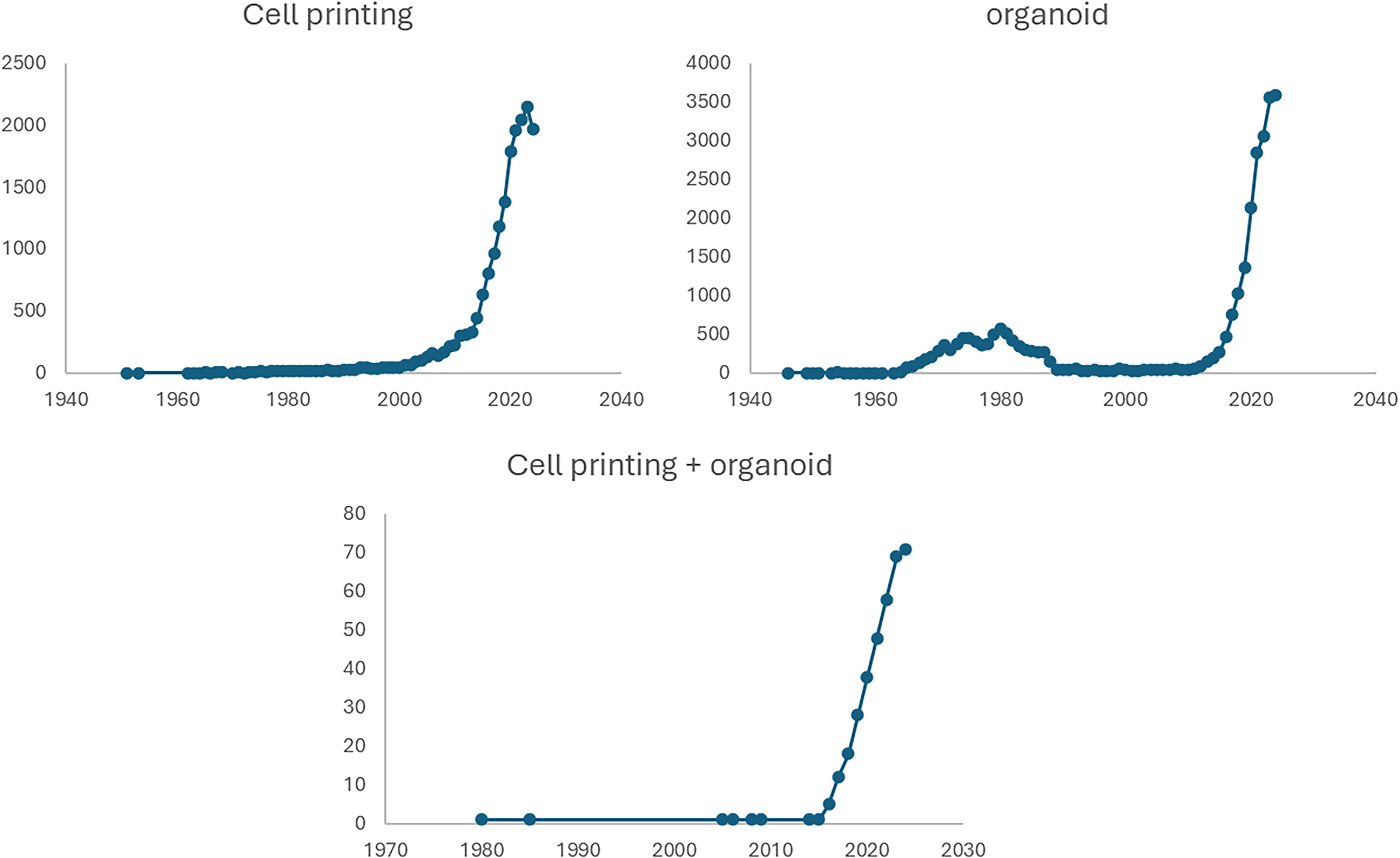
Graphs show the number of publications related to “cell printing” and/or “organoid” from the 1950s to October 2024. The numbers were acquired from PubMed by searching with specific keywords.
In the present review, we summarize recent progress in the utilization of cell printing techniques in organoid biology and discuss the advantages and limitations of this technology in controlling 3D microenvironments and reestablishing tissue structures and functions.
Applications of Cell Printing in Organoid Engineering
Features of current cell printing technologies
Bioprinting has the potential to enhance process stability, reproducibility, and precise architecture design of various tissue organoids. Recently, the application of bioprinting has demonstrated its enormous advantages in various organoid development scenarios based on different molding principles, which are classified into extrusion bioprinting, inkjet-based bioprinting, and laser-based bioprinting (Fig. 2). These diverse bioprinting technologies show specific advantages in terms of their potential applications and limitations in organoid model development (Table 1).
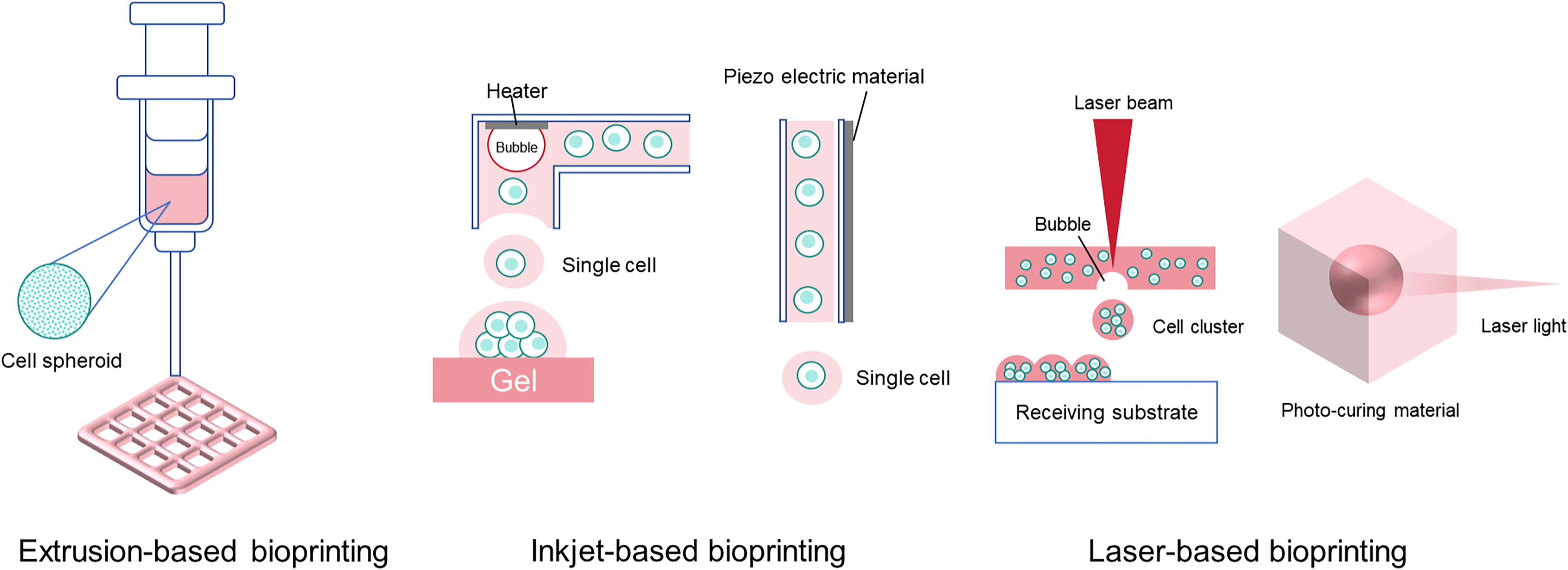
Cell printing can be classified into three types according to their molding principle: extrusion-based printing, inkjet-based printing, and laser-based printing.
Comparison of Different Bioprinting Techniques Focusing on Organoid Construction
3D, three-dimensional.
Extrusion bioprinting
Extrusion bioprinting has a robust ability to create 3D structures with biocompatible materials and has therefore been broadly utilized for in vitro organoid development. 33 One advantage of this technology is that printing 3D tissues with a high density of cells or organoids suspended in bioink allows for large-scale tissue construction. Extrusion-based bioprinting is applicable to a variety of bioink materials with high viscosity, such as hydrogels and decellularized extracellular matrices, for building complex 3D scaffolds to form large-scale tissues composed of organoids. With a coaxial nozzle design, independent solid or hollow tubular structures can be printed for the fabrication of vascular, intestinal, and urinary tract tissues. 34 The ability to fabricate in vivo-like tubular and hollow spheroid structures highlights the potential of extrusion bioprinting in tissue engineering and regenerative medicine.
The high viscosity of the bioink is well tolerated by this bioprinting method. However, extrusion bioprinting displays a low precision of spatial deposition compared with other bioprinting techniques, limiting the precise operation of micron volumes of bioinks or cell suspensions for even more intricate requirements. On the other hand, cells buried in high-viscosity bioinks accumulate into well-defined 3D structures, which can result in low viability of printed cells owing to the inescapable shear stress added to cells by the viscous bioinks.35,36
Inkjet-based bioprinting
Inkjet-based bioprinting can be classified into two types according to the principle of actuation: thermal inkjet printing and piezoelectric inkjet printing. In thermal inkjet printing, small bubbles are generated in milliseconds by momentary heating from a microheater, jetting a small volume of liquid in the picoliter out of the nozzles. In piezoelectric inkjet printing, the actuation power of liquid jetting is a narrow, long channel made of piezoelectric materials that vibrates to squeeze out a small volume of liquid from the nozzles while applying electricity. Both generate extremely small volumes of liquid droplets enveloping a single cell or a few cells that are precisely deposited at accurate positions, enabling high-precision and high-resolution cell printing. 37
One advantage of inkjet bioprinting is its high-throughput potential. Thermal inkjet bioprinting features the microstructures of inkjet printing chips. Usually, hundreds of nozzles are made on the chips, and the nozzles are available to eject simultaneously, which allows the chip to print hundreds of droplets enveloping cells simultaneously. Another characteristic of inkjet bioprinting is its lower driving force, which results in less stress on the cells and higher cell viability during the printing process. However, this same feature also limits the tolerance for bioink viscosity, making it challenging to print bioinks with high viscosity, such as parts of commonly used hydrogel materials38,39 in extrusion bioprinting. Although researchers have developed printable bioinks of hydrogels with low viscosity for inkjet bioprinting and some printable materials for inkjets have been verified,37,38 inkjet bioprinting has a lower capability for manufacturing large 3D structures than extrusion bioprinting.
Laser-based bioprinting
Laser-based bioprinting includes laser-induced photopolymerization, laser direct-write, and laser-induced transfer,12,29 which utilize laser power in two different ways: polymerization or cell transfer. Laser-induced photopolymerization, also known as stereolithography, applies 3D molding for light-curing using laser light, primarily in applications requiring high-precision patterning of cells or high-resolution tissue construction. The photopolymerization method provides a well-defined printed 3D structure; however, the structure often lacks biomimicry and has limited cell density. Additionally, the choice of photocurable materials used as cell scaffolds is restricted due to biocompatibility concerns.
For laser-induced cell transfer, a high-pressure bubble is generated at the interface between the laser-sensitive donor layer and the bioink layer, actuating a droplet of the cell-suspended bioink to the receiving substrate for cross-linking. One advantage of this method is that it prevents the bioink from contacting the dispenser directly, thereby reducing the risk of cell contamination. The viability of printed cells is relatively high due to the low stress generated by the noncontact printing process, and it is also suitable for printing bioinks with varying viscosities. However, the system is generally complex and requires high-resolution, high-intensity laser diodes to achieve efficient bioprinting, making it more expensive than other nozzle-based bioprinting methods. 29 Therefore, this technique has not been widely adopted for organoid models.
Other technique approaches
The Kenzan bioprinting method is scaffold-free 3D bioprinting, which sticks cell spheroids into a set of microneedles, called Kenzan. 13 When cells can support their structure, the needles are retracted, and the cells are nourished and grown into viable tissue. This method uses a specialized 3D bioprinter to precisely position cell spheroids in a microarray, thereby allowing cell clustering and growth.
Application of extrusion bioprinting in organoid cultures
Extrusion bioprinting has been widely utilized in organoid construction and tissue engineering because of its capability for spatially controlled deposition of cells with various biomaterials (Table 2). For example, human liver-derived epithelial organoids have been developed for toxicity studies using extrusion bioprinting. 23 These bioprinted organoids maintained high cell viability for up to 10 days and demonstrated increased expression of hepatic markers, transporters, and enzymes compared with undifferentiated controls. 17 Hong et al. presented a method for producing hepatic lobule-like microtissue spheroids using an extrusion bioprinting system that combines a precursor cartridge and a microfluidic emulsification system. 41 This method generates multiple cell-laden microtissue spheroids with a uniform diameter, exhibiting the biomimetic structure of a liver lobule with patterned hepatic and endothelial cells. The structured spheroids showed improved protein secretion, enzyme expression, and structural integrity compared with nonstructured spheroids.
Comparison of Different Bioprinted 3D Organoid and Tissue Models
ICOs, intrahepatic cholangiocyte organoids; GelMA, gelatin-based hydrogel; hPSCs, human pluripotent stem cell; HSC, human hepatic stellate cell; HUVEC, human umbilical vein endothelial cell; iPSC, human induced pluripotent stem cell.
One challenge for organoid model development is to reestablish the in vivo functions and tissue-tissue interactions of physiological organs. This is largely due to the lack of cell-type diversity and functional cells, which are typically achieved through cells with cellular pluripotency and stemness. To enhance functional emulation, syringe-based extrusion bioprinting using stem cell-derived organoids as building blocks is employed to create large-scale tissues with intricate microarchitecture, cell-type diversity, and in vivo-like functionality. 3 The organoids derived from printed cells self-organize into complex tissues, including vascular and intestinal epithelial structures with crypts and villi. 10
Extrusion-based 3D bioprinting and human pluripotent stem cells (hPSCs) have been utilized to generate kidney organoids. This approach enables rapid, high-throughput, and highly reproducible production of kidney organoids that resemble the morphology, cell composition, and gene expression of those generated manually. 24 In another study, nephron progenitor cells derived from human induced pluripotent stem cells (iPSCs) were utilized to generate kidney organoids in the 3D bioprinting system. 14 The organoids expressed markers for the major kidney cell types and contained nephron-like structures.
Extrusion bioprinting has been applied to tumor organoid development and tumor environment research. A 3D “mini-brain” model developed with extrusion printing recapitulates the interactions between glioblastoma (GBM) cells and macrophages. This model demonstrates that GBM cells actively recruit and polarize macrophages into a GBM-associated macrophage phenotype, and these macrophages, in turn, induce GBM cell progression and invasiveness. 13 An extrusion printer equipped with a switchable dual-nozzle module integrates extrusion printing with alternating viscous and inertial force-jetting techniques. This equipment was used to generate a heterogeneous tumor model containing spheroids and human umbilical vein endothelial cells (HUVECs), which maintained high cell viability and sustained growth during the culture period. In addition, lung cancer organoid arrays have been developed with a multichannel extrusion printer for anticancer drug evaluation. Hundreds of lung cancer organoids with 3D multicellular spherical structures were produced through batch culture in the hydrogel scaffolds, with higher cell activity, functional gene expression, and increased drug resistance. 30 These studies indicate that extrusion bioprinting has great potential for the development of normal and tumor organoid models.
Applications of inkjet-assisted cell printing in organoid culture
The accumulated evidence shows that thermal inkjet printing can be applied in the high-throughput preparation of size-controllable cell spheroids (Fig. 3). Zhang et al. developed and verified the capability of cell spheroid constructs in uniform size with an inkjet-based bioprinting system. 42 This system was further utilized to print cell clusters to generate mouse intestinal organoid arrays as an in vitro model for drug screening. 43
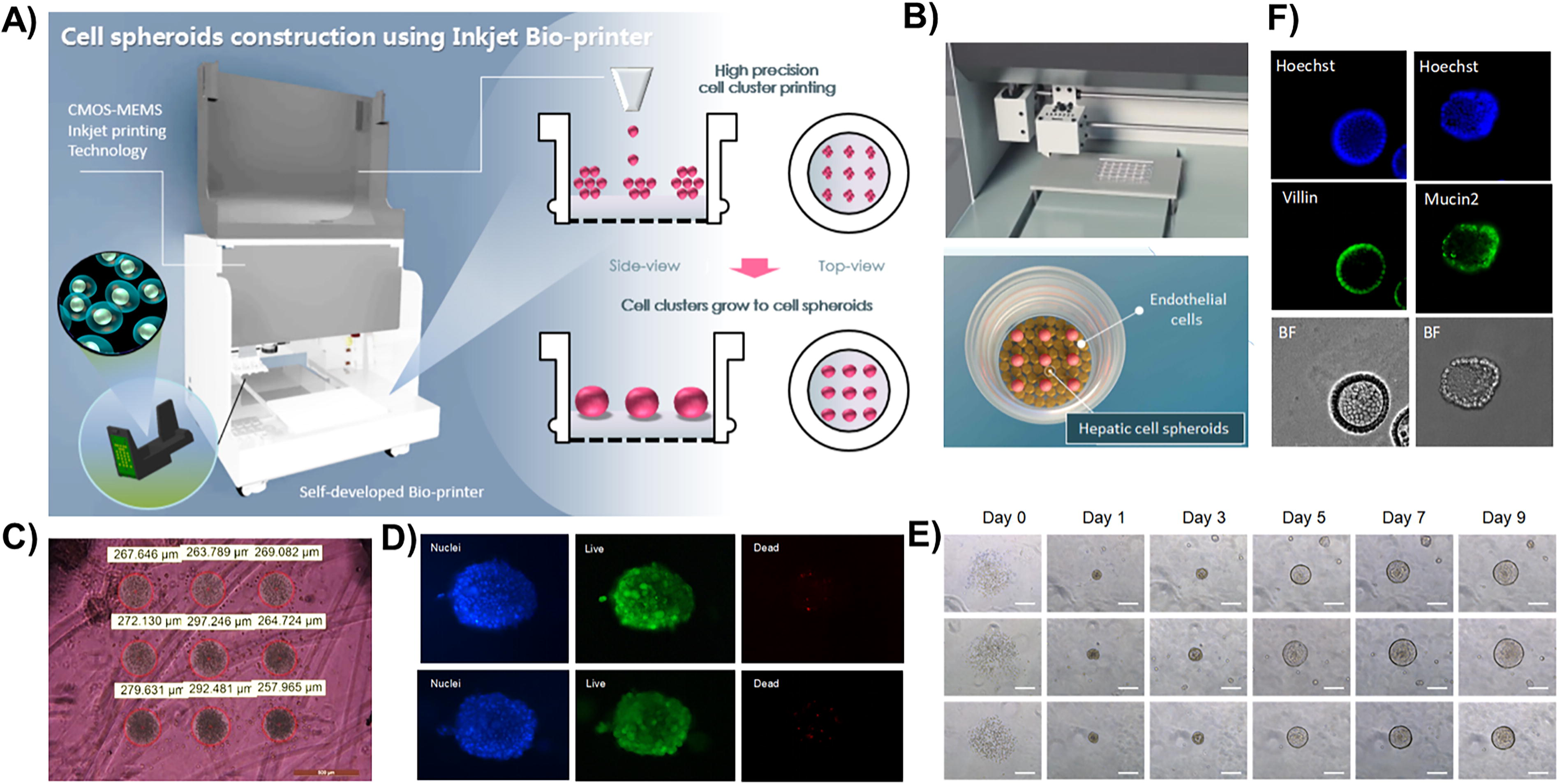
Application of inkjet-based cell printing in organoid tissue engineering.
Another application of inkjet bioprinting technology is the development of the 3D pulmonary fibrosis model. This model was created by sequentially printing lung microvascular endothelial cells, collagen type I, lung fibroblasts, and alveolar epithelial cells to form a multilayered alveolar barrier, thereby demonstrating the potential of this 3D alveolar barrier model for antifibrotic drug discovery. 44 Inkjet printing has also been applied to quantify intratumoral heterogeneity in bladder cancer. 45 In this study, patient-derived tumor organoids were dissociated into single cells and precisely printed into microwells, allowing the formation of individual organoids. 45 The organoids exhibited heterogeneous growth patterns, proliferation, and responses to chemotherapy, proving that inkjet printing is an effective tool for analyzing intratumoral heterogeneity at the single-cell level.
Application of laser-assisted cell printing
Light-driven volumetric bioprinting is a layerless printing approach capable of printing positive and negative features (channels) at high resolutions, and it has been used to generate functional liver organoids. 17 Human liver epithelial organoids were printed on a centimeter scale with designed architectures that facilitate access to metabolites with this technology. The organoids were able to undergo hepatocytic differentiation with liver detoxification function. 17 Hall et al. presented a laser-assisted bioprinting approach for transferring multicellular cartilaginous spheroids as building blocks for larger tissue structures. They successfully printed cartilaginous spheroids formed by human periosteum-derived cells (hPDCs) with high viability and the capacity for chondrogenic differentiation postprinting. 46 The laser-assisted bioprinting technology has also been utilized to generate the pancreatic spheroid array model. 40 This model aims to replicate the initial stages of pancreatic ductal adenocarcinoma development, which involves the transdifferentiation of exocrine pancreatic acinar cells into duct-like cells.
Advantages of Cell Printing in Organoid Engineering
Improved spatial control
3D cell printing can precisely place individual cells/aggregates and improve the control over the layout of tissue structure (Table 3). Inkjet printing technology achieves precise control of the placement of cells to better replicate the natural structural arrangement of tissues with high cell viability owing to the low shear stress of the printing process. By utilizing bioprinting technology to deposit stem cells, differentiated cells, or cell spheroids (such as organoids) into biomaterials, this approach facilitates cell self-assembly and tissue regeneration. It achieves precise spatial control over cell placement, mimics the embryonic development process, and promotes the formation of tissue organoids. This method enables the high-throughput, time-efficient, and automated construction of cell spheroids and organoids with exceptional precision and uniform size.42,43
Advantages of Using Cell Printing in Organoid Tissue Engineering
Laser-assisted bioprinting utilizes laser pulses to accurately deposit bioink onto a substrate, allowing precise deposition of bioink at the target location, achieving a resolution of 10–100 microns. This technology enables the fabrication of complex tissue structures, as well as the precise placement of single cells and cell aggregates. Unlike inkjet printing, laser-assisted bioprinting is suitable for bioinks with high cell density and viscosity, making it ideal for constructing large-scale tissue scaffolds with high complexity.34,47
Integrating different cell/tissue types to form functional organoids
By directly printing cell spheroids or organoids, researchers have verified the ability of centimeter-scale tissue constructs composed of different tissue cells in an arrangement built by an extrusion printer. 10 A recent study demonstrates a platform called spatially patterned organoids transfer, which employed iron-oxide nanoparticle-laden hydrogel and magnetized 3D bioprinter. Using this technology, they successfully constructed organoids composed of human iPSC-derived neural organoids and patient-derived glioma organoids. 27 Daly et al. developed cardiac microtissue models with spatially printed cardiomyocytes and fibroblasts to mimic the structural and functional features of scarred cardiac tissue. 28 In addition, an acoustic droplet printing technology has been developed, which can print multiple cells in a precise arrangement, constraining cell growth, interactions, and functions for eventually constructing an in vitro model consisting of tumor spheroid with microenvironment. 48 These studies highlight the potential of cell printing technology in constructing organoids and tissue models composed of multiple cell types.
Integration with microfluidics to generate organ-on-a-chip
Microfluidic technology benefits from laminar flow in microscale fluid dynamics, enabling more predictable and controllable fluid behavior (Fig. 4). The integration of microfluidic technology and bioprinting has brought new possibilities to the fields of tissue engineering and regenerative medicine. 49 It has been shown that integrating microfluidic systems with extrusion-based bioprinting enhances the structural and compositional properties of bioprinted tissue constructs. This integration allows for the high-resolution fabrication of 3D constructs, including tubular and vascularized structures, promoting the survival and integration of engineered tissues. 9 By combining microfluidic technology with droplet-based bioprinting technology, it is possible to achieve precise control and positioning of individual cells, thereby facilitating bioanalysis and high-throughput screening.19,20,49
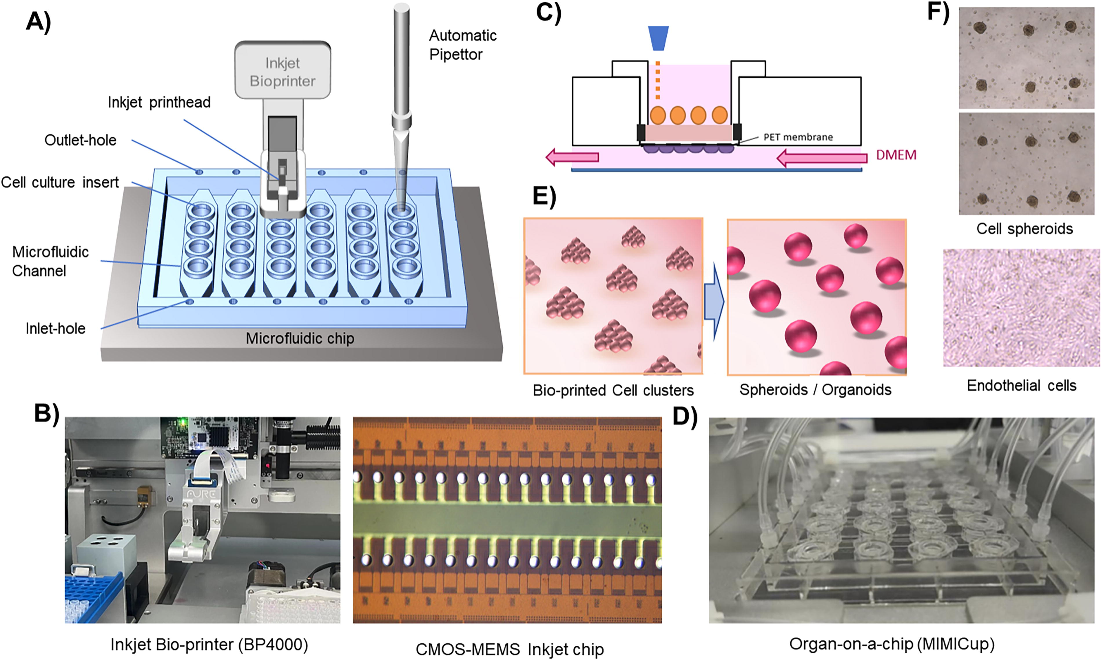
Integration of 3D bioprinting with other technologies.
Microfluidic systems and perfusion chambers have been integrated to achieve better emulations of human physiological conditions, such as continuous shear stress microenvironment in vessels, metastasis of tumor cells through different tissues, endothelial or epithelial barriers, and organ-to-organ interactions, which are commonly called organ-on-a-chip technology. 21 3D bioprinting, as a complementary technique, enables the precise deposition of cells or cell-laden matrices to build biological structures in a high-throughput, automated manner. 22
Microfluidic flow cytometric printing (μFCP) has been used to fabricate precise liver spheroids with controlled composition and uniform structure and function, 18 which can overcome the limitations of random aggregation methods that yield spheroids with variable sizes, functions, and utilities. This platform allows the systematic tuning of spheroid composition and high-throughput manufacturing of thousands of precision spheroids per hour, with improved in vitro modeling and drug screening for liver fibrosis. A combination of bioprinting techniques with a microsensor platform was developed to enable the automated deposition of single cancer cell spheroids into oxygen sensor microelectrode wells. 50 Cellular respiration rates and metabolism can be monitored in a real-time manner in this system. The combination of microfluidic devices with cell printing creates a new generation of organ-on-a-chip platforms.
Multiple organ-on-a-chip models have been developed to achieve better emulations of human organs in vitro. Extrusion bioprinting has been combined with organ-on-a-chip technology to develop GBM-on-a-chip. Using multiple channels of extrusion syringe printing and the microfluidic chip, scientists successfully reconstituted the GBM tumor model consisting of patient-derived tumor cells, endothelial cells, and decellularized ECM. 16 An airway-on-a-chip has been constructed through a similar approach in which the microchannels and 3D cell-laden structures were printed together to build the entire system. 25 3D inkjet bioprinting technology has also been utilized to develop a physiologically relevant human alveolar lung-on-a-chip model. The model integrates an inkjet-printed, micron-thick, and three-layered lung tissue within a microfluidic device that enables perfusion culture at the air–liquid interface. The bioprinted lung tissue maintained its multilayered structure and formed a tight epithelial barrier, which is a key property of the alveolar barrier. 51 Tian T. et al. utilized an inkjet-based bioprinting technique to construct liver-on-a-chip by coculturing HepG2 spheroids and human endothelial cells (HUVECs) with continuous flow. This device was used to evaluate the hepatic toxicity of acetaminophen, validating the feasibility of the bioprinting-assisted organ-on-a-chip platform for the preclinical testing of drug toxicity. 52
Cell Printing Technology in Biomedical Research
Drug screening
Organoid models are considered a powerful tool for drug screening because they represent in vivo-like tissue structure/function and mimic the progression of various diseases. However, reproducibility and quantification in the conventional organoid models remain significant challenges for high-throughput drug screening.
One feature of 3D bioprinting is its ability to generate large numbers of homogeneous organoids, overcoming the limitations of conventional organoid models in drug screening. A drop-on-demand 3D bioprinter has been utilized to generate breast cancer organoids in 96-well plates using patient-derived breast cancer cells. 53 Breast cancer organoids in this model responded to doxorubicin, EP31670, and paclitaxel treatments, with increased IC50 concentrations compared with 2D cultures.
Patient-derived GBM-on-a-chip was generated with extrusion bioprinting and GBM tumors from patients. 16 This model combined GBM cells, vascular endothelial cells, and decellularized ECM in a concentric-ring structure, successfully mimicking GBM’s pathological features of the cancer, including hypoxia-induced necrotic core, pseudopalisading formation, spatial heterogeneity of cell types, and the perivascular niche. Most importantly, this model accurately replicates patient-specific resistances to concurrent chemoradiation and temozolomide observed in clinical settings. Therefore, patient-derived tumor-on-a-chip shows great potential to identify effective treatments for patients with cancer, leading to personalized and potent cancer treatment strategies.
Toxicology studies
3D-printed organoids can be used for toxicity testing. The high-throughput feature of 3D printing allows for the simultaneous testing of multiple compounds, accelerating the process of toxicity assessment. In addition, 3D printing technology can produce homogeneous organoids, reducing the variability that may occur with traditional culture methods and thereby enhancing the reproducibility of experimental results. A high-throughput model of human liver organoids has been generated with droplet-based 3D bioprinting. 54 In this model, foregut cells were mixed with Matrigel and printed onto a pillar plate, resulting in numerous organoids with consistent morphology and function, including albumin secretion and CYP3A4 activity. The organoids showed similar responses to sorafenib and tamoxifen as in traditional Matrigel dome cultures.
3D-printed organoids can simulate the complex structure and function of human organs, including cell–cell interactions and the microenvironment at the tissue level, which is crucial for toxicity testing. For example, progenitor cells derived from the biliary tree can be cultured as organoids and differentiated into the hepatocytic lineage, acquiring mature hepatocyte functions including albumin and bile acid secretion, glycogen storage, phase I and II drug metabolism, and ammonia detoxification. Bouwmeester et al. 23 fabricated human liver-derived epithelial organoids with extrusion-based bioprinting. These bioprinted constructs enhanced the complexity of the organoids and provided a more physiologically relevant microenvironment, as evidenced by increased expression of hepatic markers, transporters, and phase I enzymes compared with nonprinted controls. Lawlor et al. used 3D bioprinting to produce kidney organoids with consistent cell numbers and viability. 24 iPSC-derived kidney progenitors were printed onto Transwell filters, forming nephrons with in vivo-like structure and function and high reproducibility. These nephrons were used for nephrotoxicity testing, showing decreased viability after exposure to nephrotoxic compounds like doxorubicin, amikacin, tobramycin, gentamicin, neomycin, and streptomycin. These studies demonstrate the great potential of 3D-printed organoids in the field of toxicity testing.
Clinical applications
A variety of preclinical models have been developed with patient-derived tumor organoids for cancer research and personalized medicine. 55 3D bioprinting technology has been employed to reconstruct in vitro hepatocellular carcinoma models for antitumor drug screening. These 3D-bioprinted tumor models revealed significant differences in drug resistance gene expression profiles compared with conventional models. 56 Furthermore, a patient-derived 3D-bioprinted HCC model has been successfully developed for personalized drug screening using primary HCC cells, demonstrating the clinical feasibility of patient-specific drug testing for individualized treatment. 57 Importantly, several active clinical trials are currently recruiting participants to assess the potential of 3D bioprinting and organoid technologies. These trials include studies focused on gastric cancer (NCT06792149), pancreatic cancer (NCT05955092), and hematological malignancies (NCT03890614).
Conclusion, Future Directions, and Challenges
The development of high-throughput and reproducible organoid models is crucial for drug screening and personalized treatment testing. Cell printing has emerged as a powerful tool for developing high-throughput and reproducible organoid models. By overcoming the limitations of traditional organoid models, such as variability and challenges in reproducibility, cell bioprinting can generate organoids with more uniform structures. The innovative use of cell printing in generating organoids, such as liver, kidney, and cancer models, highlights its potential to advance biomedical research and improve the accuracy and reproducibility of preclinical testing.
The future direction of cell printing lies in the integration of advanced technologies such as microfluidics, biosensors, and personalized tissue engineering to better mimic the in vivo tissue structure and function. Microfluidic systems can be incorporated into 3D-printed organoid platforms to precisely control the flow of nutrients, oxygen, and waste, simulating the dynamic conditions of living tissues. This will enable the creation of more physiologically relevant models, thereby enhancing their utility in drug screening and disease modeling. Additionally, the incorporation of biosensors can provide real-time monitoring of the organoid environments, further improving the analysis of their functionality and drug response. Another future direction for cell printing is personalized tissue engineering, which allows for the customization of organoid models using patient-specific cells. This approach will enable the development of organoids that accurately reflect individual genetic and disease profiles, which is critical for personalized medicine. 4D printing represents an advanced evolution of 3D printing technology, introducing “time” as a fourth dimension to the conventional 3D space.58B59 -61 This enables printed objects to undergo autonomous shape morphing or functional evolution over time in response to predefined environmental stimuli.
Despite significant advancements in bioprinting technologies, several key challenges persist in both research and industrial applications. In scientific research, a primary challenge is selecting biocompatible materials that can form desired 3D cellular architectures while ensuring high cell viability postprinting. Moreover, the lack of standardized technical protocols and regulatory guidelines remains a substantial barrier, particularly for clinically relevant applications such as drug screening with bioprinted patient-derived organoids and the transplantation of 3D-bioprinted artificial tissues. Overcoming these hurdles will pave the way for next-generation organoid models that more accurately replicate human tissues, enhancing the predictive accuracy of preclinical studies and unlocking new possibilities for tissue regeneration and precision medicine.
Footnotes
Authors’ Contributions
Y.H.H. and Y.L. collected the data and wrote the article. L.L. and T.M. wrote the article. Y.G. conceived the idea and revised the article. R.X. conceived the idea and wrote the article.
Disclosure Statement
The authors wish to declare the following potential conflicts of interest: Y.L., L.L., T.M., and Y.G. are employees of Shanghai Aurefluidics Technology Co., Ltd. This company had a role in article preparation. All other authors declare no competing interests.
Funding Information
No funding support.