
Abstract
Effective wound healing hinges on a precisely orchestrated tissue remodeling process that restores both structural integrity and functionality. This review delineates the molecular mechanisms by which chitosan-based hydrogels revolutionize wound repair. Derived from natural chitin, chitosan uniquely combines robust antimicrobial, hemostatic, and biodegradable properties with the capacity to modulate critical intracellular signaling cascades—including transforming growth factor-β, mitogen-activated protein kinase, and PI3K/AKT. These dynamic interactions drive fibroblast proliferation, stimulate the strategic transition from type III to type I collagen deposition, and finely tune extracellular matrix reorganization, thereby mitigating excessive fibrosis and minimizing scar formation. Notwithstanding its considerable therapeutic promise, clinical translation of chitosan-based hydrogels is tempered by challenges in mechanical stability and controlled degradation. We propose that advanced material engineering—encompassing precision cross-linking, nanoparticle integration, and synergistic stem cell-based strategies—could surmount these limitations. This comprehensive synthesis of current molecular insights sets the stage for next-generation regenerative biomaterials, positioning chitosan-based hydrogels as a paradigm-shifting platform for achieving superior healing outcomes in complex clinical scenarios.
Impact Statement
This review presents an in-depth discussion on the dual bioactivities of chitosan—not only as a scaffold providing mechanical support and controlled bioactive molecule release but also as a modulator of fibroblast activation and collagen deposition. By integrating recent advances in material engineering, such as precision crosslinking and nanoparticle integration, our review offers a balanced perspective on both the therapeutic potential and current challenges of these biomaterials. We believe that this work will provide valuable insights to researchers and clinicians in the fields of regenerative medicine, tissue engineering, and wound care.
Introduction
Wound healing is a multifaceted, tightly regulated process involving a cascade of events—hemostasis, inflammation, proliferation, and remodeling—that collectively restore tissue integrity and functionality. 1 In this continuum, the remodeling phase is paramount, as it governs the reorganization of the extracellular matrix (ECM), the maturation and alignment of collagen fibers, and the resolution of inflammation. 2 Even subtle perturbations during this phase can lead to chronic wounds, excessive fibrosis, or impaired tissue function. 3 Despite considerable progress in dissecting individual components of the healing cascade, clinical strategies have yet to fully recapitulate the sophisticated interplay between cellular dynamics and molecular signals required for truly optimal tissue regeneration. 4
Recent advances in biomaterial science have spotlighted chitosan-based hydrogels as transformative platforms for wound management.5,6 Chitosan, a naturally derived polymer from chitin, is endowed with unique properties—biodegradability, intrinsic antimicrobial activity, and robust hemostatic effects—that collectively enhance its clinical potential.7,8 More importantly, its ability to modulate key intracellular signaling pathways, such as transforming growth factor-β (TGF-β), mitogen-activated protein kinase (MAPK), and PI3K/AKT, underscores its role in directing fibroblast activation, regulating collagen synthesis, and orchestrating ECM remodeling.9,10 The inherent physicochemical characteristics of chitosan enable precise interactions with growth factors (GFs) and cytokines, facilitating a sustained, controlled release that can markedly improve tissue repair dynamics. 11
Nonetheless, existing studies often evaluate the benefits of chitosan’s properties in isolation, without addressing the synergistic mechanisms that underlie its full therapeutic potential during the remodeling phase. 12 There exists a critical gap in understanding how fine-tuning the mechanical stability, porosity, and bioactive molecule release of chitosan-based hydrogels can create an optimal microenvironment for cellular adhesion, proliferation, and differentiation. 13 Achieving an ideal balance is essential for promoting orderly collagen deposition and effective tissue regeneration, yet remains a significant challenge in the field. 14
In this review, we aim to bridge these knowledge gaps by synthesizing current evidence on the molecular regulation of tissue remodeling mediated by chitosan-based hydrogels. We will explore the intricate biochemical interactions between chitosan and ECM components, detailing how these interactions promote the precise deposition and structural organization of collagen fibers. 15 Furthermore, we will critically assess how chitosan’s modulation of cellular signaling cascades not only accelerates the wound repair process but also attenuates aberrant inflammatory responses that can compromise healing outcomes. By integrating molecular insights with advanced material engineering, such as optimizing cross-linking density and incorporating nanoparticle technologies, we propose strategies to design hydrogels that deliver sustained, high-efficacy therapeutic benefits in complex wound environments. 16
Ultimately, this review seeks to provide an authoritative, evidence-based perspective that not only highlights the innovative potential of chitosan-based hydrogels in regenerative medicine but also sets a new benchmark for future research and clinical translation in wound care and tissue engineering.
Overview of Wound Healing Phases
The wound healing process comprises four interconnected yet distinct stages: hemostasis, inflammation, proliferation, and dermal remodeling as shown in Figure 1, each playing a crucial role in tissue recovery. 17 Yasser H. Almadani et al. emphasized that these phases, while overlapping in time, are biologically different, underlining the complexity of the healing process (2021). 1 Deficiencies in any of these phases can hinder wound healing, preventing the restoration of both physiological function and tissue architecture. 17 The tissue repair mechanisms use a range of cell types, enzymes, cytokines, proteins, and hormones to support the recovery of tissue integrity and to prevent excessive blood or fluid loss and infection development.18,19
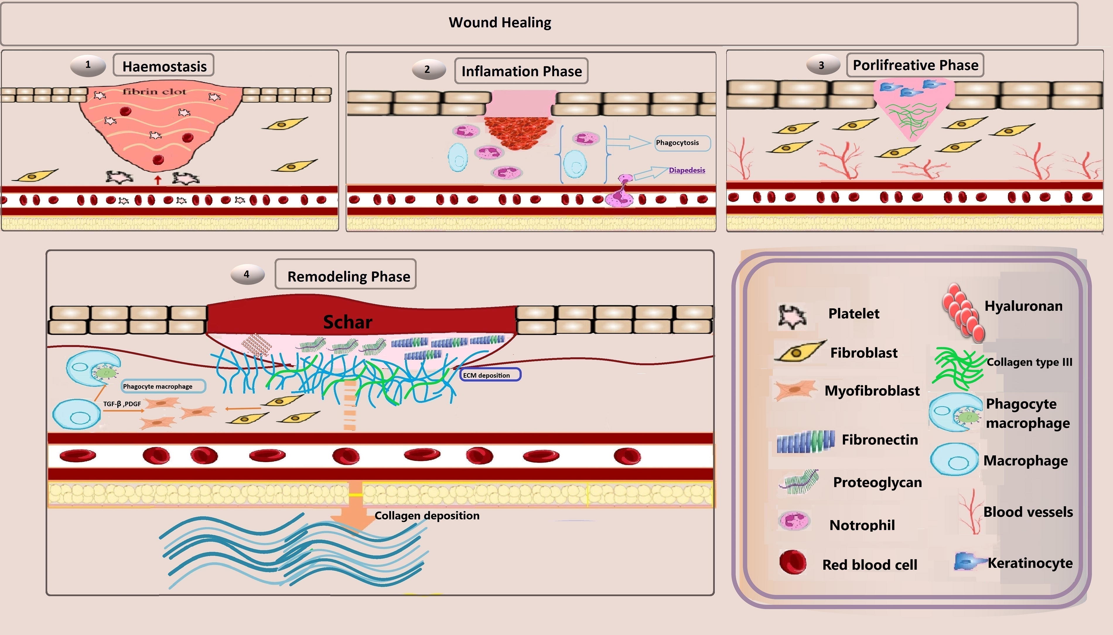
Schematic diagram of the four physiological wound healing stages. (1) Hemostasis: Platelet aggregation forms a fibrin clot. (2) Inflammatory phase: Immune cells undergo diapedesis; monocytes differentiate into macrophages with phagocytic activities. (3) Proliferative phase: Keratinocytes migrate to aid epidermal regeneration, while fibroblasts produce type III collagen, and angiogenesis occurs. (4) Remodeling phase: Type III collagen is replaced by type I, ECM is deposited, and the fibrin clot converts into a matrix rich in fibronectin, proteoglycans, and hyaluronan, supporting collagen deposition. Macrophages transition into a phagocytic phenotype, promoting fibroblast differentiation into myofibroblasts via TGF-β secretion. ECM, extracellular matrix
Hemostasis
Hemostasis, the initial phase of wound healing, is triggered by the exposure of endothelial ECM collagen following injury, a process that spans from seconds to hours. 20 Platelets bind to ECM proteins such as collagen and Von Willebrand factor through Glycoprotein (GP) VI and related receptors. This binding initiates the release of bioactive molecules, facilitating coagulation and forming a provisional fibrin matrix. The formation of an insoluble clot (eschar), composed of fibrin, fibronectin, vitronectin, and thrombospondin, effectively seals the wound and halts bleeding. Additionally, platelets release chemokines and GFs such as TGF-β and platelet-derived growth factor (PDGF), stimulating resident skin cells and attracting immune cells to the injury site.1,17
Inflammatory phase
The inflammatory phase, initiated 24–48 h postinjury, is driven by the coordinated actions of resident and recruited cells from both the innate and adaptive immune systems, marking a critical step in wound healing. 21 Platelet activation during hemostasis triggers the release of cytokines such as TGF-β and PDGF, which are pivotal in recruiting neutrophils and macrophages to the wound site. 1 Activated neutrophils combat pathogens through antimicrobial peptides and reactive oxygen species (ROS) before undergoing apoptosis, a process that macrophages regulate by phagocytosing apoptotic neutrophils. The clearance of neutrophils by macrophages stimulates the secretion of anti-inflammatory cytokines, including TGF-β and interleukin-10 (IL-10), paving the way for the transition from inflammation to tissue repair. 21
Proliferative phase
The proliferative phase involves the activation of keratinocytes, fibroblasts, macrophages, and endothelial cells, each orchestrating critical processes such as wound closure, ECM deposition, and angiogenesis. GFs and cytokines, activated within 12 h postinjury, stimulate keratinocyte migration and re-epithelialization, essential for the reformation of the epidermal barrier. Keratinocyte migration halts upon contact with opposing cells, a process regulated by contact inhibition signaling. 17 Within 2–3 days, fibroblasts migrate to the injury site, producing disorganized type III collagen in the provisional matrix. 1 They then break down this matrix using matrix metalloproteinases (MMPs), replacing it with granulation tissue rich in fibronectin and immature collagens.17,19 Angiogenesis is stimulated by the vascular endothelial growth factor (VEGF), leading to new blood vessel formation to support healing tissue. 17
Remodeling phase
The remodeling phase, the final stage of wound healing, is characterized by the wound contraction, reduced vascularity, and a decline in cellular density, signaling the transition to scar maturation. At this stage, ECM remodeling and tensile strength recovery transform granulation tissue into permanent scar tissue, a function primarily mediated by fibroblasts. Failures in this phase can worsen wound chronicity. Around 3 weeks postinjury, type III collagen in the ECM is gradually replaced by type I collagen over 6–12 months, enhancing tensile strength and tissue stability.18,19 In the absence of healing disruptions, tissue tensile strength can recover up to 85% of its original capacity as collagen remodeling, including cross-linking and fiber alignment, progresses over several months.19,20
Fibroblasts contribute significantly to ECM remodeling, converting the provisional fibrin clot into a complex matrix enriched with hyaluronan, fibronectin, and proteoglycans, which serve as scaffolds for the deposition and maturation of collagen fibrils. During the remodeling phase, fibroblasts undergo specialization into myofibroblasts, which are imperative for wound contraction and ECM remodeling through the synthesis of α-smooth muscle actin (α-SMA) and enhanced mechanical tension generation. Myofibroblasts mediate wound contracture by forming cell-cell connections through desmosomes, which enhance ECM rigidity and stabilize their presence within the wound environment. Myofibroblasts orchestrate the balance of ECM protein synthesis, promoting a shift from type III to type I collagen. This shift strengthens ECM tensile properties while optimizing its structural complexity for durable stability. 22 Macrophages partially revert to a phagocytic phenotype during this phase, adopting a “fibrolytic profile” defined by the secretion of arginase, metalloproteases, and the anti-inflammatory cytokine IL-10, which facilitate ECM remodeling. Through the exudation of TGF-β and PDGF-CC, macrophages promote fibroblast phenotypic shift into myofibroblasts, driving collagen deposition and modulating scar formation to achieve functional tissue repair. 21
Chitosan Properties Relevant to Wound Healing
Chitosan, a biopolymer derived from chitin, is extensively utilized in pharmaceutical and biomedical applications owing to its unparalleled chemical properties, biocompatibility, and powerful antibacterial activity. A 2021 study by Chih-Hsin Wang et al. highlighted chitosan’s antimicrobial effects in wound healing, demonstrating its ability to inhibit pathogenic bacteria while supporting beneficial microbial populations at the affected area. Notably, useful probiotics such as Prevotella, Lactobacillus, and Oscillibacter persisted in wounds treated with chitosan dressings for up to 6 days postsurgery, a feature absent in gauze-treated wounds. 23 Compared with conventional wound dressings, chitosan offers superior flexibility, water retention, exudate absorption, and cooling effects. These qualities make it an optimal choice for combination with other biopolymers to enhance mechanical strength and clinical performance.24-26
Biocompatibility
Minimizing complications in wound healing requires preventing adverse reactions while reinforcing the body’s natural repair mechanisms. This underscores the critical importance of physiological compatibility in wound dressings to aid effective healing. 27 Researchers have assessed the biomedical compatibility of chitosan dressings by measuring cytolysis in WS1 fibroblast cells at 24 and 48 h, finding minimal cytotoxicity comparable to control treatments. Chitosan dressings significantly inhibited cytolysis, exhibiting tissue-friendly properties comparable to Dulbecco’s modified Eagle medium and phosphate-buffered saline controls, which highlights their suitability for clinical-biological utilizations. 23 In its nanoparticle form, chitosan enhances targeted therapies by enabling precise cell targeting and ligand binding, making it ideal for advanced health-science applications. Chitosan with a low molecular weight is readily excreted, whereas higher molecular weights necessitate enzymatic degradation, a feature that allows for tailored bioscientific utilizations. When combined with zinc oxide, an eco-friendly biocide, chitosan exhibits heightened antimicrobial properties while reducing toxicity in aquatic ecosystems. 25
Biodegradability
Chitosan’s biodegradability makes it an ideal candidate for tissue engineering and regenerative medicine, as this biomaterial decomposes within the body into compounds that are harmless to tissues, minimizing long-term complications. 25 In tissue engineering, chitosan serves as a scaffold, gradually degrading within the body to provide structural support and stimulate the formation of new tissues. These properties not only mitigate long-term adverse effects but also improve the efficiency and outcomes of therapeutic interventions. 28 Chitosan’s structure is enzymatically degraded into nontoxic byproducts, with lysozyme being one of the essential enzymes involved in this step. This feature enables the body to efficiently eliminate degradation products, positioning chitosan as a prime candidate for various implementations in medical-life sciences.26,28,29
Research has demonstrated that the degree of acetylation (DA) and molecular weight significantly influence chitosan’s solubility, biodegradability, and biocompatibility, making them critical parameters for its implementations in biomedicine. The lower the DA, the higher the solubility and biocompatibility, while a lower molecular weight leads to quicker degradation. Altogether, chitosan presents a promising option for biodegradable packaging, as it decomposes over time in the environment. 29
Hemostatic properties
The initiation of hemostasis occurs through three coagulation pathways—extrinsic, intrinsic, and common. These pathways involve Factors III, VII, and calcium ions (Ca2+), which activate Factor X to promote clot formation. Chitosan enhances hemostatic efficacy via mechanisms separate from traditional coagulation pathways. It promotes platelet adhesion, aggregates erythrocytes, and inhibits fibrinolysis through interactions with platelet GPs, such as GP IIb-IIIa, and negatively charged molecules. By stabilizing clots, chitosan reinforces fibrin matrices, prolonging their stability. Its cationic nature facilitates erythrocyte aggregation, further enhancing clot stability while decreasing plasminogen activator release to limit fibrinolysis. These exceptional properties position chitosan as a promising hemostatic agent for wound care.30,31
Antimicrobial activity
Chitosan illustrates potent antimicrobial properties necessary for wound healing by effectively inhibiting bacteria and fungi. Its positively charged amino groups interact with microbial membranes, altering permeability and disrupting membrane integrity. This interaction causes intracellular leakage and subsequent cell death in Gram-positive bacteria (via teichoic acids) and Gram-negative bacteria (via lipopolysaccharides). Additionally, chitosan disrupts fungal membranes and binds to microbial DNA/RNA, inhibiting protein synthesis. Acting as a chelating agent, it binds to divalent cations on microbial surfaces, perturbing membrane stability and ultimately breaking down the cell membrane. These characteristics highlight its broad-spectrum antimicrobial efficacy, making it an essential component in advanced wound dressings.32,33
Structural properties (viscoelasticity, porosity)
Chitosan hydrogels exhibit sophisticated viscoelastic properties, which provide adaptive flexibility and mechanical support. Their porous structure eases exudate absorption, maintains optimal moisture levels, and enables gas exchange, creating an ideal microenvironment for cell migration and nutrient transport. 34 This porosity also enhances cell adhesion and proliferation, particularly in applications such as bone and cartilage repair. Reinforcing chitosan with additives such as mesoporous silica nanoparticles significantly improves the mechanical strength and durability of hydrogels, making them suitable for high-stress environments. 23 Analytical techniques such as Fourier Transform Infrared Spectroscopy and Scanning Electron Microscopy (SEM) reveal uniform structural surfaces and functional groups (C–O–C, C–N–H, O–H/N–H), which are crucial for oxygen exchange, fluid absorption, and hemostasis. These attributes position chitosan hydrogels as versatile biomaterials for wound dressing and tissue engineering. 35
Role of Chitosan in the Remodeling Phase of Wound Healing
Chitosan accelerates wound healing and reduces scarring during the remodeling phase by promoting fibroblast proliferation, collagen deposition, and increasing natural hyaluronic acid (HA) levels at the injury site (Fig. 2). 36 A study by Oliver Darwish et al. (2023) confirmed that in diabetic foot ulcers treated with chitosan-based hydrogels, inflammatory mediators such as IL-1β, tumor necrosis factor-alpha (TNF-α), and MMP-1 were downregulated, while VEGF, TGF-1β, and tissue inhibitor of metalloproteinase (TIMP-1) were upregulated. 20 This modulation enhances cell migration, ECM remodeling, and tissue regeneration. 22 The positively charged surface of chitosan adsorbs biological molecules, facilitating fibroblast growth, differentiation, and ECM synthesis, which supports effective healing and curtails fibrosis. 37
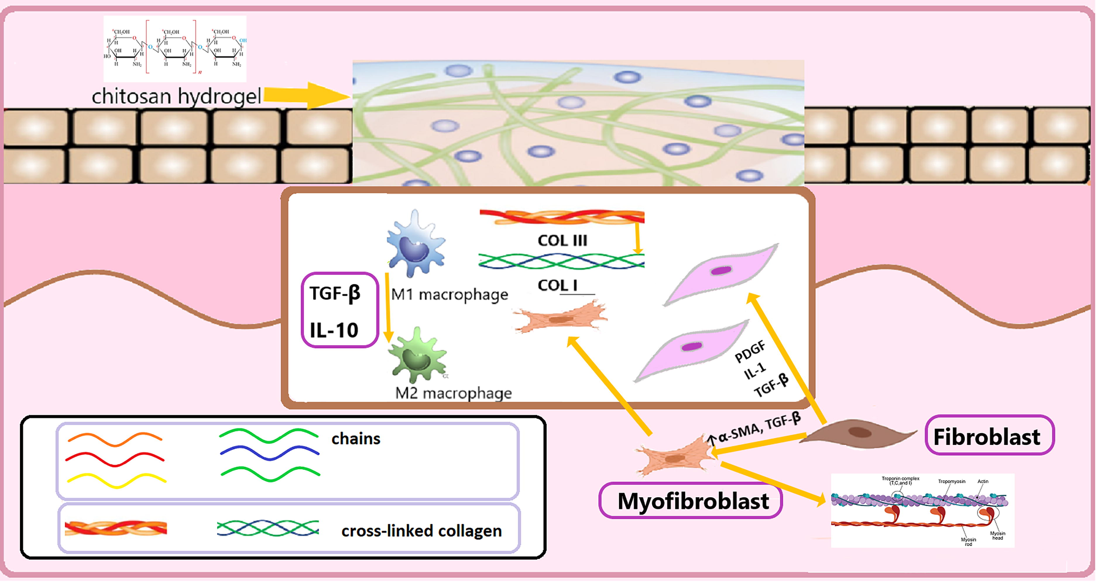
Overview of the effects of chitosan-based wound dressings on the remodeling phase of wound healing. Chitosan, composed of repeated N-acetylglucosamine units, promotes the conversion of collagen from type III to type I at the wound site and reinforces connective tissue through collagen cross-linking. Chitosan boosts α-SMA and TGF-β levels, enhancing fibroblast differentiation into myofibroblasts, characterized by actin and myosin bundles. Increased levels of TGF-β, PDGF, and IL-1 lead to fibroblast proliferation and collagen type I synthesis. TGF-β and IL-10 further stimulate the M1 to M2 macrophage transition, contributing to ECM remodeling. α-SMA, α-smooth muscle actin; IL, interleukin; PDGF, platelet-derived growth factor.
Fibroblast activation and differentiation
Fibroblasts, derived from mesoderm, are significant in tissue renewal by producing the ECM, aiding wound closure, and synthesizing collagen.38,39 Upon injury, fibroblasts proliferate rapidly and secrete ECM components to sustain tissue formation. 38 Chitosan, particularly with a high degree of deacetylation and low molecular weight, promotes fibroblast proliferation by increasing cytokine secretion, including TGF-β, PDGF, and IL-1. 40
Fibroblasts differentiate into myofibroblasts, which are integral to wound contraction due to their actin-myosin bundles. 22 A study by Abhinav Choudhary et al. (2020) found that quercetin-loaded chitosan nanoparticles increased α-SMA expression and TGF-β1 levels, inducing fibroblast-to-myofibroblast maturation and accelerating wound healing. 41
Collagen deposition and maturation
Chitosan significantly enhances collagen synthesis by activating fibroblasts and stimulating the production of GFs and cytokines. 40 It promotes the emission of glycosaminoglycans (GAGs) and type I collagen, which are paramount for wound healing. 42 Additionally, chitosan influences gene expression related to collagen synthesis and encourages pivotal GFs such as PDGF, basic fibroblast growth factor (bFGF), IL-1, and epidermal growth factor (EGF), which regulate the deposition and maturation of collagen in the ECM. 40
The synthesis and cross-linking of collagen fibers are vital for maintaining the structural integrity of connective tissues. Chitosan stabilizes the ECM by forming a strong microarchitecture for wound protection while maintaining a looser matrix that assists cell migration. 37 By promoting the conversion of type III collagen to type I collagen, chitosan contributes to the mechanical strength and stability of healing tissues. 43 Furthermore, chitosan inhibits matrix MMPs, zinc- and calcium-dependent enzymes that degrade ECM components, ensuring effective collagen deposition and minimizing the risk of chronic wounds.43,44
Scar formation and reduction
Recent research suggests that chitosan can hasten wound healing and lessen scar formation, owing primarily to the repeated subunit N-acetylglucosamine in chitosan, which is indispensable for skin tissue and scar repair. 36 Xi-Guang Chena et al. found a 130% increase in normal skin fibroblasts and a 78% decrease in keloid fibroblasts compared with controls. This indicates that chitosan has dual bioactivity: it promotes normal fibroblast growth while inhibiting keloid fibroblast proliferation, and it weakens type I collagen secretion, contributing to a lower collagen type I/III ratio45,46 (the ratio of Col I to Col III is regarded as an index for scar formation, with a higher value corresponding to raised tissue fibrosis and scar formation 18 ). Consequently, chitosan’s hemocompatibility and ability to advocate Vero cell proliferation may help minimize lesion width. 47
TGF-β acts as an essential factor in hypertrophic scar formation, regulating collagen synthesis and fibroblast behavior. Overexpression of TGF-β can cause severe scarring, making it crucial to modulate the TGF-β pathway with chitosan. 43 V. Patrulea’s examination of burn wounds in mice revealed that chitosan grows TGF-β1 and collagen production in the early postinjury phase (day 3), promoting tissue restoration, while later lowering TGF-β1 in the late postinjury phase (day 7), which would otherwise promote scar formation 48 .
Lower α-SMA levels can prevent hypertrophic scarring by lowering mechanical strain and collagen deposition. Hydrogels with lesser cross-linking show declined α-SMA levels, indicating lower fibrosis risk. Chitosan hydrogel’s high cationicity, combined with low cross-linking, can reduce fibroblast-to-myofibroblast conversion, hence inhibiting hypertrophic scar formation. 43
Mechanisms of Action: Chitosan’s Molecular Interactions
Chitosan can interact with ECM components such as GAGs and collagen, which enhances cellular adhesion and enables the controlled release of GFs. These interactions create a suitable environment for cell migration, proliferation, and tissue revival while stabilizing the ECM structure. This mechanism may improve wound healing quality and promote tissue regeneration.32,36
Interaction with ECM components
ECM is a complex network of macromolecules. Chitosan contains positively charged amino groups, which bind to ECM components such as GPs and proteoglycans, stabilizing its structure and enabling the gradual release of GFs such as hepatic growth factor from fibroblasts through the MAPK pathway. 49 In degraded ECM, such as diabetic ulcers and burns, these interactions may aid reconstruction, enhance cell adhesion, and stimulate reconstitution. By creating a supportive cell migration and proliferation environment, chitosan shortens wound healing time and improves tissue renewal quality, particularly in chronic wounds. 50 One of the significant features of chitosan is its cationic property, which enables the immobilization of enzymes, proteins, GAGs, polysaccharides, and other negatively charged molecules under mildly acidic conditions. Negatively charged molecules such as GAGs interact with chitosan, forming a stable network within the matrix. Analyses have shown that these interactions are facilitated by electrostatic bonding, maintaining the controlled release of bioactive agents, enhancing ECM structure, and aiding deep wound epithelialization. Chitosan can provide active sites for cell adhesion, cell-cell, and cell-matrix interactions, which are important for regulating cell proliferation and function. 51
One of the most significant effects of chitosan is its capability to stimulate fibroblasts, thereby increasing the production of type III collagen and enhancing ECM reorganization. This property is particularly critical in chronic wounds, such as diabetic ulcers, where ECM degradation is a major challenge. Phil et al. (2018) indicated that chitosan promotes ECM revitalization, enhances the mechanical strength of the matrix, and provides favorable conditions for cell migration and proliferation. These effects strengthen cell adhesion and facilitate cell migration, making chitosan a crucial component in addressing chronic wounds where the natural healing progression is compromised.49,52,53
Signaling pathways affected by chitosan
Chitosan activates signaling pathways of integrin, FGF, PDGF, and TGF-β. These pathways have an important role to enhance cell proliferation, ECM production, and suppressing inflammation. This activation by chitosan improves ECM structure and accelerates tissue renewal phases. This feature is highly effective in the treatment of chronic wounds, where chronic inflammation and lowered ECM production hinder healing.54,55
Chitosan is a kind of bioactive material that activates integrin-mediated signaling, enhancing fibroblast adhesion, migration, and ECM recovery. It adjusts dysregulated pathways in chronic wounds, restoring balance. Chitosan induces MAPK signaling via integrin, activating extracellular signal-regulated kinases (ERK1/2), MAPK (p38), c-Jun N-terminal kinase (JNK), which drive fibroblast migration and ECM synthesis. Transient MAPK activation peaks at 30 min before normalizing, highlighting its temporary modulatory role. Though proliferating cellular nuclear antigen expression remains initially unchanged, chitosan fine-tunes wound healing by regulating integrin signaling and promoting tissue repair. 53
Chitosan improves the treatment of chronic wounds and shortens wound healing time by addressing signaling pathway disruptions and alleviating chronic inflammation. 49 Examinations indicate that various GFs, such as PDGF and TGF-β, released from ECM, are influenced by chitosan. 56 This effect, through the activation of MAPK and PI3K/AKT pathways, enhances angiogenesis and collagen production. 49 Furthermore, this effect accelerates tissue regeneration, expands collagen production, and improves ECM structure. 31 On the contrary, FGF interacts with GAGs such as heparin in the ECM, stabilizing its activity and creating sustained cellular responses. 57 These effects strengthen ECM remodeling, tissue regeneration, and wound healing, highlighting chitosan’s pivotal role in chronic wound repair as shown in Figure 3. 56
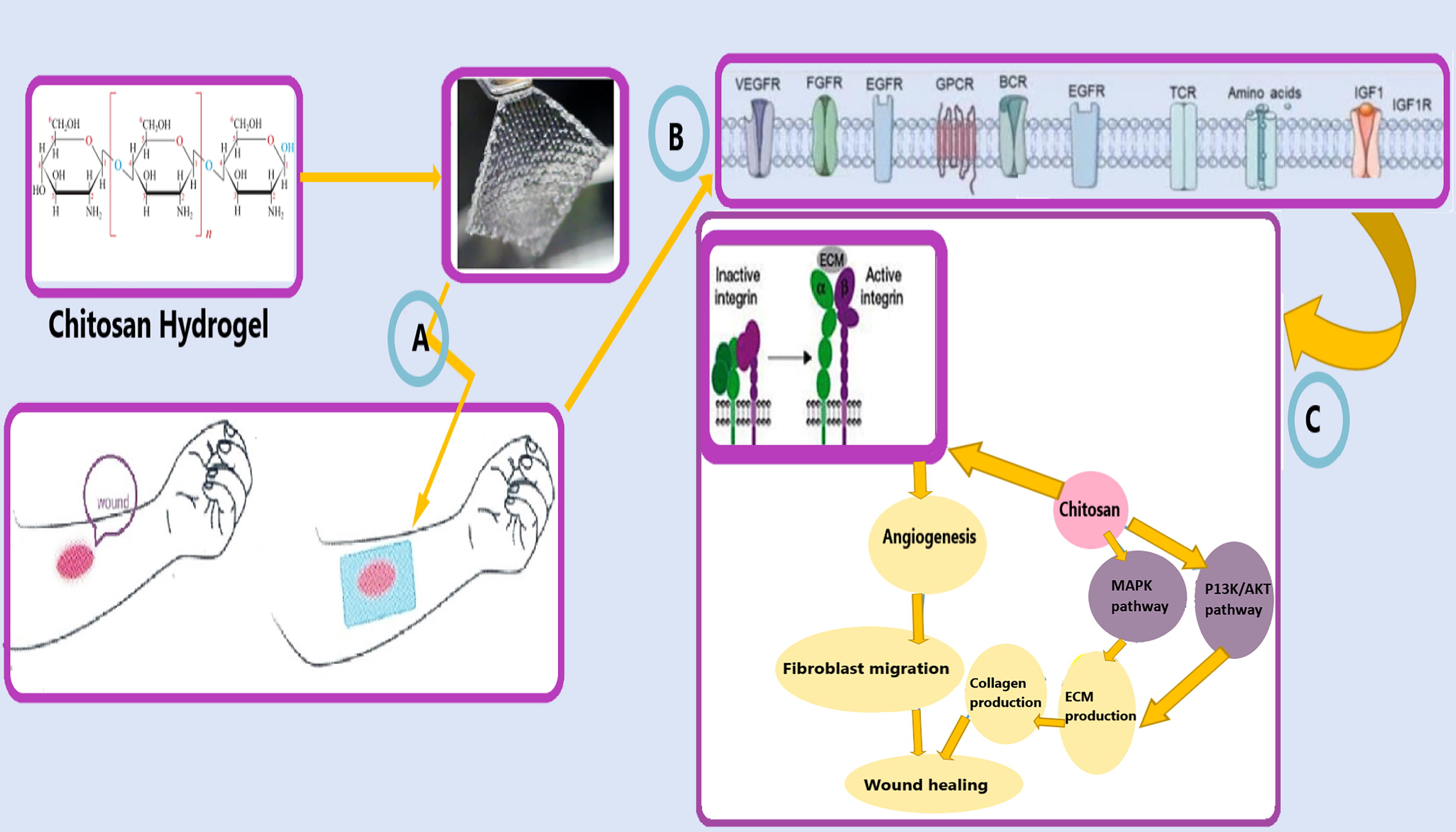
The role of chitosan-based hydrogels in wound healing: Influence on cellular signaling pathways and tissue regeneration mechanisms.
Macrophages have two roles during wound healing, proinflammatory type 1 macrophages (M1) and anti-inflammatory type 2 macrophages (M2). M1 macrophages are distinguished by markers such as CD 86 and Human Leukocyte Antigen-DR (HLA-DR) and the production of proinflammatory cytokines (IL-1β, IL-6, TNF-α) linked to inflammation and tissue damage. M2 macrophages, identified by markers such as CD206, and CD163 reduce the rate of inflammation, restore ECM, and promote repair. Chitosan shifts macrophages to the M2 cellular morphology, by decreasing M1 markers and increasing M2 markers like CD206. Researches show that chitosan decreases proinflammatory cytokines (TNF-α, IL-1β) and increases anti-inflammatory ones (IL-10, TGF-β1), promoting faster healing and ECM remodeling. Thus, chitosan supports reduced inflammation, enhanced collagen production, and improved tissue renewal, making it valuable for chronic wounds such as diabetic ulcers.56,58,59
Antioxidant and anti-inflammatory effects
Chitosan possesses powerful antioxidant properties, mitigating inflammation by neutralizing free radicals. This property is vital for preventing cellular damage in wound environments, enhancing the reconstitution stage, and preventing premature degradation of ECM. For instance, during diabetic wounds, high levels of free radicals and prolonged inflammation delay healing is available. In this situation, chitosan can accelerate the revitalization phase. By declining the expression of inflammation-related genes and inhibiting inflammatory processes, chitosan aids tissue repair. 53
Free radicals hinder wound healing by inducing oxidative stress. Chitosan, with amine and hydroxyl groups, neutralizes free radicals and reduces oxidative stress. It activates the nuclear factor-erythroid 2-related factor 2/heme oxygenase-1 (Nrf2/HO-1) pathway, boosting antioxidant genes (HO-1) and glutathione peroxidase1 (GPX1), and enzyme activity (chloramphenicol acetyl transferase, superoxide dismutase, glutathione peroxidase while lowering lipid peroxidation [MDA]) to accelerate healing. Chitosan regulates MAPK (ERK, JNK, P38) pathways to balance oxidative stress and inflammation and inhibits caspase-3 to prevent apoptosis. These properties enable chitosan to heal diabetic wounds by reducing free radicals and enhancing epithelialization, proving its efficacy in wound healing.58,59
Cytokines, such as IL-1, TNF-α, and IL-6, are important regulators of inflammation with proinflammatory effects that contribute to prolonged inflammation and delay the healing mechanism. Chitosan has shown the effectiveness to suppress proinflammatory cytokines, including ILs and TNF-α, while also inhibiting the activity of IL-1β. Additionally, it helps regulate the balance between M1 and M2 macrophages, further declining inflammation. A study by Lalita Chotphruethipong et al. exhibited that chitooligosaccharides, at a concentration of 500 µg/mL, effectively diminished the production of TNF-α, nitric oxide, and IL-6 in lipopolysaccharide-activated cells. This mechanism is indispensable for mitigating harmful inflammatory responses, thereby preventing tissue necrosis and systemic infections.58,60
Chitosan-Based Hydrogels as Scaffolds
Chitosan hydrogels are one of the most common scaffold materials because they combine the benefits of chitosan and hydrogel, allowing for adaptation to restore the damaged tissue microenvironment while also incorporating bioactive components. 61 In an animal experiment conducted by Peng Fan in 2023, chitosan-based hydrogel dressings were randomly administered to 20 rats out of 14, and it was discovered that they had a substantial impact on improving wound healing operations. 62 The reason is that the three-dimensional network architecture of chitosan-based hydrogels resembles that of a natural ECM, ensuring high water content and retention within the scaffold.61,63
3D structural support for cell infiltration and migration
Hydrogels act as scaffolds, simulating ECM and facilitating 3D tissue development. They contain functional groups and active sites, 64 which promote cell proliferation and migration while also providing a porous framework for oxygen and nutrient transport. 65 Hydrogels promote cell-matrix and cell-cell interactions by providing spatial cues and mechanical support required for cell shift to tissue-specific cells. This improves the development of organoids and their efficacy to imitate in vivo tissue functions. 66 The microenvironment and signaling molecules within hydrogels stimulate cell adhesion and migration, which are required for the creation of functional, integrated tissue substitutes. Hydrogels enable crucial cell-cell interactions and endothelialization during adhesion and migration, resulting in homogenous tissue formation. 64
Scaffolds are porous to allow for the flow of nutrients, oxygen, and waste materials, 67 as the porous channels serve as transport pathways for nutrients, metabolites, and other substances. 68 Open porosity and linked networks are imperative for cell feeding, proliferation, and migration, as well as tissue vascularization and new tissue development. 67 Furthermore, hydrogel materials’ high water content renders them highly permeable and porous, allowing oxygen and nutrients to pass swiftly and resulting in a balanced interconnected milieu that can stimulate a directed cellular fate. 69
Controlled release of bioactive molecules
Functional groups in hydrogels interact with bioactive substances to provide prolonged release. Improved cross-linking, structural aspect, pore size, and electrostatic charges allow for long-term distribution. Enzymatic breakdown in the body further aids in a regulated release. 64 Adding bFGF to chitosan-based hydrogels inhibits inflammation and accelerates healing in skin burns, according to a 2024 study by Xueyan Che et al. 70 FGF promotes collagen deposition by increasing fibroblast proliferation and differentiation, which are necessary for healing. 71 Additionally, persistent VEGF release controls blood vessel growth, 72 facilitating vascular and epithelial reanimation. 61 This promotes angiogenesis, fundamental for tissue engineering, and accelerates healing by supplying oxygen and nutrients to the trauma site. 71
Incorporating antifibrotic chemicals into hydrogels allows for controlled release, preventing TGF-β synthesis and signaling in fibroblasts. This inhibits Smad activation, which prevents myofibroblast development. Moreover, sustained release of these agents can target GF pathways such as fibroblast growth factor receptors (FGFRs), vascular endothelial growth factor receptors (VEGFRs), and platelet-derived growth factor receptors (PDGFRs), causing the decreased total collagen, inflammatory chemokines, and profibrotic factors,73,74 attenuated excessive deposition of disorganized collagen types I and III, and inhibited hypertrophic scar formation, resulting in improved dermal wound repair and fibrosis reduction. 75
Hydrogel mechanical properties
Mechanical properties of chitosan hydrogels include stiffness, pore size, viscoelasticity, and degradability, which are regulated by molecular weight, cross-linking density, and other components.68,71 They attach tightly to the skin and promote wound closure through self-contraction. Their volume contraction and tissue adhesion properties are critical in early wound healing. The hydrogels’ pore size decreases with increasing temperature, demonstrating remarkable thermoresponsive self-contraction capabilities that improve wound closure via a biomechanical approach. 76
Hydrogel stiffness affects several cell functions, including morphology, proliferation, migration, and differentiation. 68 Soft hydrogels improve fibroblast activity, whereas stiffer ones boost cell proliferation while causing cell cycle arrest in softer ones. Stiff hydrogels promote fibrotic transformation and expanded contractility, which lowers cell mobility. 77 Furthermore, they stimulate myofibroblast development by expanding the signaling of α-SMA markers. 68 Chitosan hydrogels have a considerable impact on collagen deposition because they stimulate fibroblast activity and collagen production. They also alter fibroblast gene expression associated with collagen formation and grow GFs that govern collagen synthesis, hence improving overall tissue healing and matrix deposition. 71
Comparative Insights: Chitosan Versus Other Biomaterials in Remodeling
In the evaluation and comparison of various materials used for scar tissue reduction and regeneration, chitosan displays superior effectiveness in prolonged functions when compared with alginate, collagen, and HA because of its cationic polysaccharide structure with amino and hydroxyl functional groups. 78 These groups create strong electrostatic interactions, enhancing mechanical stability, prolonging the contact time with the wound surface, and extending the drug delivery duration, thereby accelerating tissue repair. 28 Its potential to stimulate tissue recovery and singular molecular signaling properties further emphasize its efficacy. 78 In this comparison, the lower stability of alginate in aqueous environments, 79 collagen’s dependency on reinforcing agents,80,81 and the limited moisturizing function of HA 82 render these materials less effective during the regeneration phase. The advanced properties of chitosan, including improved stability, extended contact duration, and amplified tissue repair efficiency, address these challenges, making it a prominent biomaterial for use in biomedical fields.78,83 Therefore, among the evaluated materials, chitosan emerges as a superior option for scar tissue restoration.
Comparison with alginate, collagen, hyaluronic acid
The remodeling phase is indispensable in wound healing, involving ECM reconstruction and restoration of tissue structure. Biomaterials like alginate, collagen, HA, and chitosan contribute significantly to this mechanism, each offering distinctive advantages. However, compared with other biomaterials, chitosan demonstrates exceptional benefits in the remodeling phase. 59
Alginate dressings are biomaterials that have a positive effect on the remodeling sequence. A study observed that the rate of wound closure in the alginate group significantly increased from the fourth day compared with the control group (the group that received only Vaseline). This study showed that these materials have a faster epithelialization process and increase the expression of total collagen in the new skin tissue, which is why they accelerate wound closure. Also, Western blot analysis showed a significant growth in the expression of type I and III collagen in wounds treated with calcium alginate compared with the control group. 84 However, alginate degrades rapidly owing to water solubility and pH sensitivity, limiting its endurance during remodeling, often necessitating reinforcement with other materials. 85
Collagen, a fundamental ECM protein, regulates inflammation and tissue healing by binding to cytokines like IL-1β, IL-6, and IL-8. Its interaction with type I collagen and integrins α1β1 and α2β1 on endothelial cells enhances angiogenesis and granulation tissue formation, strengthening wounds and modifying type III collagen into type I. This pathway forms mature scars and restores function. However, collagen’s hydrophilicity and enzymatic degradation reduce its effectiveness during remodeling. 86
Another factor supporting tissue remodeling through its water-binding capacity and bioactive properties is HA. High molecular weight HA creates hydrated, stable microenvironments ideal for tissue repair, retaining water up to 1,000 times its weight. 87 HA interacts with cellular receptors such as CD44 and receptor for hyaluronic acid-mediated motility, triggering signaling cascades for cytoskeletal reorganization and cellular motility. This supports fibroblast migration and ECM synthesis, as keratinocytes aid epidermal reconstruction. These mechanisms highlight HA’s fundamental role in advancing tissue repair at cellular and molecular levels.88,89
Among these, chitosan, as a cationic and biodegradable biopolymer, plays a vital role in remodeling. In fact, chitosan, with its chemical structure, is effective in tissue re-epithelialization and collagen production during wound remodeling. The active amine groups in chitosan establish strong electrostatic interactions with the ECM, facilitating the rearrangement of type I and III collagen. This feature enhances the strength and flexibility of the tissue. Additionally, by modulating reactive oxygen species levels and reducing free radicals, chitosan prevents collagen degradation and provides a stable environment for long-term wound repair. All in all, the cationic properties of chitosan also allow it to bind with GFs such as TGF-β and PDGF, ensuring their controlled release. This contributes to the stimulation of fibroblast proliferation and increased ECM synthesis. 90
Advantages of chitosan for prolonged remodeling phase
Tissue remodeling, often constituting a third of the wound healing process, can extend up to a year or longer. 91 Notably, fetal, mucosal, and genital wounds tend to heal with minimal or no scarring, while the endometrium regenerates scar-free. 92 During remodeling, collagen is deposited in a well-organized network, with type III collagen transforming to type I. Furthermore, collagen turnover remains balanced, maintaining a constant total collagen content. 93 Moreover, MMPs activity regulated by pH is crucial for this phase of tissue repair. Their activity is regulated by TIMPs, whose production by fibroblasts is stimulated by TGF-β and IL-6. The delicate balance between MMPs and TIMPs is indispensable for wound healing. Imbalances can result in chronic wounds, hypertrophic scars, or keloids. During the formation of hypertrophic scars, unlike the normal wound healing sequence, abnormal ECM accumulation and excessive collagen synthesis have been observed.93,94
Chitosan-based dressings provide an optimal environment for the remodeling phase of wound healing. 90 By forming bioactive hydrogels, chitosan enhances the mechanical strength of ECM by increasing collagen production, balancing between MMPs and TIMPs and inhibiting the formation of hypertrophic scars.83,95 In a study conducted by Kazuo Kojima et al., it has been proved that these hydrogels can enhance the interaction of fibroblasts, stimulating the expression of genes associated with collagen production, thereby promoting increased collagen synthesis by fibroblasts. 96 Furthermore, chitosan hydrogels can modulate wound remodeling by influencing the balance between MMPs and TIMPs. Studies have indicated that wound healing with chitosan gels is associated with extended expression of TIMP-1. The upregulation of MMP-12 gene expression by chitosan gels are cross-linked chitosan hydrogels (700 kDa) (Ch700-G) chitosan gels suggests a potential mechanism for this effect (Fig. 4).
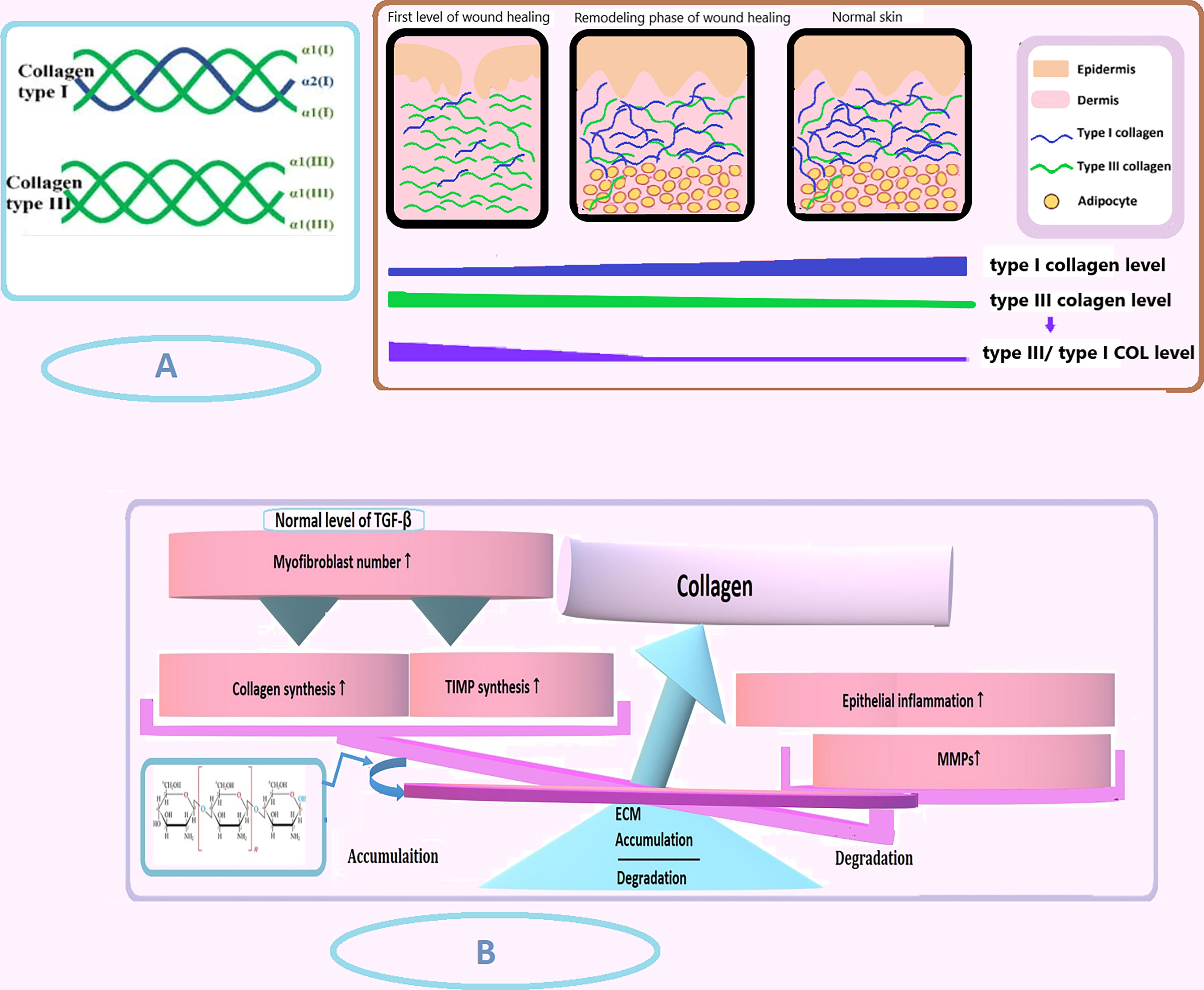
The role of collagen dynamics and chitosan in wound healing. (
Moreover, chitosan-based hydrogels have been shown to modulate the cationicity of chitosan and inhibit the formation of hypertrophic scars during wound healing. Hydrogels loaded with varying concentrations of genipin (2.5–15%) suppressed the expression of α-SMA and induced matrix metalloproteinase-1, thereby inhibiting hypertrophic scarring. 83
Challenges and Future Perspectives
Although Chitosan and its derivatives have numerous benefits for wound healing and tissue engineering, they have limitations such as low mechanical stability and potential immunogenic responses, which restrict their usage in load-bearing utilizations. Ongoing research strives to overcome these issues and maximize their promise in regenerative medicine. 97
Current limitations of Chitosan-Based hydrogels
Chitosan’s intrinsic brittleness and poor elasticity might compromise hydrogel stability and structural integrity, particularly under mechanical stress. 98 In 2024, Rahman and Mondal presented that chitosan can be affected by a variety of degradation methods, including acid hydrolysis, oxidative-reductive depolymerization, ultrasonic degradation, or enzymatic degradation using specific enzymes. 99 Chitosan, lacking protein structure, illustrates low biostability and trouble creating matrixes for wound healing, resulting in scar contraction and scar formation. 98 Chitosan hydrogels can swell in high humidity, resulting in faster active compound release while reducing mucoadhesive properties. Chitosanases and lysozymes (a nonspecific protease in all mammalian tissues) quickly degrade chitosan into nontoxic oligosaccharides, limiting their long-term stability and stability in bioscientific utilizations.98,99 Furthermore, it is important to note that their breakdown rate depends on their molecular weight, polydispersity, degree of deacetylation, purity, air temperature, and moisture content.
In particular, chitosan hydrogels are a critical component of tissue remodeling and restructuring through drug and GF release, 61 but their low stability limits their application for boosting the remodeling phase. For example, chitosan-based hydrogels cannot deliver GFs and cytokines for an extended period of time, despite their necessity for fibroblast activation, collagen deposition, and macrophage differentiation, all of which take place during the wound healing remodeling phase. 21 Moreover, improper fibroblast activation will prevent correct wound reconstruction, 100 resulting in failure or problems during wound repair.
Chitosan may also give rise to allergic reactions, making it contraindicated for its use in some patients. According to research, it can induce an immunological response (allergy) in certain individuals. Furthermore, alterations of chitosan may result in varied levels of toxicity. 99 Shengxin Peng et al. in 2022 reported anaphylaxis and an acute allergic reaction 5 min after an intra-articular chitosan injection. Additionally, the route of administration impacts the speed and severity of allergic reactions, 101 emphasizing the importance of comprehensive human hazard testing for chemical hydrogels to ensure biosafety. 70 Furthermore, chitosan can bring about toxicity through cell membrane damage, and its integration into medications might alter pharmacokinetics and biodistribution, impacting cellular distribution on account of the charge interactions. 99
Future research directions
To solve the limitations of chitosan-based hydrogels, additional polymers must be added. 70 Despite improvements, research gaps remain, especially in understanding how structural modifications affect material properties and performance. Future studies should correlate chemical structures, such as deacetylation degree, molecular weight, and functional groups, with mechanical, biological, and physicochemical qualities to optimize chitosan derivatives for particular operations. Advanced biomedical applications, such as controlled drug delivery and tissue engineering, require further exploration to improve drug release administration, tissue epithelialization capabilities, and long-term extended biointegration. Thus, future research is recommended to focus on innovative approaches like nanotechnology-based platforms and biofunctionalized scaffolds. 34
Chitosan hydrogels can be improved through changes like Schiff’s base reaction, carboxymethylation, acetylation, and sulfonation. 102 These functionalizations improve chitosan’s chemical and physical features, increasing resistance and decreasing biodegradability. These hydrogels are capable of encapsulating bioactive substances for targeted distribution. Chemical alternations, such as as cross-linking, improve rigidity and mechanical strength, resulting in frames that are less degradable, immunogenic, and toxic, as well as more biocompatible. Their water-soluble nature, along with antibacterial, antimutagenic, and antioxidant aspects, contributes to effective bioadhesion and mucoadhesion. 103
Nanoparticles properly incorporated into hydrogels lead to modest swelling rates, steady degradation, and excellent antibacterial activity, promoting wound repair. 104 Adding Ca2+ and silver nanoparticles improves stretchability, toughness, tissue adhesion, antimicrobial efficacy, angiogenesis, and collagen deposition, making hydrogels ideal for wound healing. 71 Silver nanoparticles also have adhesive, conductive, self-healing, and antibiofilm indicators. 104 Metallic salts like Ag+, Cu +, and Zn + strengthen antimicrobial activity against bacteria, fungi, and microbes. 71 Increasing nanoparticles in hydrogels heighten mechanical features, stability, tensile strength, and physicochemical biostability while maintaining nontoxicity. 59 However, more in-depth analysis and in vivo testing are needed to examine nano-formulation biodistribution, pharmacokinetics, and off-target effects.70,97 GFs have also been utilized as cross-linkers, contributing to both hydrogel bioactivity and mechanical properties while increasing biological stability. 105 For example, bFGF derives epithelialization, collagen synthesis, hair follicle development, and the formation of new blood vessels. VEGF165/TGF-β1 also boosts inflammation and healing in fibroblast gene expression. 71
Chronic wound repair is challenging as a result of persistent inflammation. Evaluations seek to enhance the anti-inflammatory and antioxidant trait of chitosan-based hydrogels, 104 which have high mechanical strength, injectability, biocompatibility, and biosafety, as well as significantly lowering inflammation markers in chronic conditions and overcoming limitations such as thermal instability and rapid clearance. 106 Biomembrane-based nanostructures such as liposomes and exosomes are used for chronic wound treatment because of their unique features. Liposomes promote solubility and sustained release, and exosomes boost hydrogel biological activity and drug targeting. Despite exosomes’ short half-life and injection-based administration, combining them with hydrogels mitigates drawbacks and improves therapeutic benefits. Future advances in nanotechnology and cell membrane encapsulation will improve these materials for chronic wound therapy. 75 This novel wound dressing aims to provide multipronged therapy by effectively controlling infections and exudates while also speeding up wound healing. Positive in vitro results highlight its potential as a next-generation wound dressing, but more in vivo research is needed to confirm its efficacy. 97
To make chitosan more effective and efficient for remodeling, introducing stem/stromal cells would be effective as they provide cells and GFs/cytokines at the defect site for a long period of time. 61 For instance, hydrogels containing umbilical cord stem cell factor can stimulate neovascularization and skew toward M2 macrophages, or the umbilical-cord-derived mesenchymal stem cell exosomes combined with hydrogel can increase the TGF-β1 producing, which is essential for fibroblast proliferation and consequently wound tightening by enhancing α-SMA expression.41,107 Encapsulating neural stem cells in chitosan-based hydrogels will also allow the long-term development of cell globules in a 3D environment. Moreover, using carboxymethyl chitosan/hyaluronic acid-dopamine in hydrogel might be helpful during the remodeling phase by considerably affecting collagen deposition and cell proliferation at the injury site. Interestingly, melatonin has been shown to raise the expression of collagen III, alpha-SMA, and TGF-β1 proteins, making this hormone a promising candidate for accelerating wound healing by loading them into hydrogels. 61
Conclusion
This comprehensive review unequivocally demonstrates that chitosan-based hydrogels represent a groundbreaking advancement in the remodeling phase of wound healing. Their unique physicochemical attributes—encompassing exceptional biocompatibility, tailored biodegradability, intrinsic antimicrobial efficacy, and robust hemostatic properties—establish a highly favorable microenvironment that meticulously orchestrates the complex interplay of cellular and molecular processes essential for effective tissue regeneration.
Critically, the hydrogels’ capacity to modulate key signaling cascades, such as TGF
Despite these compelling advantages, current limitations—including suboptimal mechanical stability, variable degradation kinetics, and potential immunogenicity—necessitate further refinement. Future research should be rigorously directed toward advanced material engineering strategies, such as precision cross-linking, nanoparticle integration, and synergistic incorporation of stem cell-based therapies. These innovative approaches are expected to overcome existing challenges and significantly augment the clinical efficacy of chitosan-based hydrogels.
In conclusion, the strategic deployment of chitosan-based hydrogels heralds a paradigm shift in wound management and regenerative medicine. Their multifaceted capacity to modulate cellular behavior and ECM architecture offers a robust and clinically translatable platform for achieving superior healing outcomes in complex wounds. Continued interdisciplinary efforts and rigorous clinical investigations will be critical to fully harnessing their transformative potential and establishing these hydrogels as a cornerstone in high-impact therapeutic applications.
Footnotes
Acknowledgments
The authors acknowledge the invaluable discussions and constructive criticisms from anonymous reviewers, which significantly augmented the article’s comprehensiveness and clarity.
Authors’ Contributions
R.M.: Investigation, Data Curation; Visualization, Reference Collection, Writing Original Draft, Review Final Manuscript; A.H.: Investigation, Data Curation; Visualization, Reference Collection, Writing Original Draft, Review Final Manuscript; M.S.: Supervision, Conceptualization, Methodology, Editing Original Draft, Review Final Manuscript.
Ethics Approval and Consent to Participate
The study was conducted following the highest ethical standards. The data presented in this article are accurate and authentic.
The Availability of Data and Materials
The datasets collected and/or analyzed during the current study are available from the corresponding author on reasonable request.
Author Disclosure Statement
During the preparation of this work the author(s) used ChatGPT 4o only to improve language and readability. There are no conflicts of interest for the authors.
Funding Information
The authors received no financial support for the research, authorship, or publication of this article.