
Abstract
We report the x-ray microtomographic imaging of three-dimensional (3D) structure of soft tissues. The transparency of biological tissue to hard x-rays enables radiographic analysis of tissue entrails. However, biological tissues are mainly composed of light elements, which produce little contrast in a hard x-ray transmission image. Tissue structures were visualized by contrasting biological constituents with heavy elements. Efficient x-ray absorption by heavy-element dyes allowed the radiographic visualization of microstructures of soft tissues. The high-resolution computed tomography analysis provided the 3D microstructure of these microcontrasted tissues. Element-selective visualization of the stained tissue using x-ray absorption edges revealed the specific architecture of internal components. The structures obtained were used for rapid prototyping, giving 3D copies of human capillary vessels and fruit fly body.
Introduction
The primary method for visualizing 3D structures of soft tissues is confocal optical microscopy. 4 Optical computed tomography and ultramicroscopy have recently been applied to the 3D analysis of biological tissues,5,6 although the internal structure of opaque or chromatic samples cannot be observed using visible light. Magnetic resonance imaging provides non-invasive mapping of neural activity, 7 whereas cellular structures cannot be visualized because of the limited spatial resolution.
In contrast, the transparency of biological tissue to hard x-rays enables radiographic analysis of tissue entrails. However, biological tissues are mainly composed of light elements, which produce little contrast in a hard x-ray transmission image. We recently reported that the structure of the Drosophila larvae central nervous system (CNS) can be visualized by contrasting every neuron using a metal impregnation method. 8 In conjunction with high-resolution x-ray computed tomography,9,10 hereafter called micro-CT, metal microcontrasting has revealed the 3D structures of the Drosophila larvae and adult CNS11,12 and the neuronal structure of the human frontal cortex. 13
Histological staining methods using heavy elements are known as conventional techniques for the optical observation of biological structures. Efficient x-ray absorption by heavy-element dyes should allow the radiographic visualization of microstructures of any soft tissues. Therefore, the 3D structure of biological tissues can be determined using microcontrasting x-ray analysis. Here, we report an application of microtomographic imaging to human capillary vessels stained by Golgi impregnation and to fruit fly Drosophila melanogaster and zebrafish Danio rerio stained by Bodian impregnation. Element-selective visualization revealed 3D architecture of visceral components in the fruit fly body. The structures obtained were used for 3D prototyping, giving microfabricated copies of biological objects.
Materials and Methods
Human cerebral cortex
Human samples were obtained with informed consent, using protocols approved by the Clinical Research Review Board of Tokai University Hospital. Cortex of normal brain tissue (44 years old, male) was dissected at autopsy and fixed with 10% formaldehyde for 7 days. Anatomical analysis found no abnormality in the brain tissue. The tissue sample was further dissected into a 5-mm block and subjected to Golgi impregnation, as reported previously. 13 Potassium dichromate and silver nitrate treatment steps were repeated three times. A prismatic sample with dimensions of 0.5 mm × 0.5 mm × 2.5 mm was prepared from the stained tissue block using razor blades.
Fruit fly
Wild-type Drosophila melanogaster Canton-S were raised on cornmeal-molasses fly food and kept at 20°C. Reduced-silver Bodian impregnation was performed, as described previously. 11 Fly food containing 1% (w/v) ammonium tetrachloroplatinate was used for raising flies for the visceral staining. Adult flies were fixed for 24 h in a formaldehyde solution. The fixed sample was rehydrated and subjected to modified reduced-silver staining. The silver impregnation was performed for 65 h at 37°C. The sample was then toned with 1% hydrogen tetrachloroaurate for 180 min.
Zebrafish
Wild-type Danio rerio were purchased from Trust Aqua Service (Tokyo, Japan) and raised using standard fish food at 25°C. Juvenile fish 1 week old was fixed for 16 h in a formaldehyde solution and impregnated as described previously. 11 The silver impregnation was performed for 20 h at 37°C. The sample was then toned with 1% hydrogen tetrachloroaurate for 120 min.
Tissue embedding
The stained samples were sequentially immersed in ethanol, n-butylglycidyl ether, and Petropoxy 154 epoxy resin (Burnham Petrographics, Rathdrum, ID). The human and zebrafish samples were transferred into a borosilicate glass capillary (W. Müller Glas, Berlin, Germany) filled with epoxy resin. The outer diameter of the capillary was 0.7 mm. The fruit fly samples were placed in silicone tubing filled with epoxy resin. The inner diameter of the tubing was 3 mm. The samples were then incubated at 90°C for 16 h to rigidify the epoxy resin.
Micro-CT
The microtomographic analyses were performed as described previously.11–13 The human and zebrafish samples were analyzed at the BL20XU beamline 14 of the synchrotron radiation facility SPring-8 (Hyogo, Japan). The samples were mounted on the goniometer head of the microtomograph using a brass fitting specially designed for the glass capillary sample. Transmission radiographs were recorded usng a CCD-based x-ray imaging detector (AA50 and C4880-41S, Hamamatsu Photonics, Hamamatsu, Japan). The field of view and effective pixel size of the detector were 1.00 mm × 0.65 mm and 0.50 μm × 0.50 μm, respectively. A total of 1800 images were acquired with a rotation step of 0.10° and an exposure time of 300 ms. The spatial resolution of the 3D structure was estimated to be 1.0 μm in each direction. 9
The fruit fly samples were analyzed at the BL20B2 beamline 15 of SPring-8. The sample pellets were mounted on the goniometer head of the microtomograph using adhesive tape. Transmission radiographs were recorded using a CCD-based x-ray imaging detector (AA40P and C4880-41S, Hamamatsu Photonics). The field of view and effective pixel size of the detector were 5.5 mm × 3.6 mm and 2.75 μm ×2.75 μm, respectively. Each image was acquired with a rotation step of 0.10° and an exposure time of 600 ms. The spatial resolution of the 3D structure was estimated to be 8 μm.
The convolution back projection method was used for tomographic reconstruction. 9 The reconstruction calculation takes approximately 30 h on a Windows PC equipped with the Core 2 Duo processor operating at 2.1 GHz. Reconstructed images were further processed using the program suite SLICE. 16 Element-specific visualization was performed using Au LIII and Pt LIII x-ray absorption edges. The gold distribution image was prepared by subtracting the 3D distribution of linear absorption coefficients at 11.910 keV from that at 11.930 keV. The platinum image was prepared by subtracting the 11.550-keV image from the 11.570-keV image. All other images were prepared by rendering linear absorption coefficients at 12.000 keV. Volume-rendered figures of the 3D structures obtained were produced using the program VG Studio MAX (Volume Graphics, Heidelberg, Germany).
3D printing
The continuous density was extracted and converted into the Standard Triangulated Language (STL) format using the program suite SLICE. 16 Extraction of the capillary vessel density was performed by applying a mask that covered a cylindrical volume (diameter: 12 μm) along the capillary axis. The 3D fabrications were performed using a rapid prototyping apparatus (D-MEC, Tokyo, Japan) using SCR950 oxetane resin (JSR, Tokyo, Japan).
Results and Discussion
The tomographic analysis of the entire fruit fly visualizes the 3D structure of the whole body system. In a cutaway section of the cephalon region (Fig. 1A), the structure of the cerebral ganglion and compound eyes were visualized as head entrails. The structure of the fruit fly administrated with the platinum compound was also analyzed. Three-dimensional distributions of platinum and gold were selectively observed using x-ray absorption edges (Fig. 1B). The platinum absorption was mainly found in the trophi and visceral components, whereas gold was nonspecifically observed from the overall body. In the zebrafish, the metal impregnation deeply stained the lenticular cortex and sclerotic coat, as shown in Figure 1C. The pigment macules exhibited affinity for metal dye, giving clear images of raised skin blotches on the dorsal surface of the fish body. Although the biological systems of the fruit fly and zebrafish are different from those of mammals, Bodian staining has been used for optical observation of mammal tissues. Therefore, these tomographic analysis methods can be applied to mammal tissues in which the 3D structures are crucial for their function.

Three-dimensional (3D) structure of fruit fly and zebrafish. Computed tomography (CT) densities were rendered by the scatter HQ algorithm. Scale bars: (
The blood vessel network in the CNS is essential for maintaining brain functions. We have reported the visualization of the 3D structure of the frontal cortex of human brain. 13 The neurons exhibited the layered structure characteristic of the neocortex. In the present study, brain tissue containing capillary vessels was analyzed to obtain a structural template for fabricating the capillary architecture. A region of the internal pyramidal layer is shown in Figure 2A. The capillary blood vessels and pyramidal neurons were visualized as positively stained regions. Blood cells in the capillary vessel displaced metal dye and appeared as a negatively stained image.

Three-dimensional (3D) microstructure of human frontal cortex. The brain surface is to the top. (
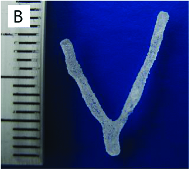
The structures of the capillary blood vessels and the adult fruit fly were used for rapid prototyping (Fig. 1D and 2B). These models were prepared by piling up 100-μm layers of resin, giving a 200-fold magnified model of the capillary and an 18-fold model of the fruit fly. Although the resin and its piling thickness used in the present prototyping is not appropriate for the reproduction of biological functions, these models indicate that the microtomographic structure of soft tissues can be used as a 3D template for the artificial fabrication of a biological architecture. It has been proposed that bioprinting techniques can be used to fabricate biomimetic structures. 17 Therefore, the microtomographic analysis of tissues and organs can provide the 3D microstructural basis that allows the reproduction of biological tissues.
Optical sectioning microscopy including confocal microscopy is the primary method for visualizing 3D structures of biological systems. Although the maximum spatial resolution of an optical microscope is estimated to be approximately half the observation wavelength (200–300 nm), the observation depth is typically limited to 100 μm in confocal microscopy because of absorption and refraction effects. The resolution along the depth direction is worse than the in-plane resolution, resulting in the anisotropic feature of the 3D image. These cause difficulties in 3D reconstruction of the entire optical image of thick samples. Thus, optical sectioning microscopy is mainly used for imaging sectioned samples labeled with highly selective probes.
The energy of x-rays is several hundred or thousand fold higher than the visible light used in optical microscopy. Therefore, the diffraction-limited resolution of x-ray microscopy is much higher than that of optical microscopy. We have discussed that the viewing field and spatial resolution of the micro-CT analysis can be varied depending on the pixel size, image dimensions, and x-ray optics. 13 In the present study, the 3D structure of the human brain in a cylindrical region 1.00 mm in diameter × 0.65 mm in height was reconstructed from the 2000 × 1300 pixel projection images with effective pixel size of 0.5 μm. Although the spatial resolution of the reconstructed structure from projection images has been estimated to be 1.0 μm, 9 application of the zone plate optics 10 would enable structure determination at a higher resolution. We have reported a radiographic analysis of nerve tissue at 160-nm resolution 11 visualizing the neuronal structures. Although the metal staining protocols should be further investigated, selective staining along with micro-CT analysis at this higher resolution would enable functional analysis of the biological tissues from the 3D microstructure.
Heavy-element microcontrasting in conjunction with micro-CT analysis can be applied to any biological systems. The present results indicate that the conventional metal staining method facilitates the 3D tomographic visualization of soft tissue structures. Other histological staining methods, including reticulin silver impregnation, phosphotungstic acid staining, and osmic acid fixation, can also be used as microcontrasting procedures. Because uniform and precise staining of the tissue structure is essential for tomographic visualization, a variety of staining methods and dyes should be examined. Along with the microcontrasting method, micro-CT analysis can shed light on the underlying structural basis of biological functions.
Footnotes
Acknowledgments
The synchrotron radiation experiments were performed at SPring-8 with the approval of the Japan Synchrotron Radiation Research Institute (JASRI) (Proposal Nos. 2006B1716, 2007A1844, 2007A2072, 2007B1102, and 2007B1894).