
Abstract
Recent in vitro studies with electrospun nanofibers have used a range of techniques. The in vitro system presented in this article describes electrospun fibers deposited onto chemically reactive substrates to provide fiber adherence and surface chemistry control of the substrate. Fibers of poly(ɛ-caprolactone) (PCL) or of a blend of PCL and collagen type I (C/PCL) were electrospun directly onto collectors coated with isocyanate-terminated star (polyethylene glycol) (sPEG). Alternatively, parallel electrospun fibers were collected on dual collectors in “dilute” quantities and transferred onto sPEG-coated substrates. The initial reactive nature of the substrates allows the collection of very few fibers, which adhere well during frequent washes. Furthermore, the sPEG layer transforms into protein-repellent substrates with the additional potential to include specific cellular mediators such as glycine-arginine-glycine-aspartate-serine (GRGDS) peptides to promote cell adhesion. Therefore, the fiber and substrate chemistry can be modified independently, which is particularly useful for in vitro studies of guided migrating cells. In the present work, dissociated cells of dorsal root ganglia seeded onto the substrates were investigated to assess the influence of different combinations of fiber material, fiber orientation, and surface functionalization. Cell adhesion was observed predominantly on the nanofibers, except when the sPEG layer on the substrate contained GRGDS. On the cell-repellent sPEG substrates, neurites were aligned in direct contact with parallel C/PCL fibers and less so with PCL fibers. In contrast, neurite alignment showed less guidance effect with C/PCL electrospun fibers on the GRGDS/sPEG-coated substrates. Therefore, the combination of oriented biologically active fibers on cell-repellent surfaces enhanced the guidance of such cells. These reactive substrate systems provide a multitude of in vitro combinations for providing cells with specific mediators and, in turn, defining the optimum environment of regenerating devices for in vivo studies.
Introduction
Mechanical stabilization of the electrospun fibers is an important technical prerequisite for cell culture studies, in which frequent media changes are required; studies of single electrospun fibers and cells are therefore still relatively limited in the literature. Several methods with specific limitations have been described to adhere electrospun fibers on substrates for further cell experiments. Glue, 2 biocompatible adhesives, 5 or disposable bandages 6 secure the fibers at the edge of the substrate. Complete attachment of the fibers in a one-step procedure can be achieved by electrospinning onto collectors with adhesive coatings, for example, with thin films of poly(ethylene terephthalate). 7 Another possibility is using initially chemically reactive layers of isocyanate-terminated 6-armed star-poly(ethylene glycol) (sPEG). These sPEG-coated substrates can chemically adhere nanofibers before being transformed into a protein-repellent surface by reaction of the isocyanate functionalities with water to form amino groups, which form urea crosslinks with residual isocyanate groups. 8
In this article, we present a two-dimensional in vitro system to test conditions needed for neurite guidance in nerve regeneration experiments with oriented fibers deposited onto functionalized substrates. Two different materials, poly(ɛ-caprolactone) (PCL) and a blend of PCL with collagen type I from calf skin (C/PCL) were electrospun into randomly and aligned single fibers and deposited on reactive sPEG substrates. Collagen activates integrin receptors at the surface of axons and glia and therefore offers binding sites for cell adhesion and migration.9–11 The inclusion of collagen should, therefore, result in better viability and cell attachment to the nanofibers than pure PCL fibers. Recent publications have indicated that collagen degrades to gelatin during the electrospinning process, especially when fluoroalcohols are used as solvents.12,13 The loss of collagen was described to be between 45% 12 and 99%. 13 However, the positive effect of including collagen has already been confirmed in several in vitro experiments with fibers of PCL/collagen blends, resulting in greater cell proliferation than pure PCL fibers.4,14,15
The freshly prepared sPEG-coated substrates promote fiber fixation before being transformed into a protein- and cell-repellent surface, permitting in vitro investigations with frequent media changes without fiber lift-off. Cell-adhesive substrate surfaces are achieved by adding specific peptide sequences, e.g., repetitions of glycine-arginine-glycine-aspartate-serine (GRGDS) to the sPEG surface of the substrate. Protein adsorption on the substrates was investigated because it was expected to play a key role in altering the chemistry of the surface.
The combination of a reactive substrate system (e.g., sPEG) with electrospinning allows us to investigate the influence of biologically active extracellular matrix (ECM) molecules in the configuration of orientated fibers on cell behavior and neurite alignment.
Materials and Methods
Materials
Unless otherwise stated, all chemicals were purchased from Sigma-Aldrich (Munich, Germany). Toluene and tetrahydrofuran (THF) were dried by distillation over lithium aluminum hydride. SPEG (star-shaped polyethers with a backbone of 80% ethylene oxide and 20% propylene oxide and isocyanate end groups, Mw = 12,000 g/mol) was synthesized from hydroxyl-terminated star polymer (DOW Chemicals, Terneuzen, Netherlands) by reaction with isophorone diisocyanate according to the literature. 16 All other chemicals were used as received.
Substrate preparation
Reactive substrates were produced by surface modification of cover glass (No. 1, diameter-12 mm, Marienfeld, Lauda-Königshofen, Germany) with sPEG as described elsewhere. 17 Briefly, the cover glasses were cleaned in ethanol (KMF, Lohmar, Germany) and activated using ultraviolet-ozone treatment for 12 min. Then the cover glasses were transferred into the glove box and aminosilanisated with a solution of 0.3 mL of N-[3-(trimethoxysilyl)propyl] ethylene diamine (97%) in 50 mL of dry toluene for 2 h. After being washed with dry toluene, the cover glasses were stored in toluene in a nitrogen atmosphere. Before spin-coating, a solution of 20 mg of sPEG in 0.2 mL of THF was prepared under a nitrogen atmosphere. At a normal atmosphere, 1.8 mL of deionized water was added and crosslinking allowed for 5 min. Subsequently, the cover glasses were placed onto the spin-coater and covered with the filtered solution (0.2 μm, Whatman, Maidstone, Great Britain). The cover glasses were accelerated within 5 s to 4000 rpm for 40 s. The prepared cover glasses are termed “sPEG-substrates” in this study. Silicon wafers were treated according to the same procedure with sPEG, and these substrates are called “sPEG-sil.” Additionally, sPEG substrates were surface functionalized with the cell adhesive peptide sequence GRGDS (H-Gly-Arg-Gly-Asp-Ser-OH) (Bachem, Bubendorf, Switzerland). GRGDS was dissolved in deionized water (500 μg/mL) and mixed with the sPEG/THF solution. After crosslinking for 5 min, this solution was spin-coated to the cover glass. These substrates are termed “GRGDS-sPEG-substrates.”
Electrospinning and fiber deposition
Poly(ɛ-caprolactone) (PCL) (Mw = 65,000 g/mol) was dissolved in chloroform/methanol (75/25 v/v). A blend of PCL with 25 wt% type I collagen from calf skin (C/PCL) was prepared by suspending the polymers in 1,1,1,3,3,3-hexafluoro-2-propanol overnight followed by a 2 h ultrasonic treatment. Electrospinning was performed with concentrations of 3 to 9 wt%. A 1-mL syringe with a stainless steel 20G flat-tipped spinneret (Hamilton, Bonaduz, Switzerland) was filled with the suspension, which was then pumped with flow rates between 0.05 and 0.5 mL/h while voltages of 15 or 20 kV were applied. The distance between spinneret and target was 15 or 20 cm. Random fibers were collected on single targets while parallel fibers were collected suspended between dual collectors with a gap of 4 cm, using the gap method for alignment.18,19 These strategies typify the two main collection strategies for electrospun fibers cited in the literature. 1 Figure 1 shows a scheme of both electrospinning systems. Varying the collection time between 10 s and 6 min allowed different densities of fibers to be collected. Fibers were deposited onto different substrates for use in further experiments.
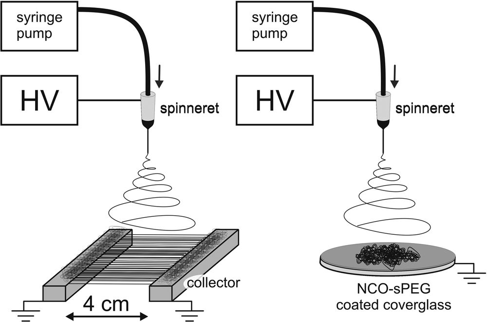
Schematic of electrospinning system with a high-voltage supply (HV), syringe pump, spinneret, and grounded collector. With these configurations, parallel fibers are collected and suspended in the air between two parallel bars (left), or random fibers are collected directly onto a collector with a chemically reactive nanolayer (right).
SPEG-coated cover glasses were used for in vitro experiments and antibody staining, and substrates were produced as described before. 4 Electrospun fibers were deposited onto the sPEG-substrates 30 min after spin-coating the sPEG; samples prepared in this way are termed “sPEG sub-fib.” The same procedure was applied to GRGDS-sPEG substrates (“GRGDS-sPEG sub-fib”). Figure 2 shows a diagram of the preparation of the substrate-fiber system used in cell experiments; electrospun fibers of different materials were deposited onto differently modified cover glasses. Cell experiments and the analysis of substrates and fiber surfaces were performed with these samples.
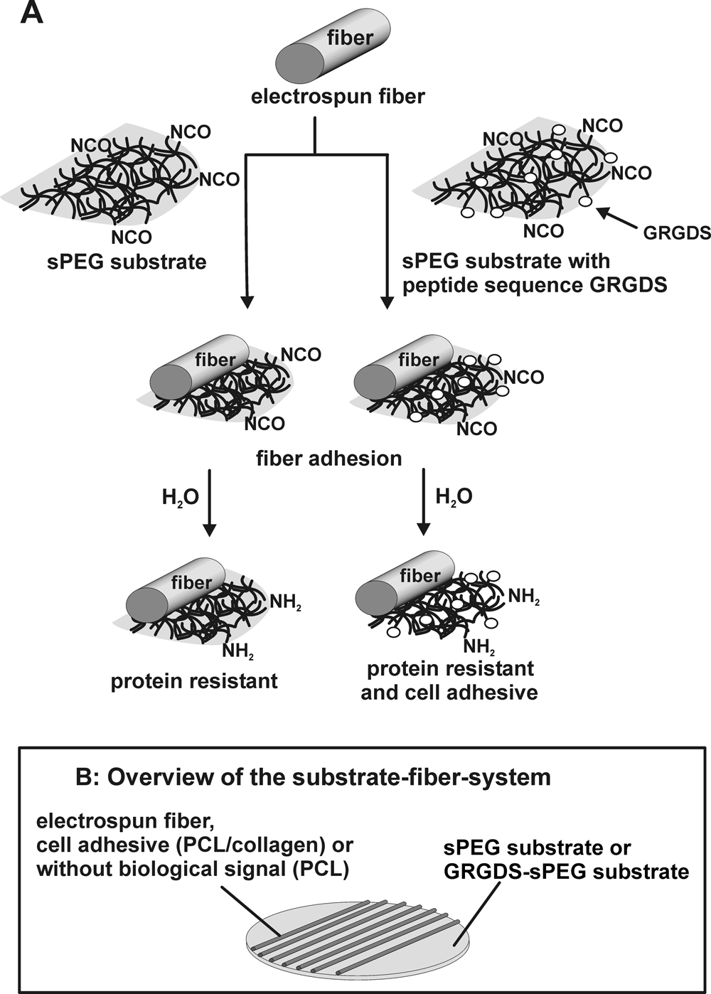
Substrate-fiber system used for surface characterization and cell experiments. (
Reactive substrate characterization
Fiber characterization
The electrospun fibers were visualized using scanning electron microscopy (SEM) (S360 Zeiss NTS, Oberkochen, Germany; Hitachi S3000 N, Hitachi High Technology Europe GmbH, Krefeld, Germany), using 15 kV and working distances of 5 to 15 mm after sputtering with gold for 90 s (S150B, Edwards, Crawley, Great Britain). The SEM images were used to measure fiber diameters, which are presented as mean ± standard deviations (n = 2, counting 90 fibers for random PCL and C/PCL fibers; 57 fibers for parallel PCL fibers; 57 fibers for parallel C/PCL fibers). The distances between the fibers were also determined using SEM images.
Antibody staining
Parallel PCL and C/PCL fibers were placed onto sPEG-coated cover glasses, pre-wetted in phosphate buffered saline (PBS) for 5 min, and blocked for 1 h in PBS containing 10% normal goat serum (NGS) and 1% bovine serum albumin (BSA). The substrates were consequently incubated for 4.5 h at room temperature with the monoclonal anti-collagen type I antibody (Sigma C2456, ascites fluid, clone COL-1). This antibody has been reported to have no cross-reactivity with other collagen types (i.e., II, III, IV, V, VI, VII, IX, X, and XI) and does not react with the thermally denatured molecule. The antibody was diluted in antibody diluent (ABD) (PBS containing 10% NGS and 2% BSA; 1:100). The samples were washed three times with PBS followed by 1 h incubation with a fluorescent secondary antibody Alexa Fluor™ 488 nm (Molecular Probes, Invitrogen, Paisley, Great Britain) diluted in ABD (1:500) at room temperature in the dark. Negative controls were prepared by omitting primary antibodies. Afterwards, all samples were washed three times with PBS and then mounted upside-down onto microscope slides using Fluoroprep (bio-Mérieux, Marcy l'Etoile, France). Epifluorescent microscopy was performed with a Zeiss Axioplan microscope. Images were taken with a constant exposure time of 50 s. Because the microscope gave a green background even without any samples, a background image was taken as blank with the same exposure time. This blank was then subtracted from the images of the samples using Adobe Photoshop software (adobe software manufacturer, San Jose, California, U.S.).
Protein adsorption
Protein adsorption of sPEG-coated silicon wafers with PCL and C/PCL fibers were determined with fluorescently labeled BSA, as previously described. 20 Briefly, the prepared sPEG-sil fibs were incubated with rhodamine red–labeled BSA diluted in PBS (50 μg/mL) for 20 min followed by three incubations in PBS for 20 min each. Afterwards the samples were incubated for 60 min in PBS and washed thoroughly with distilled water. The samples were kept in the dark during incubation and washing. The sPEG-sil fibs were visualized using fluorescence microscopy (Axioplan 2 imaging, Zeiss), and images were taken with a constant exposure time of 20 s. Silicon wafers coated with GRGDS-functionalized sPEG were also tested for protein adsorption.
Cell culture experiments
Chicken embryos (day 10) were removed from the egg, decapitated, and dissected in ice-cold PBS. The skin was removed with forceps to access the lumbar dorsal root ganglia (DRGs), closed forceps were inserted on both sides of the spinal cord and opened to cut off the limbs. Three to four DRGs from each side were transferred into a 15-mL centrifuge tube with ice-cold PBS. For dissociation of DRGs, the PBS buffer was removed, and 1 mL of 0.25% trypsin/ethylenediaminetetraacetic acid (EDTA) (Invitrogen) was added for 5 to 8 min on ice, followed by 5 to 8 min at 37°C. After washing with PBS, the tissue was triturated in 1 mL of Dulbecco's modified Eagle medium (DMEM)/F12 + 10% fetal calf serum (FCS). Then 10 mL of PBS was added and the suspension centrifuged at 130 g for 10 min. The supernatant was removed, and the cells were re-suspended in 1 mL of medium and counted in a Thoma chamber.
Cells were cultivated at 37°C and 5% carbon dioxide on prepared sPEG- or GRGDS-sPEG substrates with electrospun fibers in 24-well plates under sterile conditions. SPEG sub-fib and GRGDS-sPEG sub-fib were sterilized with ultraviolet light for 2 h and cells seeded at a density of 2 × 10⁴ cells/500 μL DMEM/F12 (1:1) containing 10% FCS and 2% B27 (growth promoting factor) supplement (GIBCO, Invitrogen, Paisley, Great Britain) per well. Nearly 20,000 cells per well adhered, and this number was stated as 100% in the calculation of the proliferated cells.
After 4 days of incubation, the cells were fixed with 4% paraformaldehyde and processed for immunocytochemical staining. The primary antibodies included anti-S100 to mark Schwann cells (1:200; polyclonal rabbit antibody; S2644 Sigma) and anti-NF 200 (1:500; neurofilament 200 kDa, monoclonal mouse antibody; N0142 Sigma) to stain neurons and axons. The antibodies were diluted in tris-buffered saline and Triton X (TBS-T) containing 1% normal goat serum. Incubation was overnight at 4°C. For secondary staining, Alexa Fluor™ 488 goat anti-mouse immunoglobulin IgG (MoBiTec; A-11001) and Alex Fluor™ 546 goat anti-rabbit IgG (MoBiTec; A-11010) were diluted 1:1000 in TBS-T and incubated for 1 h at room temperature. Cell nuclei were stained with 4’6-diamidino-2-phenylindole (DAPI; 1:1000) for 5 min. Evaluation was done with an epifluorescence microscope (Zeiss Axiophot). To determine cell numbers, cells were counted from five randomly positioned pictures (area 0.15 mm2) from every cover glass. For every condition, experiments were repeated at least four times.
Measurement of neurite alignment was performed on fluorescence images with Image J software. The total length of every neurite was measured, as well as the proportion of its length that was in direct contact with a fiber, which was unambiguously identified with a 40× objective. For every neurite, the ratio of its length in contact with an electrospun fiber to its total length was calculated. For every condition, at least 15 axons, all longer than 100 μm, were randomly selected and evaluated. Data are presented as means with standard errors of the mean and evaluated using analysis of variance (ANOVA) and Tukey-Kramer test for multiple pairs (JMP 4.0 software, SAS Institute, Inc., Cary, NC).
Results
Electrospinning and fiber characterization
In this study, we electrospun PCL and C/PCL. Two different collection configurations were used to achieve random or parallel fibers (Fig. 1).
Several governing parameters, such as voltage, collection distance, and flow rate, were investigated to obtain high-quality fibers. However, the solution concentration played the most important role in achieving uniform fibers and the required concentrations were determined for PCL and C/PCL. Figure 3 shows SEM images of C/PCL fibers produced with different solution concentrations. Low solution concentrations of 3 and 5 wt% resulted in beaded fibers (Fig. 3A, B) and a suspended collection of oriented fibers was not obtained. Increasing the concentration to 7 wt% improved the fiber quality and resulted in homogeneous continuous fibers when collected on a single collector (Fig. 3C). Collection of parallel fibers was also possible at this concentration (Fig. 3D).
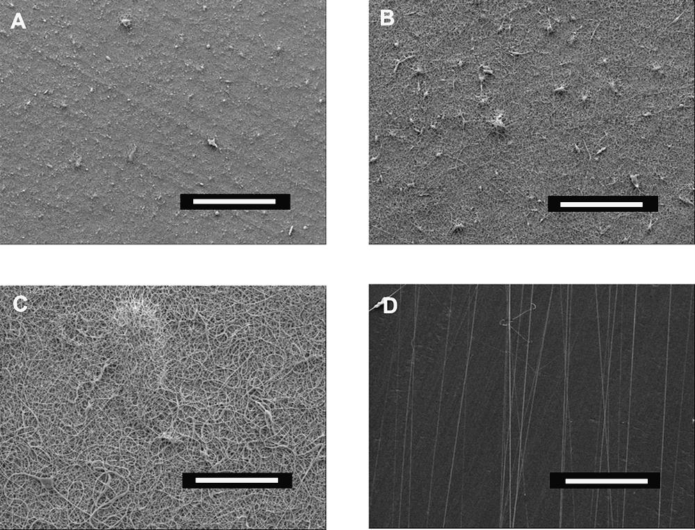
Scanning electron microscopy images of electrospun fibers made of a blend of collagen I and poly(ɛ-caprolactone) produced with different solution concentrations showing different fiber qualities and orientations. (
Figure 4 shows examples of PCL and C/PCL random and parallel fibers, which were used for all following experiments. Electrospinning of pure PCL at 9 wt% resulted in fibers with diameters of 0.49 ± 0.24 μm for random collection, which was similar to the diameter of parallel fibers (0.56 ± 0.29 μm) (Fig. 4A, B). For random and parallel collection, the highest-quality fibers were achieved with an applied voltage of 20 kV, a distance of 20 cm between spinneret and target, and a flow rate of 0.5 mL/h.

Scanning electron microscopy images of random and oriented fibers of poly(ɛ-caprolactone) (PCL) and a blend of collagen I and PCL (C/PCL) produced under optimum conditions for the achievement of good-quality fibers. (
PCL and collagen type I from calf skin were blended to obtain mechanically stable fibers with better cell recognition properties than pure PCL fibers. As with pure PCL, a voltage of 20 kV, a distance of 20 cm, and a flow rate of 0.5 mL/h resulted in the highest-quality fibers. The random fibers had a diameter of 0.74 ± 0.18 μm (Fig. 4C), whereas parallel fibers had a diameter of 0.73 ± 0.31 μm (Fig. 4D).
Fiber density of PCL and C/PCL parallel fibers depended on the collection time. For standard samples, fibers were collected between 45 s and 60 s, resulting in diluted fibers with distances of 10 to 20 μm between them (Fig. 3D, 4B, D). It was also possible to shorten or prolong the collection time and collect fibers with densities of 40 fibers/mm to 450 fibers/mm (data not shown).
Characterization of in vitro substrates
Substrates were prepared using surface modification of silicon wafers or glass cover slips with a sPEG or a cell-adhesive GRGDS-sPEG layer. Electrospun fibers of PCL and C/PCL were deposited onto the activated surfaces in a second step. Electrospinning directly onto unmodified cover glasses was also attempted. In this case, the random fibers adhered to the glass surface and were not readily washed off, probably because of residual solvent in the fibers that improved adhesion to the collector. Surprisingly, suspended arrays of parallel fibers that were placed onto unmodified cover glasses lifted off the substrate at first contact with water. It is likely that the residual solvent in the suspended fibers had evaporated before collection, thus preventing fiber adhesion. In contrast, parallel and random fibers deposited onto the sPEG layer adhered to this surface and were not washed off, irrespective of the method of collection.
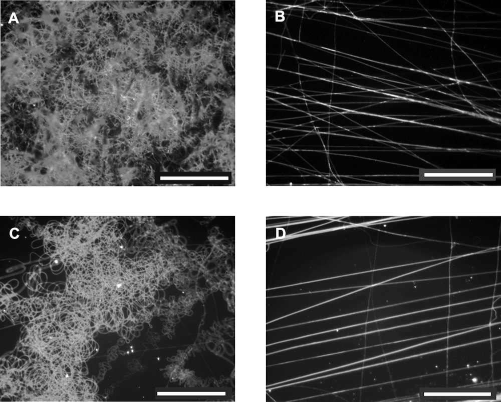
Collagen was introduced into the fiber to improve cell attachment because it mediates cellular recognition via biochemical signals. The presence of collagen at the surface of the nanofibers was investigated using anti-collagen I antibody staining of PCL and C/PCL fibers. Figure 5 presents fluorescence images of stained C/PCL (Fig. 5A, B) and PCL (Fig. 5C, D) fibers. The C/PCL fibers (Fig. 5A) showed more or less equally distributed green fluorescence, indicating the presence of collagen, whereas no fluorescence was seen with the PCL fibers (5C). Non-stained C/PCL fibers did not auto-fluoresce, and second antibody staining without the first antibody was also negative for C/PCL (Fig. 5B) and PCL (Fig. 5D) fibers.
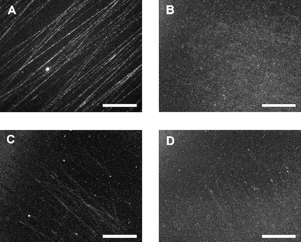
Fluorescent images of fibers stained with collagen-I antibody. (
Protein adsorption of biomaterial influences the response for in vitro and in vivo experiments. Therefore the protein adsorption of the sPEG-substrates with fibers was qualitatively determined by incubating the samples with the rhodamine red–labeled model protein BSA. Figure 6 shows fluorescence images of the substrates. All fibers showed strong fluorescence, indicating protein adsorption, whereas the sPEG-coated silicon wafer underneath, lacking the fluorescent dye, demonstrated the protein-resistant properties of the sPEG coating. GRGDS-sPEG substrates also did not show any fluorescence, whereas non-incubated controls with PCL and C/PCL fibers did not auto-fluoresce (not shown).
Fluorescence images of random and parallel poly(ɛ-caprolactone) (PCL) and a blend of collagen I and PCL (C/PCL) fibers, stained with rhodamine red–labeled bovine serum albumin (BSA) on star (polyethylene glycol)
In vitro tests
In vitro experiments with the electrospun fiber/sPEG substrates were performed to test substrate durability, cell adhesion, survival, and axon guidance. SPEG substrates were covered with PCL and C/PCL fibers in a random and in a parallel orientation. Additionally, parallel C/PCL fibers deposited on cell-adhesive GRGDS-sPEG substrates were prepared. Dorsal root ganglia contain glial cells (Schwann cells, satellite cells) and sensory neurons of the peripheral nervous system. Using dissociated dorsal root ganglia, cell numbers and neurite alignment along fiber direction (random and parallel) were measured to investigate the influence of fiber material and orientation in combination with the effect of cell-repellent (sPEG substrate) or cell-adhesive surface coatings (GRGDS-sPEG substrate).
The substrates were robust during culture, in that fibers did not lift off during media exchanges and sequential washing for immunocytochemisty. In this respect, the reactive sPEG substrate was an excellent and simple method to adhere electrospun fibers. After 4 days incubation in vitro (DIV), DAPI-stained cells that adhered to the substrates were counted. Because of proliferation of glial cells, more than twice as many cells were counted on electrospun fibers after 4 DIV, without significant differences between materials (PCL vs C/PCL) or orientation (random vs parallel) of the electrospun fibers (Fig. 7). Cells did not adhere to or survive on the sPEG substrates between the fibers or on sPEG substrate without fibers. However, with GRGDS-sPEG substrates, cells attached and proliferated on this surface, an observation previously seen and documented. 8

Effect of electrospun nanofibers on peripheral nervous system cell survival and proliferation. Cells from chick dorsal root ganglia (DRG) were dissociated and seeded on randomly orientated (ran) or parallel oriented (par) nanofibers consisting of poly(ɛ-caprolactone) (PCL) or a blend of collagen I and PCL (C/PCL). The numbers of viable cells after 4 days in vitro were counted and expressed as a percentage of the initial population seeded on the fibers (n = 8 explants, error bars indicate standard error of the mean). The more than doubling of cell numbers indicates that both types of fibers, irrespective of orientation, supported survival and proliferation of DRG cells. Cells grew on star (polyethylene glycol) (sPEG)–glycine-arginine-glycine-aspartate-serine substrate with or without fibers but not on pure sPEG.
Orientated nanofibers also directed the growth of regenerating axons from DRG neurons in vitro. After 4 DIV, regenerating sensory axons were fixed and stained immunocytochemically against 200 kD neurofilament protein. Immunostained axons in contact with C/PCL fibers are shown in Figure 8A. The lengths of axon sections that were in direct contact with or that had detached from electrospun fibers were measured, and the percentage of neurite length in direct contact with nanofibers was calculated for every axon (Fig. 8B). Contact guidance on orientated C/PCL fibers was more pronounced (84%) and significantly higher (t-test, p < 0.01) than on PCL fibers (35%) or on randomly orientated C/PCL fibers (57%). Similarly, when C/PCL fibers were attached to the sPEG-GRGDS surface, smaller stretches of neurites were in contact with the fibers (40%). Thus, PCL fibers exhibited a weaker ability to guide axonal growth than C/PCL fibers, and randomly collected C/PCL fibers, where axons frequently encountered nanofibers perpendicular to their path, were less effective than parallel fibers. When the surface beneath the electrospun fibers was modified with GRGDS peptide, which competed for integrin binding on the cell surface, the guidance effect of C/PCL fibers was reduced.
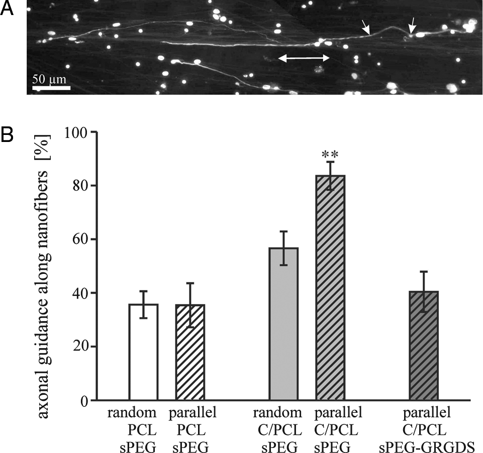
Axon guidance along electrospun nanofibers on different substrates after 4 days in vitro. (
Discussion
Electrospun fibers
The aim of this study was the development of a system that allows investigation of interactions between cells and electrospun nanofibers. Nanofibers with diameters in the range of small-diameter axons and ECM fibrils were produced by electrospinning. High-quality electrospun fibers are homogeneous and bead-free and have a low-diameter distribution. The most important parameter for these high-quality electrospun fibers was the concentration of the polymer solution. Concentrations of at least 7 wt% were needed for the production of suspended parallel fibers, because lower concentrations resulted in beaded fibers (Fig. 3 and 4), as reported previously. 21 Greater concentrations and viscosities correspond with greater interactions between solvent and polymer (and a reduction of free solvent molecules). Thus, when the solution is stretched under the influence of charges, fewer beads occur. 22 Different fiber densities of parallel fibers were possible, by simply varying the collection time. Although we did not test this parameter in detail, our system offers the possibility of investigating the influence of diluted single fibers on single cells as well as cell behavior on highly dense fiber mats.
In vitro substrates
The reactions forming the protein-resistant surface during preparation are shown in detail in Figure 2. The isocyanate end groups of the sPEG molecules react with water to form amino groups, and these react with residual isocyanate groups to form an urea-crosslinked sPEG nanolayer on the substrate. Thus, the initially chemically reactive layer transforms into an unreactive layer, which additionally prevents unspecific protein adsorption due to the PEG backbone. Modification with GRGDS creates a cell-adhesive but still protein-resistant surface. Experiments with the model protein BSA confirmed the protein-resistant properties. PCL and C/PCL fibers showed protein adsorption; in contrast, neither the underlying sPEG substrates nor the cell-adhesive GRGDS-sPEG substrates demonstrated protein adsorption (Fig. 6). GRGDS was used in small amounts (500 μg/mL) and was randomly bound to the sPEG. Approximately 1.2 GRGDS molecules are bound to one star molecule, which has been shown to be enough for cell adhesion 8 but allows the formation of a protein-resistant network upon crosslinking of the remaining sPEG arms. (Bio)materials used in in vivo and in vitro experiments typically come in contact with proteins as long as no special conditions are chosen. Within seconds, proteins interact with the biomaterial and adsorb to its surface depending on the proteins' affinity to the biomaterial surface. 23 As cells interact with these adsorbed proteins, it is important to restrict protein adsorption on the desired material for the successful development of surface-modified materials for in vitro systems and implants. The protein-resistant properties of sPEG-coated substrates allow the investigation of the cellular response specifically on the fibers. In contrast, when oriented fibers have been directly placed onto glass slides, it has been reported to be difficult to distinguish the influence of fibers on cells because the cells attached all to the glass as well to the fibers. 2
Isocyanate endgroups of sPEG molecules additionally react with amine and hydroxyl groups at the fiber surfaces and bind the fibers covalently to the substrate. Potentially, a range of different electrospun materials, not only PCL or collagen, can be readily bound to different targets without being washed off when coming in contact with water. These adherent fibers are strongly bound to the substrate surface and can readily be tested in cell cultures in which frequent media changes are required. No further fixation of the fibers as in other cell experiments2,5,6 is required, and even investigations of single cells and nanofibers are possible. Only a few experiments have been reported using single-cell interactions with individual fibers,2–4 probably because of the difficulties of single-fiber preparations for cell culture. The present investigation demonstrates a method using chemically reactive substrates that is extremely robust for the adhesion of electrospun fibers for in vitro investigations.
In vitro experiments with substrate-fiber systems
Cell culture experiments with dissociated DRG demonstrated that the configuration of electrospun fibers on cell-repellent sPEG surface was well suited to investigate interactions between nanofibers, glia cells, and growing axons.
Cell numbers (Schwann cells and neurons) counted after 4 DIV were not significantly different between the various types of electrospun fibers and confirmed biocompatibility of the substrates. 4 Proteins adsorbed to collagen-containing and pure PCL fibers and played a role in cell adhesion.
Collagen was included in the fibers to provide cell-activating signals9,10 and enhance the positive effects on cell behavior. Recent publications have demonstrated the partial degradation of collagen to gelatin during electrospinning.12,13 We could show the existence of collagen type-I immunoreactivity at the surface of C/PCL fibers using antibody staining (Fig. 5A). Although the staining was weak, a clear difference between collagen-containing C/PCL fibers and pure PCL fibers was observed, and at least some non-degraded collagen seems to be present at the fiber surface. Furthermore, collagen-mediated improvement of bioactivity (cell viability and attachment) of human coronary artery endothelial cells has been demonstrated when using collagen-functionalized poly(
In our experiments, stronger neurite alignment of dissociated DRG cells along C/PCL fibers than along pure PCL fibers indicated biochemical interactions between cell membranes and collagen contained in the fibers and showed the positive effect of collagen. Neurite alignment was significantly higher for parallel C/PCL fibers on a sPEG substrate than on all other samples. Corey et al., who investigated neurite alignment of outgrowing neurites of DRG explants on random and aligned poly-
The surface modification of the sPEG coating with GRGDS altered the axon-guiding properties of the nanofibers, and more than half of the neurites grew without direct contact with oriented C/PCL fibers. There is a competition between the fiber–substrate surface chemistry and the inherent topological guidance of the fiber. 25 These results demonstrate the need for a system in which surface material, fiber material, and fiber orientation can be varied independently because cells react on the combination of all three parameters.
The results demonstrate the potential of combining adhesive oriented fibers with anti-adhesive substrates or matrices. To translate this into in vivo guidance of neurons within tissue engineered devices, we hypothesize that the inclusion of adhesive fibers within a non-adhesive matrix will be likely to assist the guidance of neurons, rather than the inclusion of adhesive fibers within adhesive matrices. However, for nerve regeneration, not only is neurite outgrowth important, but also other cell populations such as Schwann cells, which myelinate axons in the peripheral nervous system and initiate neurite outgrowth and axon regeneration after injuries.26,27 The surrounding matrix should, for example, support Schwann cell proliferation while preventing neurite outgrowth away from the fibers at the same time. With our fiber deposition system, it is possible to collect fibers of different material or with different functionalities and deposit them next to each other on the same samples. These materials can be chosen in such a way that they support the different needs for nerve regeneration (e.g., regrowth of different fiber systems) in addition to the matrix. These requirements can be tested with our fiber substrate system in two dimensions. In a feasible three-dimensional system, fibers could be imbedded into a matrix that has the same properties as the substrate surface in two dimensions. The sPEG system offers an opportunity for this because not only thin surfaces, but also thick hydrogels 28 can be prepared, which may be functionalized in a similar manner. However, such predictions require in vivo validation to correlate the in vitro results with the in vivo performance. Furthermore, the use of collagen IV and other ECM molecules that support the regeneration of damaged peripheral nerve fibers might enhance the guidance. 29 An alternative would be to bind peptide sequences to the fibers, to specifically activate integrin receptors of regenerating neurites. The incorporation of recent technologies for functionalized, protein-resistant fibers will be likely to provide greater insight into combinations that might be successful in guiding neurite outgrowth. 20 In such a scenario, surface chemistry of fiber and substrate can be controlled independently and without non-specific protein adsorption.
Conclusion
An in vitro system was developed to test cellular interactions with biologically functionalized nanofibers in which the chemistry of fibers and the underlying substrate can be varied independently. The system was designed to comprise a chemically reactive coating of the substrate that transformed into a protein resistant sPEG or GRGDS-sPEG layer. Fibers adhered strongly to the surface because of the chemical reaction between sPEG molecules and functional groups at the fiber surface, allowing in vitro experiments with frequent media changes. Culture of dissociated DRGs demonstrated cell behavior with regard to the competitive functionality of the fiber and substrate surfaces. The combination of aligned C/PCL fibers on non-adhesive surfaces was most efficient in guiding neurite outgrowth. The results demonstrate the benefit of having available a system in which different parameters can be varied independently to evaluate competitive parameters regarding cellular response on electrospun fibers.
Footnotes
Acknowledgments
We wish to thank Prof. Hoecker for his assistance with the manuscript. This work was supported by a grant from the Interdisciplinary Centre for Clinical Research “BIOMAT” within the faculty of Medicine at Aachen University (RWTH) (TV B111) and by DFG-Graduiertenkolleg 1035 “Biointerface.”