




Abstract
Fragmin/protamine microparticles (F/P MPs) can be stably coated onto plastic surfaces. A capability of F/P MP–coated plates was investigated to immobilize fibroblast growth factor (FGF)-2 as a substratum to expand human bone marrow–derived mesenchymal stem cells (BMMSCs). FGF-2 molecules in low (2%) human serum (HS) medium were immobilized onto F/P MP–coated plates, and the FGF-2 was gradually released into the medium with a half-releasing time of 4–5 days. BMMSCs adhered well to the F/P MP–coated plates, and grew at a doubling time of about 28 h in low (2%) HS medium with FGF-2 (5 ng/mL), while the cells grew at a doubling time of about 30 and 38 h in high (10%) HS medium and in low (2%) HS medium with FGF-2, respectively, without F/P MP coating. The expanded BMMSCs on the F/P MP–coated plates in low (2%) HS medium with FGF-2 maintained their multilineage potential for differentiation into adipocytes and osteoblasts.
Introduction
Most protocols for the expansion of BMMSCs include high concentrations (10–20%) of serum as a nutritional supplement. In some cell cultures, this involves multiple supplies of culture medium including fetal bovine serum (FBS), which raises concerns over possible infections as well as immunological reactions caused by medium-derived FBS proteins, sialic acid derivatives, and the like.8,9 Thus, patients may experience problems when undergoing autologous cell-based therapies if a serum other than an autologous serum is used during the culturing of the cells. On the other hand, it would be difficult to obtain large amounts of autologous serum from the patient for large-scale autologous cell culture.
Growth factors such as fibroblast growth factors (FGFs), hepatocyte growth factor, and vascular endothelial growth factor are especially immobilized on the extracellular matrix via binding to heparinoid (heparin, low-molecular-weight heparin, heparin sulfate, etc.).10,11 Fragmin (low-molecular-weight heparin) has pharmacological and practical advantages compared to heparin. The lower protein binding affinity of fragmin produces a low, stable, and predictable anticoagulant response, thereby obviating the need for laboratory monitoring to adjust the dosage. 12 On the other hand, protamine, a purified mixture of proteins obtained from fish sperm, neutralizes heparin and fragmin by forming a stable complex that lacks anticoagulant activity. 13 Protamine is also in clinical use as an antidote of heparin to reverse heparin's anticoagulant activity. 13 In this study, we used fragmin as a heparinoid and protamine to prepare fragmin/protamine microparticles (F/P MPs). 14 The purpose of the present study was to evaluate the expansion of BMMSCs on F/P MP–coated plates in low human serum (HS) medium with FGF-2. This BMMSC culturing procedure on F/P MP–coated plates in low HS medium with FGF-2 would be very useful for cell-based therapies, since such a procedure would make it possible to use autologous serum for large-scale autologous cell culture.
Materials and Methods
Preparation of F/P MPs and F/P MP–coated plates
The preparation of F/P MPs was previously described. 14 Briefly, 0.35 mL of protamine solution (10 mg/mL; Mochida Pharmaceutical, Tokyo, Japan) was added drop by drop to 0.7 mL of fragmin solution (6.4 mg/mL; Kissei Pharmaceutical, Tokyo, Japan) with vortexing for approximately 2 min. The mixed F/P MPs, which constituted a milky solution, were then washed twice with phosphate-buffered saline (PBS) to remove nonreactants using centrifugation (6000 rpm, 10 min), and the precipitates finally resuspended 1 mL PBS. More than 7 mg of dry F/P MPs was obtained from 1 mL of the resuspended F/P MPs solution.
Forty-eight-well tissue culture plates (well area, 0.65 cm2) (Sumitomo Bakelite, Tokyo, Japan) were coated for 1 h at room temperature with 0.2 mL of 0.35 mg/mL F/P MPs in PBS solution. The used F/P MPs solutions were removed from the wells by pipetting, and the plates were gently washed with PBS. It was estimated that about 20 μg of added F/P MPs (70 μg) was immobilized onto the each well of the 48-well tissue culture plates by the 1,9-dimethylmethylene blue method using a sulfated glycosaminoglycan assay kit (Blyscan™; Biocolor, Newtownabbey, Northern Ireland). The F/P MPs were stably coated to the plastic surface.
Cell growth assays
BMMSCs were purchased from Takara Bio Corporation (Tokyo, Japan). BMMSCs were cultured in Dulbecco's modified Eagle's medium (DMEM; Life Technologies Oriental, Tokyo, Japan) supplemented with the indicated concentrations of HS (SER019029 from human adults; Biopredic International, Rennes, France) and FGF-2 (Fiblast; Kaken Pharmaceutical, Tokyo, Japan), and antibiotics (100 U/mL penicillin G and 100 μg/mL streptomycin) on F/P MP–coated plates. The BMMSCs used in all experimental analyses were between fifth and seventh passages.
For cell growth assay, BMMSCs were plated at an initial density of 10,000 cells/mL on 48-well tissue culture plates in 0.3 mL of DMEM supplemented with antibiotics and the indicated concentrations of HS and FGF-2 on F/P MP–coated plates, and the cells were cultured for the indicated time periods. After incubation, 200 μL of the used medium including 22 μL of WST-1 reagent (Cell counting kit; Dojindo, Kumamoto, Japan) was added to each well. In here WST-1 is reduced to a soluble formazan dye by dehydrogenase in the cells. The OD values of the produced formazan were then read at 450 nm using an Immuno Mini Plate Reader (Nunc InterMed, Tokyo, Japan) after 1 h incubation. The OD450 values were exactly proportional to the number of BMMSCs (data not shown).
Adsorption of FGF-2 onto F/P MP–coated plates
An ELISA for FGF-2 using F/P MP–coated plates was performed to evaluate the adsorption of FGF-2 onto the F/P MP–coated plates. Low (2%) HS DMEM (100 μL) including the indicated concentrations of FGF-2 was added to the F/P MP–coated 48-well tissue culture plates, and they were incubated with shaking for 2 h at 37°C. Subsequently, the used medium containing the indicated concentrations of FGF-2 was transferred into separate F/P MP–coated plates and incubated again for 2 h at 37°C. The plates were then washed thoroughly with PBS/bovine serum albumin (0.1%) (BSA; Wako Pure Chemical Industries, Osaka, Japan) three times with shaking. Diluted (1:500 with PBS/BSA) anti-FGF-2 (R&D Systems, Minneapolis, MN) was added to the plates and incubated with shaking for 30 min at room temperature. The plates were again washed thoroughly with PBS/BSA, and 200 μg/mL of anti-IgG horseradish peroxidase conjugate (diluted 1:1000 with PBS/BSA) (Bio-Rad Lab, Hercules, CA) was added to the plates and incubated for 30 min with shaking at room temperature. Each well was again washed thoroughly with PBS/BSA, and the color was developed with shaking by adding 300 μL/mL of horseradish peroxidase substrate solution (Bio-Rad). The plates were mixed for 30 min, and 50 μL of sulfuric acid (1 M) was finally added to each well to stop the reaction. The plates were read at 450 nm using the Immuno Mini Plate Reader (Nunc InterMed).
Induction of adipocyte and osteoblast differentiation and staining methods
Adipocyte differentiation and osteoblast differentiation were induced by placing the expanded BMMSCs in each defined medium (defined adipogenic induction medium, defined adipogenic maintenance medium, and defined osteogenic induction medium) (Lonza Walkersville, Walkersville, MD). For adipocyte differentiation, the BMMSCs were cultured in the defined adipogenic induction medium for 4 days followed by 3 days of culture in the defined adipogenic maintenance medium. After three complete cycles of induction/maintenance, the BMMSCs were cultured for 7 more days in the defined adipogenic maintenance medium. For osteogenic differentiation, the cells were cultured with the defined osteogenic induction medium for 14 days.
The adipocyte-induced cells were stained with oil red O (Wako Pure Chemical Industries). 15 Alkaline phosphatase (ALP) cytochemistry for osteoblast-induced cells was carried out by using the substrate for ALP composed of bromo-chloro-indolyl phosphate and nitro-blue-tetrazolium chloride (Sigma Fast; Sigma Aldrich, Stenheim, Germany). 15
Statistical analysis
Data were subjected to statistical analysis by analysis of variance (F-test), followed by Student's t-test. The statistical analyses were carried out using StatMate (Atms, Tokyo, Japan). The level of significance was determined at p < 0.001.
Results
BMMSC growth on FGF-2-bound F/P MP–coated plates
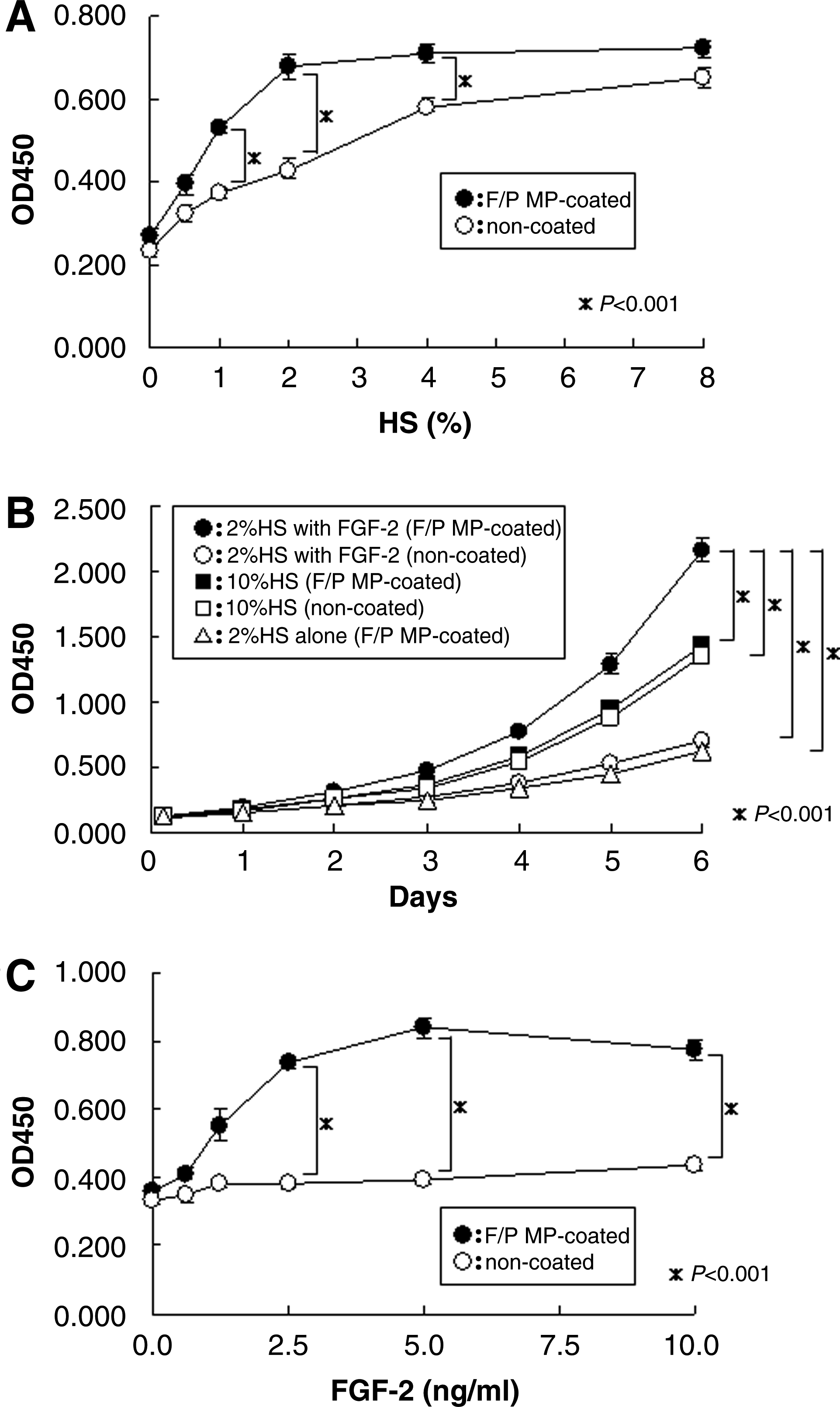
The growth rate of BMMSCs on F/P MP–coated plates was higher than that on noncoated plates in various concentrations (8%, 4%, 2%, 1%, 0.5%, and 0%) of HS with 5 ng/mL FGF-2 (Fig. 1A). BMMSC growth was optimally stimulated on F/P MP–coated plates in low (2%) HS DMEM with 5 ng/mL FGF-2 with a doubling time of about 28 h (Fig. 1B). Yields of BMMSCs after 6 day culturing on F/P MP–coated plates in low (2%) HS DMEM with 5 ng/mL FGF-2 were always two- to three fold higher than that of BMMSCs cultured on noncoated plates with 10% HS, or with low (2%) HS DMEM with 5 ng/mL FGF-2 (in five experiments).
(
BMMSCs were cultured in low (2%) HS DMEM without FGF-2 on F/P MP–coated plates or on noncoated plates onto which the indicated concentrations of FGF-2 was preimmobilized at 4°C for 18 h (Fig. 1C). The growth of BMMSCs was stimulated in a concentration-dependent manner by preimmobilized FGF-2 on F/P MP–coated plates, while cell growth on noncoated plates was not stimulated by similar pretreated FGF-2. Thus, preimmobilized FGF-2 onto the F/P MP–coated plates appeared to be bioactive for proliferation of BMMSCs, since those cells grew well on the FGF-2-immobilized F/P MP–coated plates (Fig. 1C).
BMMSCs cultured on F/P MP–coated plates with 5 ng/mL FGF-2 in low (2%) HS DMEM had similar fibroblast-like morphology as compared to noncoated plates in high (10%) HS DMEM without FGF-2 (data not shown).
Adsorption of FGF-2 onto F/P MP–coated plates
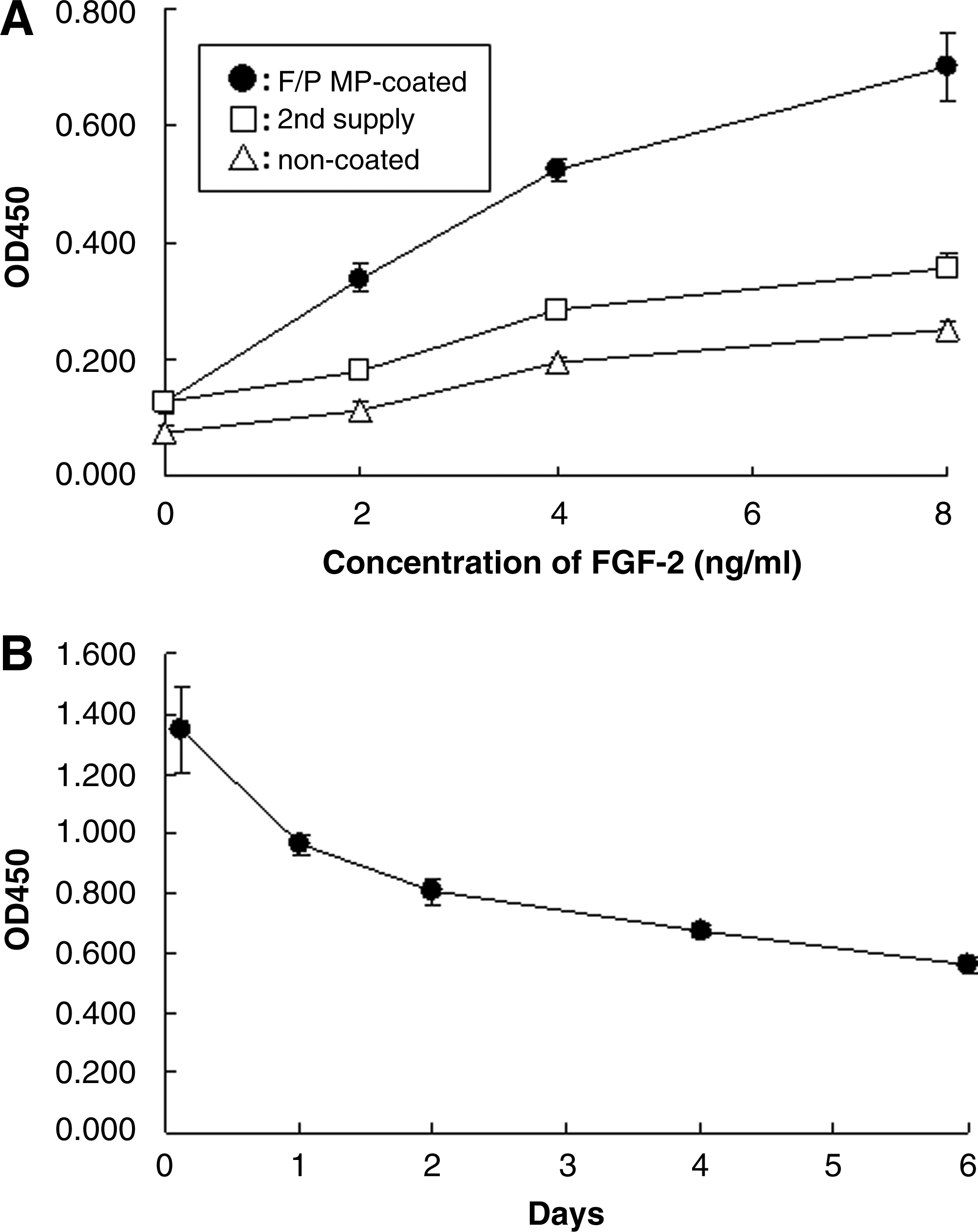
FGF-2 is known to specifically bind to heparinoids.10,11 When 200 μL of various concentrations of FGF-2 in low (2%) HS DMEM was added to F/P MP–coated 48-well plates and incubated at 37°C for 2 h, FGF-2 was detected on the plates in a concentration-dependent manner (Fig. 2A). When the used medium was applied to a second group of plates, the FGF-2 levels of the latter plates decreased to about 53% of the former plates as measured by enzyme-linked immunosorbent assay (ELISA). These results indicated that roughly 0.37 ng of FGF-2 was immobilized onto the initial F/P MP–coated 48-well plates using a concentration of 5 ng/mL FGF-2 in low (2%) HS DMEM (Fig. 2A).
Immobilization and release of FGF-2 onto F/P MP–coated plates. (
When the FGF-2 that was preimmobilized with 5 ng/mL FGF-2 on F/P MP–coated plates at 4°C for 18 h was incubated in low (2%) HS DMEM at 37°C with gentle shaking, approximately 30% of the incorporated FGF-2 was released from the F/P MP–coated plates within 1 day, followed by further gradual release afterward (Fig. 2B). Subsequently, approximately 37% of incorporated FGF-2 remained in the F/P MP–coated plates after incubation for 6 days (the culture medium was changed everyday). Thus, the immobilized FGF-2 was gradually released into the medium over 6 days.
Differentiation assays
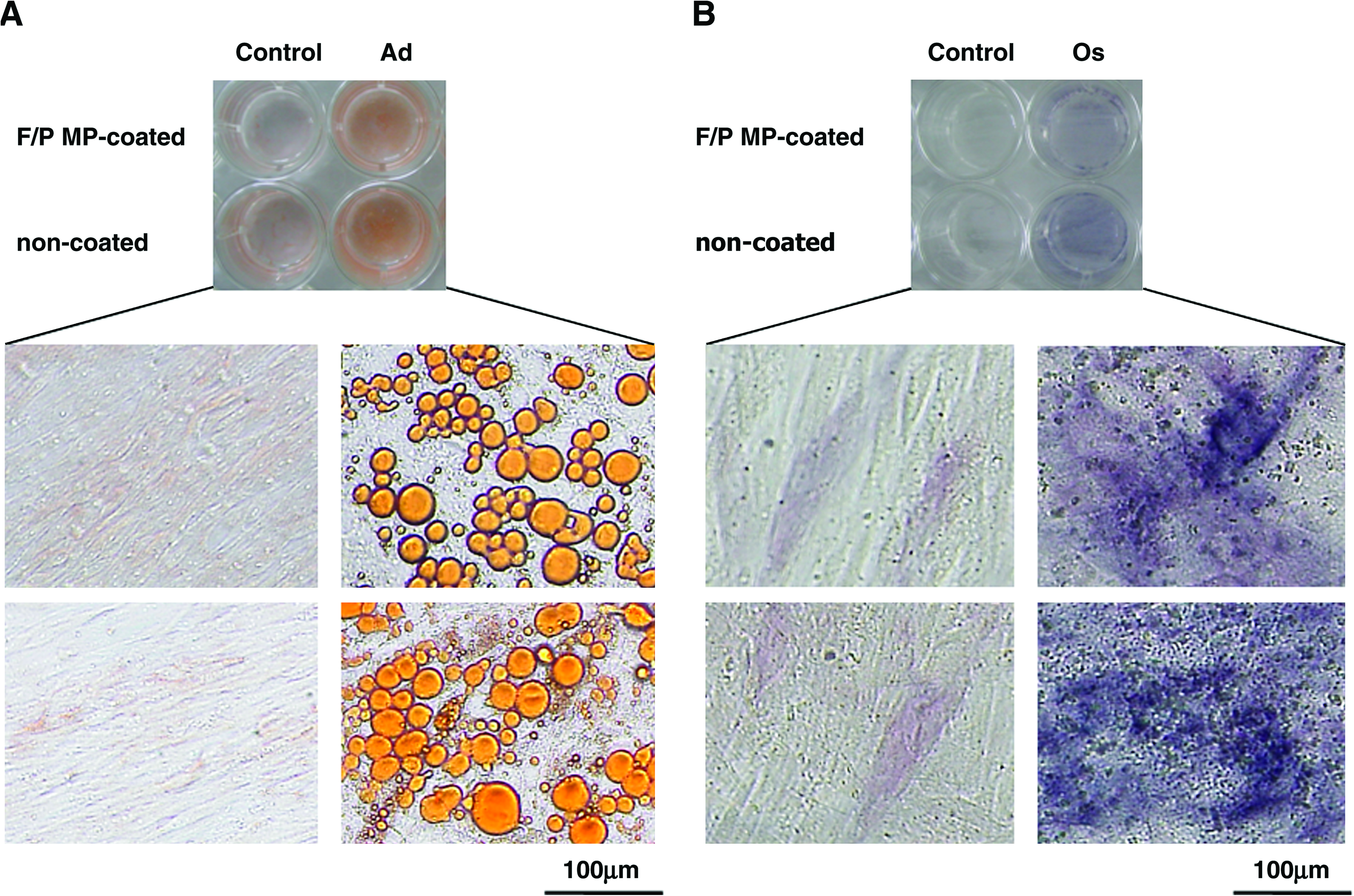
To determine the potential ability of expanded BMMSCs to form adipogenic and osteogenic phenotype cells, BMMSCs were cultured under specific conditions for each type of differentiation. 15 For the adipogenic assay, after cyclic induction with the defined adipogenic induction and maintenance medium, BMMSCs had cytoplasmic lipid droplet accumulation and became adipogenic phenotype cells. Histological examination revealed the formation of small lipid droplets in some cultures after three cycles of treatment. During the culturing, the number of lipid droplet–containing cells increased. The lipid droplet–containing cells were stained with oil red O after 28 days (Fig. 3A). For the osteogenic assay, the expanded BMMSCs were cultured in the defined osteogenic induction medium. ALP activity was observed after 14 days (Fig. 3B).
(
Discussion
We previously produced F/P MPs as a carrier for the controlled release of heparin-binding growth factors and cytokines. 14 F/P MPs can be stably coated onto plastic surfaces. In this study, we investigated the capability of F/P MP–coating as potential substrate for FGF-2 immobilization to stimulate the growth of human BMMSCs. We were also able to effectively expand BMMSCs using low (2%) HS medium on F/P MP–coated plates with FGF-2. In addition, the expanded BMMSCs still maintained their multipotent differentiation ability for adipocytes and osteoblasts.
Fragmin is a low-molecular-weight heparin with anticoagulant activity much lower than that of native heparin, 12 and it was controllable to produce small size of microparticles with fragmin other than heparin (data not shown). Therefore, fragmin was used to prepare the F/P MPs in these studies. Basic protamine molecules complexed with acidic molecules (fragmin) form microparticles through ionic interactions. 14 It seems likely that polypeptides such as FGF-2, once bound to F/P MPs, are gradually released from the particles in vivo (decreasing by half within 5 days). 14
In this study, we showed that F/P MPs are easily coated onto plastic surfaces and exhibit an ability to immobilize heparin-binding growth factors such as FGF-2. 14 The growth of BMMSCs could be stimulated on F/P MP–coated plates with FGF-2. Our results show that the growth of BMMSCs on F/P MP–coated plates in low (2%) HS medium with FGF-2 was optimally stimulated, and that the growth rate was higher than that with high (10%) HS medium without FGF-2 on either F/P MP–coated plates or noncoated plates.
When an autologous serum is used in the autologous cell-based therapies, it would be difficult to obtain large amounts of serum from the patient. It should be noted that similar stimulation was observed for cultured BMMSCs on F/P MP–coated plates in low (2%) HS medium with FGF-2 as that with FBS. The results thus suggest that effective expansion of human BMMSCs may be possible using the present system in low (2%) HS medium with FGF-2.
It is important to evaluate whether the multidifferentiation potential of expanded BMMSCs using the presented method is maintained. Our results clearly show that expanded BMMSCs on F/P MP–coated plates in low (2%) HS medium with FGF-2 did keep their ability to differentiate into adipocytes, and osteoblasts in vitro. The presented method for culturing BMMSCs may provide a safe and effective expansion of cell source, especially in the preparation of large amounts of BMMSCs that are needed for cell-based therapies in several clinical fields.
Footnotes
Disclosure Statement
No competing financial interests exist.