
Abstract
In the present study, we applied and optimized a heparin-based hydrogel system, formed by thiolated heparin and diacrylated poly (ethylene glycol), for three-dimensional chondrocyte culture. Encapsulation by the heparin-based hydrogel did not affect the chondrocyte viability (better than calcium-induced alginate gel), and the heparin-based hydrogel promoted chondrocyte proliferation, while maintaining chondrogenic nature. Phenotypic analyses, such as glycosaminoglycan accumulation and histological staining, also supported the proper role of the heparin-based hydrogel for cartilage regeneration; a continuous increase in glycosaminoglycan amount was observed during the culture period. At the transcriptional level, the gene expression of type II collagen and Sox-9 was maintained, whereas type I collagen expression was not observed. The chondrocyte expansion was affected by the gel strength, and there existed an optimum gel concentration for it. Based on the results, the heparin-based hydrogel is a promising material for chondrocyte culture, potentially applicable for cartilage regeneration.
Introduction
Previously, we reported the development of an in situ–forming heparin-based hydrogel formed by a Michael-type addition reaction between thiolated heparin (Hep-SH) and diacrylated poly (ethylene glycol) (PEG-DA). 20 Heparin is a highly negatively charged natural polysaccharide, composed of heterogeneous mixture of sulfated D-glucosamine and D-glucuronic acid. Based on their specific binding with various growth factors, heparin-containing systems have been studied for the controlled release of growth factor and the regeneration of injured tissue based on it.21,22 However, application of heparin as a main component of tissue regeneration scaffold has been relatively limited.23,24 Here, we characterized and optimized the chondrocyte culture in the in situ–forming heparin-based hydrogel, suggesting this hydrogel as a new system for cartilage regeneration.
Materials and Methods
Materials
Heparin (sodium salt, from porcine intestinal mucosa, Mw 12 kDa) was purchased from Cellsus Ins. (Cincinnati, OH). PEG-DA (Mw 6 kDa, degree of substitution 98%) was purchased from Sunbio (Anyang City, Korea). Sodium chloride, potassium chloride, potassium phosphate monobasic, sodium phosphate dibasic, agarose, chloroform, isopropanol, acridine orange (AO), propidium iodide (PI), and ethanol were from Sigma Chemical (St. Louis, MO). Phosphate buffered saline (PBS, 0.01 M PBS solution with 0.138 M NaCl and 0.0027 M KCl, pH 7.4) was prepared with potassium phosphate monobasic and sodium phosphate dibasic. As a cell culture medium, Dulbecco's modified Eagle's medium (DMEM) with 4500 mg/L of D-glucose and glutamine, fetal bovine serum (FBS) (certified) (10%), penicillin G, and streptomycin sulfate was used (all from Gibco, Grand Island, NY). Trypsin–ethylenediaminetetraacetic acid (EDTA) (0.25%) was also purchased from Gibco. For cell viability assay, AO and PI were from Molecular Probes (Eugene, OR). Safranin-O and hematoxylin were purchased from Sigma (St. Louis, MO).
Preparation of the heparin-based hydrogel
Heparin-based hydrogels were prepared by a Michael-type addition reaction between Hep-SH and PEG-DA, as previously reported by us. 20 Thiol derivatives of heparin (Hep-SH) with 40% conversion of COOH group to thiol group were prepared. Briefly, heparin was dissolved in double-distilled, deionized water at a concentration of 10 mg/mL. About 1.75-fold molar excess of 1-Ethyl-3-[3-dimethylamino]propyl] Carbodiimide (EDC) and 1-hydroxy-benzotriazole hydrate (HOBT), and 2-fold molar excess of cysteamine (per COOH of heparin) were added to this solution, and pH was adjusted to 6.8 with 0.1 M NaOH and/or HCl solution. After 5 h reaction at room temperature, the reaction solution was thoroughly dialyzed using a dialysis cassette (slide-A-Lyzer Dialysis Cassette, 7.5 kDa Mw-cutoff; Pierce, Rockford, IL). A 10-fold molar (moles per COOH of heparin) excess of dithiotreitol (DTT) was added to reduce the oxidized disulfide groups to free thiol groups for 3 h at pH 7.5, and then pH was adjusted to 3.5 by the addition of 1.0 N HCl. The acidified solution was dialyzed against dilute HCl (pH 3.5) containing 100 mM NaCl, followed by dialysis against dilute HCl at pH 3.5. Hep-SH was further purified by running a PD-10 desalting column (Amersham Biosciences, Piscataway, NJ) and lyophilized. Previously, we reported that the molar ratios among reactants were diversified to control the modification of carboxylic groups with an excess of dithiotreitol (DTT). 20 After attachment of cysteamine to heparin, H in two methylene units in cysteamine were clearly observed in 1H NMR spectra. 25 The amount of thiol group attached to heparin was measured using the molar absorptivity of Ellman's reagent at 412 nm.
Then, heparin-based hydrogels were made by dissolving Hep-SH and 6 kDa PEG-DA (1:1 molar ratio of thiol group and acrylate group) in cell culture medium to make 5%, 10%, or 15% (w/v) solution and filtered through a 0.2 μM sterile filter. In all cases, hydrogels were made within 10 min at 37°C and pH 7.4. More detailed characterization on the gelation kinetics was reported previously. 20 In comparison, the PEG-based hydrogels were also prepared by reacting tetra-functional poly (ethylene glycol) sulfhydryl (10 kDa) and 3.4 kDa PEG-DA (1:1 molar ratio of thiol group and acrylate group) to make the same concentration (10% [w/v]) hydrogels with similar mesh size and mechanical properties of the heparin-based hydrogel. The PEG-based hydrogel and the heparin-based hydrogel prepared at this condition were characterized to have the similar mesh size, based on the gelation kinetics, gel strength, swelling ratio, and the theoretical estimation of the mesh size, as reported previously by us. 25 Alginate hydrogels were also prepared at 1.2% alginate solution with 0.15 M NaCl, 0.025 M HEPES, and 0.10 M CaCl2.
Mechanical properties of the heparin-based hydrogels
Moduli of the unswollen and swollen heparin-based hydrogels at various concentrations were measured (Table 2) using a rheometer (Gemini; Malvern Instruments, Worcestershire, United Kingdom), equipped with a temperature controller at 37°C and a solvent trap. Samples were analyzed with sandblast parallel plate geometry. An angular frequency of ω = 1 rad/s and strain of γ = 0.1% were selected to ensure a linear regime of oscillatory deformation. 20 Moduli of unswollen gels were measured after gelation in the sample holder. In the case of swollen gels, the preformed gels were swollen in the cell culture media overnight at 37°C, and were cut to fit the sample holder geometry of the rheometer and measured.
Isolation of articular chondrocytes
Two-week-old New Zealand White rabbits were purchased from Samtako Bio Korea (Osan, Korea). Rabbits were maintained and treated in accordance with the National Institutes of Health Guide for the Care and Use of Laboratory Animals with the institutional animal care and use committee approval. They were housed in a room with controlled temperature and photoperiod (10 h of dark and 14 h of light, with lights on from 06:00 to 20:00 h). Rabbit articular chondrocytes were isolated from the cartilage of rabbits as described previously. 26 Cartilage slices were dissociated enzymatically for 4 h in DMEM with 0.2% collagenase type II (Sigma). Individual cells were suspended in DMEM supplemented with 10% (v/v) FBS, 50 μg/mL streptomycin, and 50 units/mL penicillin. Then, cells were plated on culture dishes at a density of 5 × 10⁴ cells/cm2 at 37°C in a standard cell culture condition (humidified atmosphere of 5% CO2).
Chondrocyte culture in the heparin-based hydrogel
For chondrocyte culture in the hydrogel, chondrocytes (passage 1) were encapsulated into the hydrogel by adding cells to the precursor solution during gelation. About 50 μL of the gel precursor (1:1 molar ratio of thiol group and acrylate group of Hep-SH and PEG-DA) solution at various concentrations (5%, 10%, or 15% [w/v]) containing cells (2 × 10⁶ cells/mL or 6 × 10⁶ cells/mL) was poured into a 96-well plate with an ultralow attachment surface (Corning, Midland, MI), and incubated for 30 min at 37°C for cell-encapsulated gel formation. Then, 200 μL of cell culture medium, composed of DMEM supplemented with 10% (v/v) FBS, 50 μg/mL streptomycin, and 50 units/mL penicillin, was added to the gel-formed culture wells and cultured in a standard cell culture condition. The medium was replaced with the fresh one for every 2–3 days.
Viability and proliferation of encapsulated cells
The viability of chondrocytes inside the hydrogel was determined by double-staining procedure using AO and PI. 27 Stained cells were imaged using a fluorescence microscope (DM-IRB; Leica Microsystems, Wetzlar, Germany).
The proliferation of chondrocytes inside the hydrogel was characterized by measuring cell metabolic activity with Cell Proliferation Reagent WST-1 assay (Roche, Basel, Switzerland). 28 At each measurement point, WST-1 was added for 2 h, and the colorimetric absorbance of the produced formazan at 450 nm, directly proportional to the total number of viable cells, was measured using a scanning multiwell spectrophotometer (FL600; Bio-Tek, Winooski, VT) with hydrogel containing no cells as a control. Cell numbers were calculated based on a standard curve.
RNA isolation and reverse-transcriptase polymerase chain reaction analysis
RNA extraction of the chondrocytes encapsulated in the hydrogel was done using TRI REAGENT (Molecular Research Center, Cincinnati, OH) and reverse-transcribed with ImProm-IITM reverse transcriptase (Promega, Madison, WI), according to the manufacturer's protocol. 29 RNA pellets were dissolved the RNase- and DNase-free water, and the quality of RNA was ensured by the 260/280 absorbance ratio. The quantity of RNA was estimated by absorbance at 260 nm. RNA was also checked by the electrophoresis of 1% agarose gel. About 1 μg/μL of RNA was reverse-transcribed with MoMLV-RT (Invitrogen, Purchase, NY) for 5 min at 25°C for annealing and for 60 min at 42°C for extending the first strand, as described previously. 30 About 1 μg/μL of cDNA was amplified in a total volume of 10 μL polymerase chain reaction (PCR) containing 20 mM Tris-HCl (pH 8.4), 50 mM KCl, 1 μM of the respective primers, 200 μM dNTP, and 2.5 units of Taq DNA Polymerase (Takara Bio, Kyoto, Japan). PCR primers and conditions are summarized in Table 1. The magnesium chloride concentration was 1.5 mM. Amplifications were carried out in an Eppendorf master cycler under the following conditions: denaturation for 3 min at 94°C, followed by cycles of 20 s denaturation at 94°C, 20 s annealing at the primer-specific temperature, and 15 s elongation at 72°C. PCR products were electrophoresed on 1% agarose gels and viewed after ethidium bromide staining.
GAPDH, glyceraldehyde 3-phosphate dehydrogenase.
Quantification of sulfated glycosaminoglycans
The amounts of sulfated glycosaminoglycans (GAGs) were estimated using dimethylmethylene blue metachromatic assay (Sigma). 31 In brief, the color reagent was prepared by dissolving 16 mg of dimethylmethylene blue in solution (1000 mL, pH 3.0) containing 3.04 g of glycine (Sigma), 2.37 g of NaCl, and 95 mL of 0.1 M HCl. The chondrocyte-cultured hydrogels were immersed in a digestion solution with 500 μg/mL papain (Sigma) in 0.1 M sodium phosphate (pH 6.2) with 5 mM Na2EDTA (Sigma), and 5 mM cysteine-HCl (Sigma). After overnight incubation at 60°C for digestion, they were centrifuged at 17,000 g for 10 min. The supernatant was collected and stored at 4°C until the GAG assay was performed. The total GAGs were determined using a UV–Visible spectrophotometer at 525 nm by comparing with a standard curve of shark chondroitin sulfate (Sigma) in the range of 0–50 μg/mL. The absorbance at 525 nm from the heparin-based hydrogel itself was considered as a baseline; therefore, the GAG amount was estimated using the additional intensity from the deposited GAG in the hydrogel.
Histology
The characteristics of chondrocytes cultured in the heparin-based hydrogel for 2 weeks were analyzed colorimetrically by Safranin-O staining. Briefly, samples were embedded in Optimal Cutting Temperature (OCT) compound (Tissue-Teks; Sakura Finetek, Kyoto, Japan) and frozen. The cell-cultured hydrogels were cut into 10-μm-thick sections at –20°C, and mounted on glass slides. The Safranin-O staining was done by the modified protocol as previously described. 32
Immunohistochemistry and immunofluorescence staining
Evidence for chondrogenesis was also assayed based on the immunohistochemical detection of collagen type II and aggrecan, the primary matrix proteins of articular cartilage, and collagen type I as a negative control. Cryo-sectioned samples (10 μm) after 4-week culture were placed in methanol, acetone, and formalin (19:19:1) solution for 30–40 min, and were fixed on the slide. After washing with PBS, the samples were blocked with 3% H2O2 for 10 min. The sections were then placed in 1% bovine serum albumin for 30 min at 37°C. The primary antibodies (Chemicon, Temecula, CA), such as type II collagen, aggrecan, and type I collagen, were applied and allowed to react overnight at 4°C. The sections were then incubated with the secondary biotinylated anti-mouse/anti-goat antibodies and peroxidase-labeled streptavidin (LSAB2 System; Dako, Carpinteria, CA). After color development using the Histonstain-SP with AEC (Dako), the cover slips were applied and the slides were optically monitored (TE2000-U; Nikon, Tokyo, Japan).
Immunofluorescence staining was also done for the chondrocyte-cultured heparin-based hydrogel after 14 or 28 days of culture. The samples were incubated with 1% bovine serum albumin to block unspecific binding sites, rinsed, and then incubated with specific primary antibodies for 1 h at 37°C. After binding of the primary antibodies, the samples were washed with PBS and then incubated for 1 h at room temperature with the secondary fluorescence-labeled antibody, Alexa-Fluor 488 Goat Anti-Mouse IgG (Invitrogen). Finally, the samples were counterstained with 4′,6-diamidino-2-phenylindole (DAPI) to see the location of cells and viewed by fluorescent microscopy (TE2000-U; Nikon). In both immunohistochemical and immunofluorescence staining, heparin-based hydrogel without cells was used as a negative control. In the case of collagen I staining, collagen type I–coated slides were also used as a positive control.
Statistical analysis
Data were statistically evaluated by Student's t-test. The minimum level of significance was set at p < 0.05.
Results
Initial cell viability and proliferation after encapsulation
Cell viability after encapsulation in the heparin hydrogel was analyzed, and compared with PEG hydrogel and alginate hydrogel (Fig. 1). Fluorescence staining revealed a uniform distribution of the cells throughout the heparin hydrogel and high cell viability (above 90%) in 2 h after encapsulation, which indicates that the present gelation was nontoxic to chondrocytes during encapsulation. PEG hydrogel showed similar distribution and viability (above 90%) to the heparin hydrogel. In contrast, a significant cell death was observed in the case of alginate gel (less than 70% of viability),33–35 which is the most commonly used hydrogel for chondrocyte culture. Thus, the present gelation method used for the heparin hydrogel and PEG hydrogel provide a better environment for initial cell encapsulation than calcium-ion induced gelation of alginate for chondrocytes.
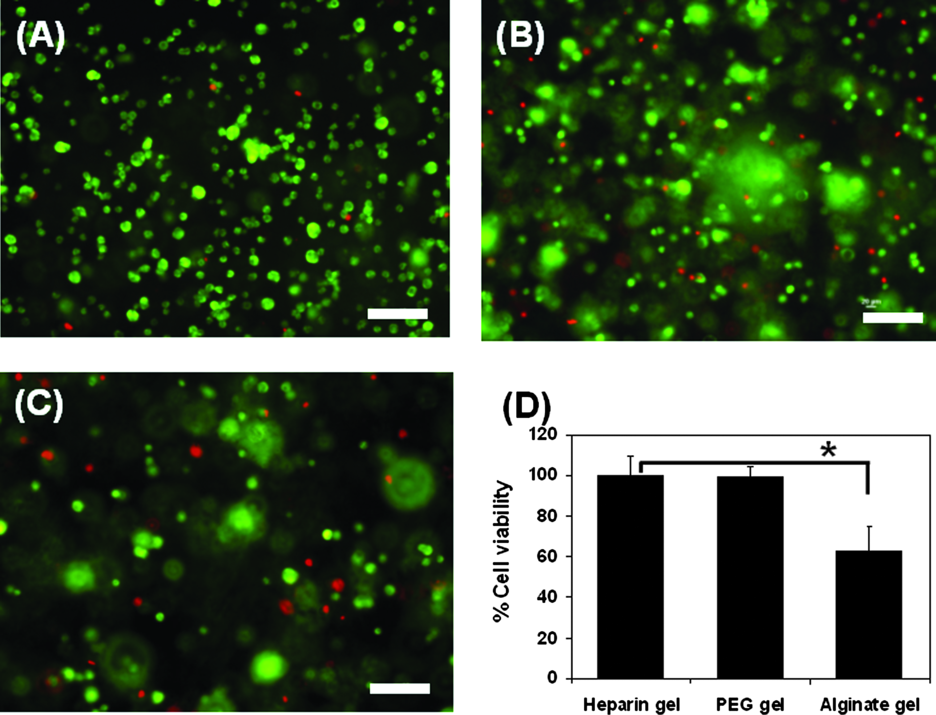
Chondrocyte viability in 2 h after encapsulation. (
In hydrogel constructs, the initial cell density and the mechanical strength of the hydrogel have been shown to affect the cell proliferation, and the extracellular matrix (ECM) content of collagens and GAGs.36,37 Proliferation of chondrocytes at two different cell seeding densities (2 × 10⁶ and 6 × 10⁶ cells/mL) was analyzed in the heparin hydrogels prepared at various precursor concentrations to control the mechanical properties. Modulation of the total precursor concentration resulted in the systemic control of the gel strength. By increasing the total concentration from 5% to 15% (w/v), the elastic modulus after gelation increased more than fourfold (from less than 3 × 10³ to over 104 Pa) (Table 2). Since the hydrogels remained swollen in culture medium during chondrocyte culture, the fully swollen hydrogels in cell culture medium at 37°C were also characterized. After swelling, the strength of the gel decreased significantly, ∼25% of the initial strength.
Chondrocytes proliferated well in the heparin-based hydrogels in all cases with several gel precursor concentrations and two different cell seeding densities (Fig. 2). In terms of cell seeding density, chondrocytes in the hydrogels with the higher initial cell seeding density showed better proliferation than those with the lower initial cell seeding density. With the higher initial cell seeding density, cells proliferated continuously for 14 days (Fig. 2B), whereas the saturation in cell number was observed in 10 days for the hydrogels with the lower cell seeding density, and even a decrease was observed in 14 days for 10% (w/v) hydrogel (Fig. 2A). Thus, to achieve a large cell population in culture, it was necessary to use a high initial cell seeding density.
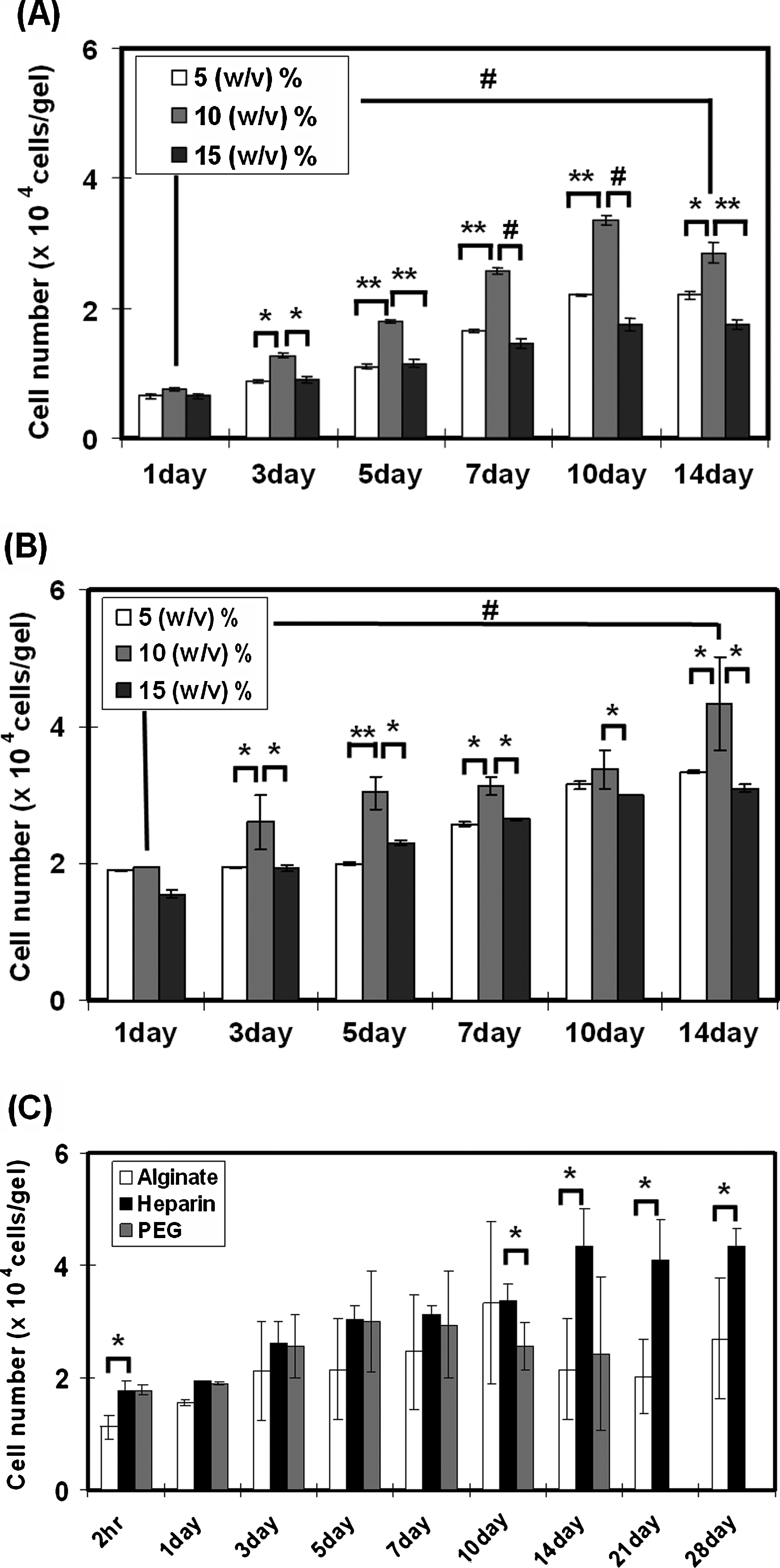
Chondrocyte proliferation in the hydrogel, measured by WST-1 assay. (
The effect of gel strength was more significant (Fig. 2A, B). Among three different precursor concentrations of the hydrogels, 10% (w/v) hydrogel revealed the best proliferation of chondrocytes compared to other concentrations at both cell seeding densities, and the differences were statistically significant, which suggests that there exists optimum gel strength for chondrocyte proliferation in the heparin hydrogel. This result is somewhat unexpected since it has been reported that the stronger gel is better for chondrocytes culture and the gel strength at 10% (w/v) is much weaker than that of natural articular cartilage.2,37 The proliferation of chondrocytes and the maintenance of chondrogenic phenotype inside the hydrogel were also confirmed by light microscopic images at 1, 7, and 14 days of culture (Fig. 3). After 14 days, cell number increased significantly (as quantitatively analyzed in Fig. 2B), and maintained their round morphology inside the heparin hydrogel.

Cell morphology inside the heparin-based hydrogel after culture for up to 14 days, observed by the contrast microscope. Scale bar: 100 μm.
As shown in Figure 2C, chondrocytes proliferated moderately in all kinds of hydrogels. However, the chondrocytes in the heparin hydrogel proliferated better than in the PEG hydrogel and the alginate hydrogel. Cell proliferation inside the PEG hydrogel started to decrease after 7 days, and the difference between PEG hydrogel and heparin hydrogel became significant in 10 days. In addition, the chondrocyte-cultured PEG hydrogel did not maintain its integrity, but was degraded in 14 days. Chondrocytes cultured in the alginate hydrogel also showed a much lower proliferation than those in the heparin-based hydrogel. These cell proliferation results demonstrate that the heparin-based hydrogel is suitable for chondrocytes culture, and in terms of proliferation, it might be better than the alginate hydrogel. Also, the results show that the gel strength is an important parameter for chondrocyte proliferation in the heparin-based hydrogel.
Reverse-transcriptase PCR analyses
The expression of chondrogenic marker genes in the heparin hydrogels was analyzed by reverse-transcriptase PCR (RT-PCR). The initial state of chondrocytes used for culture inside the hydrogels revealed a relatively good chondrogenic state (strong expression of collagen II and Sox-9 vs. weak expression of collagen I); thus, cells were rather close to a primary state (Fig. 4). Data also illustrate evidently the higher expression levels of Sox-9 and collagen II, and the almost complete suppression of collagen I expression from cells cultured in all heparin hydrogels at all time points regardless of precursor concentration. Among three concentrations of the hydrogel, the faint expression of collagen I might be observable until day 14 in the 5% (w/v) hydrogel, whereas no type I expression was observed in the 10% or 15% in day 14. However, more detailed analyses will be necessary to characterize the effect of hydrogel concentration on the gene expression of cultured chondrocytes in the hydrogels. In any case, Figure 4 shows that the effect of hydrogel concentration was not significant, and further it shows that 10% and 15% gels are at least equal or better than 5%. This result did not conflict with the results of cell proliferation (Fig. 2) and GAG production (Fig. 5, will be discussed later); thus, further analysis, for example, real-time PCR, will not affect finding the optimal concentration of the hydrogel for chondrocyte culture. Although cell-to-cell interaction of chondrocytes is known to be crucial to inhibit the dedifferentiation into fibroblast-like cells, the poor proliferation rate in hydrogel is also one of the major problems for in vitro chondrocyte culture, often resulting in dedifferentiation. 38 The results of cell proliferation and RT-PCR analysis for the present system suggest that the heparin hydrogels at all precursor concentrations enabled the expansion of chondrocytes in vitro while maintaining the chondrogenic phenotype.
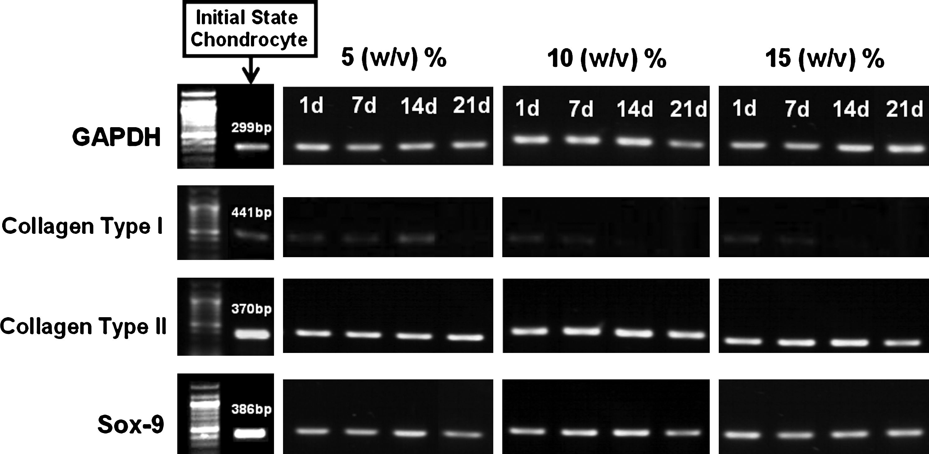
mRNA expression of the heparin hydrogels with different total precursor concentrations.

Glycosaminoglycan (GAG) contents of the heparin hydrogels produced by chondrocytes. (
GAG contents produced by chondrocytes in the heparin hydrogel
The GAG contents inside the chondrocyte-cultured hydrogels were estimated. As shown in Figure 5, the heparin hydrogels at all concentrations showed the increase in GAG amounts by culturing. Noticeably, 10% (w/v) hydrogel possessed the significantly higher GAG content than other concentration hydrogels (Fig. 5A). At day 28, the GAG contents increased more than 22-fold for the 10% (w/v) hydrogel, compared to eightfold increase for the 5% (w/v) hydrogel and 12-fold increase for the 15% (w/v) hydrogel. This result well coincides with the highest cell proliferation for the 10% (w/v) heparin hydrogel (Fig. 2). Figure 5B shows the continuous increase (up to 28 days) in the GAG contents even after normalizing to the cell numbers for the 10% (w/v) heparin hydrogel. Therefore, after cell proliferation of chondrocytes was somewhat saturated in 14 days, the cells produced GAGs continuously during further culturing. Thus, the cellular activities of chondrocytes in the heparin hydrogel were proven by the produced GAGs, and the effect of gel strength on them was also confirmed by the quantitative estimation of GAG amounts.
Histological analyses and immunostaining of the chondrocyte-cultured heparin hydrogel
During the regeneration of cartilage tissue, individual cartilage-specific morphological properties and structural characteristics, such as lacunae, and collagen type II and aggrecan expression are generally observed. 39 In Figure 6, round cells with a diameter of 8–10 μm in lacunae, generally present as a single or clusters containing two or more cells, were observed after 14 days of culture, even though the cryo-sectioning of the cell-cultured hydrogel caused the expansion and the fragmentation of the hydrogel. This image also indicates the preservation of the chondrogenic phenotype of cells cultured in the heparin hydrogel.
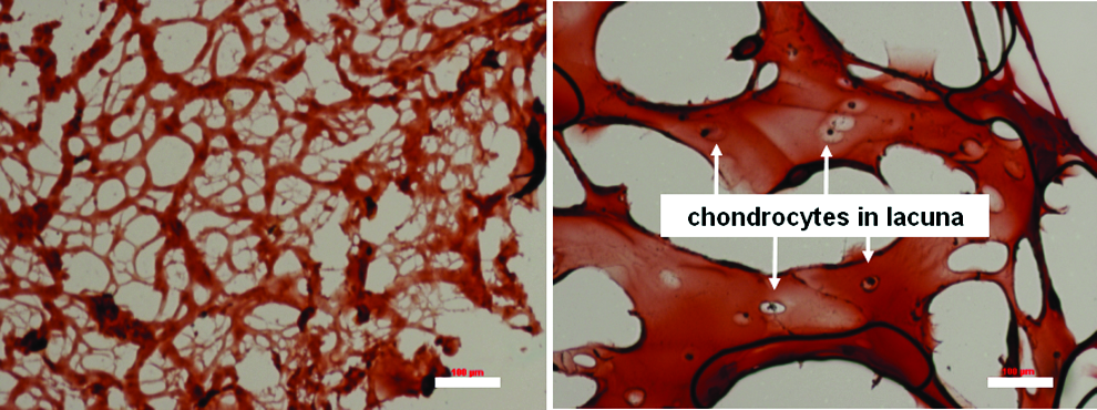
Optical microscopy image of lacunae at 14 days (right) with Safranin-O staining after cryo-sectioning, compared to heparin hydrogel without chondrocytes (left). Scale bar: 100 μm. Color images available online at
In addition to the gene-level expression of the proper ECM (Fig. 4), the actual deposition of the proper ECM by the cultured chondrocytes in the heparin hydrogel was also confirmed by immunohistochemical and immunofluorescence staining. In general, immunostaining illustrates the morphology and the distribution of chondrocytes, and the distribution and the intensity of extracellular components. Although the cryo-sectioning of the hydrogel construct made the expansion and the fragmentation of the system, thus resulting in the lower densities of cells and ECMs compared to those in culture condition, it was evident that a progressive increase in the expression of type II collagen and aggrecan, major characteristic marker proteins of hyaline and elastic cartilage tissue, was observed from 2 to 4 weeks (Fig. 7). In addition, chondrocytes were distributed throughout the whole hydrogel with similar density. Under the cryo-sectioned state of the hydrogel construct, chondrocytes were surrounded by a layer of cell-associated matrix with intense staining of type II collagen, but little or no type II collagen staining was found in the interterritorial matrix, in accord with previous reports.40,41 The interterritorial space between chondrocytes seemed to mainly consist of heparin hydrogel itself, similar to the case of alginate gel. 39 Upon immunofluorescence staining (right side of Fig. 7), the cells were also stained positively for collagen II and aggrecan (green color), and the most intense signal was found in intracellular space surrounding the nuclei (blue color by DAPI). Also, the intensities of collagen II and aggrecan staining increased significantly from 2 to 4 weeks. These staining results also showed a good agreement with the quantitative estimation of GAG amounts (Fig. 5). In contrast, collagen type I was not detected in heparin hydrogels at any culture time (only blue color in immunofluorescence staining from nuclei of cells by DAPI, but no green color from collagen type I). Overall, histological observations of ECM production by the encapsulated chondrocytes were consistent with the results from the biochemical analyses as well as immunohistochemical and immunofluorescence staining, all supporting the good expression and secretion of proper ECM materials by chondrocytes cultured in the heparin hydrogel.

Immunohistochemical (left) and immunofluorescence (right) staining for type II collagen, aggrecan, and type I collagen of cryo-sectioned, chondrocyte-cultured heparin hydrogel (10% [w/v]) for 2 and 4 weeks. Control group: N, negative control stained with heparin hydrogel without cells; P, positive control stained with collagen type I–coated slide. Scale bars: 500 μm. Color images available online at
Discussion
For three-dimensional culture of chondrocytes in vitro, various kinds of hydrogels, including alginate gel,42,43 agarose gel, 44 fibrin gel,45–47 chitosan gel, 48 various peptide gels,49,50 and synthetic polymer gels,51–53 have been studied. Fibrin gel cultured with bovine chondrocytes showed enhanced cell proliferation and GAG production using a high seeding density (125 × 106 cells/mL) compared to a low density (12.5 × 10⁶ cells/mL) in 5 weeks. 45 Agarose gel (2%) also showed the higher GAG production from a high cell density (60 × 106 cells/mL) than from a low cell density (10 × 106 cells/mL) in 7 weeks. 44 So, typically, a cell seeding density above 15 × 106 cells/mL was used for chondrocyte culture. Bovine chondrocytes cultured in fibrin gel with 125 × 106 cells/mL showed enhanced cell proliferation (<2-fold) and GAG production (4-fold) in 4 weeks. 46 In addition, bovine chondrocytes cultured in photo-polymerized PEG hydrogel with 75 × 106 cells/mL resulted in twofold increases both in DNA and GAG amounts from 2 to 4 weeks. 51 In another case of a PEG-based hydrogel (20 × 106 bovine chondrocytes/mL), the DNA content increased over twofold, but the GAG amount decreased from 3 to 6 weeks. 52 Bovine chondrocytes cultured in alginate gel with 15 × 106 cells/mL showed a threefold increase in cell number for 6 week culture. 43 In contrast, with a low cell density (2 × 10⁶ cell/mL), no cell proliferation was observed in alginate gel. 42 In the case of chitosan gel with 50 × 106 bovine chondrocytes/mL, no increase in GAG content was observed from 2 to 4 weeks. 48 Therefore, compared to previous reports, the present result of the heparin-based gel showing a more than twofold increase in cell number and over 20-fold increase in GAG amount in 2 weeks using 6 × 10⁶ cell/mL suggests the excellent chondrogenic potential of the heparin-based hydrogel. An apparently good chondrogenic characteristic of the heparin-based hydrogel is not clearly understood here. Some suggested reasons include (1) an anionic nature and relatively similar structure of heparin with other well-studied polysaccharides for chondrocytes, such as hyaluronic acid and alginate, and (2) the potential role of heparin-based hydrogel as a reservoir for secreted growth factor by chondrocytes 54 inside the hydrogel, thus affecting chondrocyte culture, in turn. However, more rigorous experiments will need to validate these effects.
The present system, composed of heparin as a main component and PEG as a cross-linker, potentially can combine the advantage of tissue compatibility and biological activities of the natural polysaccharide component, which aids integration with cells or biomolecules, with the use of synthetic cross-linker that enables more reproducible and finely tunable hydrogel preparation, contrary to typical naturally sourced hydrogels. Previously, we also proved the biocompatibility and the inertness of the heparin-based hydrogel; no sign of inflammation or bleeding was observed from the in vivo implantation experiment in mice. 55 So, the heparin-based hydrogel can be an interesting candidate for cartilage regeneration material. However, it should be also noted that the mechanical properties of the present hydrogel are much weaker than the natural cartilage, similar to other hydrogel systems. Therefore, the chondrocyte-encapsulated heparin gel alone will not be appropriate to substitute the cartilage defect in terms of both mechanical and biological functions. But, the present system as a cell-seeded, in situ–forming gel is applicable to autologous chondrocytes transplantation therapy as a chondrocyte-injecting system. Application of the present system would increase the cell seeding efficiency, and may eliminate the need of using periosteum during autologous chondrocyte transplantation therapy. In addition, by combining with a solid, porous scaffold, the weak mechanical properties of the hydrogel could be overcome. The future direction of the work is to explore the composite system composed of a porous scaffold having similar mechanical properties with the natural cartilage and the present hydrogel, which can provide the efficient cell seeding into a porous scaffold as well as a good chondrogenic environment.
Conclusion
A heparin-based hydrogel, formed by Michael-type conjugate addition, was applied for chondrocyte culture. In situ–forming heparin-based hydrogel could encapsulate the chondrocytes with minimal cytotoxicity. Also, it was shown that the heparin hydrogel could support the chondrocyte proliferation and maintenance. The expansion and ECM production of chondrocytes cultured in the heparin hydrogel were significantly affected by the precursor concentration (the strength) of the hydrogel. Specifically, among different precursor concentrations of the hydrogels, 10% (w/v) hydrogel encapsulating chondrocytes showed the best proliferation and the GAG production rate. The biochemical analyses, histological staining of Safranin-O, and histological observations of collagen II and aggrecan all demonstrated that the heparin hydrogel provides the proper environment for the cartilage tissue formation by chondrocytes. Therefore, the heparin-based hydrogel system is potentially applicable for cartilage regeneration as a cell-seeded, injectable hydrogel.
Footnotes
Acknowledgments
This work was supported by the Korea Research Foundation Grant funded by the Korean Government (MOEHRD) (KRF-2007-313-D00226).
Disclosure Statement
No competing financial interests exist.