




Abstract
Composites of bone marrow stromal cells (BMSCs)/β-tricalcium phosphate (β-TCP) have been increasingly used as bone substitutes and studied as a bone graft model for bone tissue engineering. The number of seeded cells in the composites is a crucial factor for achieving successful bone tissue regeneration. In this study, we showed that the actual number of cells in BMSC/β-TCP composites 24 h after seeding at densities of 1.0 × 10⁶, 1.5 × 10⁶, 2.0 × 10⁶, and 1.0 × 10⁷ cells/mL was 2.8 ± 1.5 × 10⁵, 3.4 ± 2.3 × 10⁵, 3.7 ± 1.0 × 10⁵, and 3.7 ± 1.8 × 10⁵, respectively, indicating that even when one regular cell-seeding concentration was applied to the β-TCP, the actual number of cells in the individual BMSC/β-TCP composites varied considerably. In clinical setting, it is important to choose composites containing an appropriate number of cells before implanting them to patients. In an attempt to searching for the practical tools that can nondestructively evaluate the actual number of cells in β-TCP after cell seeding, we looked into ultrasound system and developed a nondestructive and quantitative ultrasound device. We successfully demonstrated for the first time that ultrasound amplitude effectively responded to the quantity of BMSC/β-TCP composites after 24-h cell seeding, and was well correlated to the actual number of cells contained (r = 0.903). Using this ultrasound device, orthopedic surgeons can choose composites that contain favorable number of cells before implantation. Our device could be a valuable, convenient, and nondestructive tool for future bone tissue engineering.
Introduction
Composites of BMSCs and β-TCP have been previously used as a bone graft model for bone tissue engineering,10–14 and various methods of cell loading into β-TCP have been examined.11–13 β-TCP has small pores that allow the seeding of a large number of cells with a uniform distribution throughout the porous scaffold. The number of seeded cells in the composites is a crucial factor for achieving a uniform distribution of cells throughout β-TCP blocks, which is important for successful bone tissue regeneration. Therefore, the efficacy of cell-seeding procedures into β-TCP has been studied.11–13 In clinical setting, it is important to choose composites containing an appropriate number of cells before implanting them to patients. Unfortunately, there is a possibility that the actual number of cells contained in the BMSC/β-TCP composites may vary in different composites, even when cells are seeded into different β-TCPs at the same cell density. There have been a few studies that evaluated number of cells in the BMSC/β-TCP composites after cell seeding.14–16 In these previous studies, scanning electron microscopy and 3-(4,5-dimethylthiazol-2-yl)-2,5-diphenyltetrazolium bromide (MTT) staining were used.14,16 However, to evaluate it with these techniques, BMSC/β-TCP composites must be destroyed. Since β-TCP block cannot be destroyed before implantation into the human body, it is impossible to apply these techniques in the clinical situation. Thus, the practical tools that can nondestructively evaluate actual number of cells in β-TCP after cell loading have to be developed for the successful bone tissue engineering.
In an attempt to searching for the nondestructive evaluating tools, we looked into ultrasound system. In general, ultrasound amplitude through homogeneous material is larger than through heterogeneous material, because of effects such as scattering and reflection, which can cause attenuation of ultrasound amplitude occur at the boundaries of the heterogeneous material.17,18 Additionally, ultrasound system has many other advantages in that it provides dynamic imaging, is compact, and has low cost. As a result, nondestructive ultrasound testing has been used for evaluating degradation of insulating oil, 19 or estimating tissue elasticity. 20
BMSC/β-TCP composites might increase the amount of solid contained in the β-TCP block as a result of BMSCs adhering to the walls of pores after seeding, thus changing the ultrasound wave compared with original β-TCP blocks without seeding BMSCs. We hypothesized that ultrasound system could be a useful method for nondestructively and quantitatively evaluating the actual number of cells inside β-TCP blocks after cell seeding. In this study, we (1) investigated the relationship between various seeding densities into β-TCP blocks and the actual number of cells inside cell/β-TCP composites 24 h after cell seeding, and (2) developed a nondestructive and quantitative ultrasound device and evaluated the relationship between the ultrasound amplitude and the actual number of cells inside the composites 24 h after cell seeding.
Materials and Methods
Scaffolds
Porous ceramic blocks (Olympus, Tokyo, Japan) composed of β-TCP were used as scaffolds in this study (β-TCP porosity, 75%; average pore size, 100–400 μm diameter, with interconnecting paths of 100–200 μm. β-TCP block dimensions, 10 × 10 × 10 mm). Before being seeded with BMSCs, the β-TCP blocks were sterilized in a dry heater at 180°C for 4 h.
BMSC isolation and culture
Japanese Government's guidelines for the care and use of laboratory animals were strictly followed throughout this study. The bone marrow of 15-week-old Sprague-Dawley rats was aseptically obtained from their femurs. After cutting both epiphyses of the femurs, the contents of the bone marrow cavity were harvested by flushing the cavity with minimal essential medium (Sigma, St. Louis, MO), containing 15% heat-inactivated fetal bovine serum (Sigma) and antibiotics, on a 100-mm culture dish. The cultures were incubated at 37°C with 5% humidified CO2. Five to 7 days after initial incubation, the culture medium was changed and thereafter twice weekly. Approximately 2–3 weeks later, the adherent cells were harvested with 0.05% trypsin–0.02% EDTA (Wako, Osaka, Japan) and passaged into noncoated 150 cm2 culture flasks for further expansion. Three- to five-passage cells were used in the following experiments.
Methods of cell seeding into β-TCP block
Seeding with simple droplets of a cell suspension onto β-TCP block is inefficient, primarily due to interference by air remaining in the pores. Recently, researchers reported that a low-pressure environment enhances cell-seeding efficiency.11–13 We modified this under low-pressure loading method. Briefly, the β-TCP block was soaked in a 50 mL syringe with control medium and pulled by the power of 0.7N. Each β-TCP block in the syringe was decompressed to extract the air and thus extracted air was easily removed from the syringe. For cell seeding, each treated β-TCP block was put in one well of a 24-well plate and soaked with 1.0 mL of different concentrations of BMSC suspension. In the syringe system described in this study, a force of 0.7 N was adapted, because some β-TCP blocks broke when a force of more than 0.7 N was applied. After soaking, the 24-well plate (Falcon, Tokyo, Japan) was incubated for 24 h at 37°C.
Ultrasound device
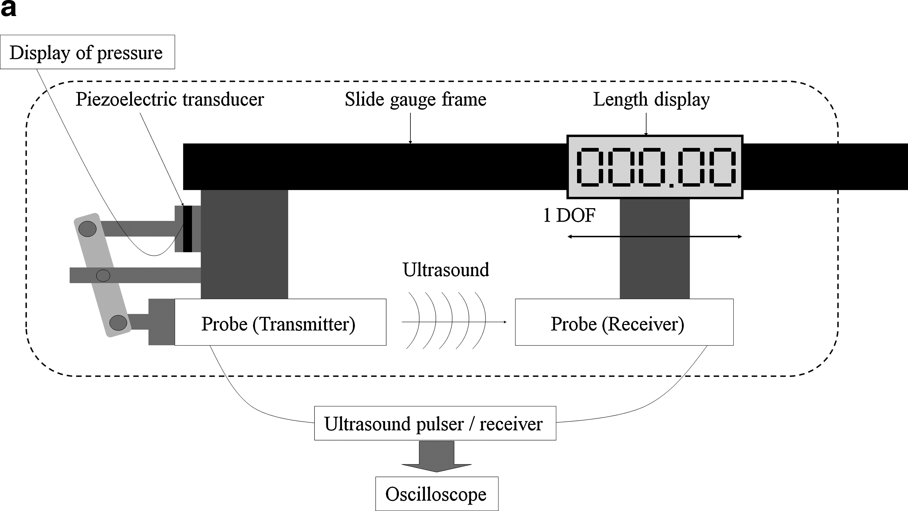
An ultrasound device was developed for this study to evaluate the number of cells in the BMSC/β-TCP composites. Figure 1a shows the ultrasound device system and Figure 1b shows photograph of probes and slide gauge frame. The device has a transmitting probe and a receiving probe (NSI Corp., Kawanishi, Japan) attached to the slide gauge frame (Absolute Digimatic; Mitutoyo Corp., Kanagawa, Japan). Both probes are set to be collinear at opposite sides to propagate the ultrasound wave from the transmitting probe to the receiving probe. One of the probes can be moved along the frame to adjust the size of the material to be inserted between them. The interval of the probes was accurately measured and observed on a digital display. The ultrasound wave data were recorded by an oscilloscope (DL-1720; Yokogawa Electric, Tokyo, Japan) via an ultrasound pulser/receiver (NSI-2000; NSI Corp.). Two hundred volts was applied to the transmitting probe with a sampling frequency of 200 MHz. The applied pressure for pinching material was set to a constant (3.5 N) and monitored by a pressure gauge attached to the end point of the frame. The distance of the ultrasound device was zeroed where the distance of the probes was shortest while the pressure gauge indicated a zero value. 21
(

Measurement: Ultrasound amplitude
After the seeded β-TCP blocks were incubated at 37°C with 5% humidified CO2 for 24 h, we evaluated the BMSC/β-TCP composites with the ultrasound device. Figure 2 shows the data obtained at β-TCP in the culture medium (a) and BMSC/β-TCP composite (concentration: 1.0 × 10⁶) in the culture medium (b). In these figures, the difference between the maximum and minimum voltages is calculated as the amplitude at 0.40 V (a) and 1.43 V (b). Characteristically, the received ultrasound wave amplitude decreases in heterogeneous material containing a cavity such as β-TCP due to a scattering and attenuating phenomenon. Hence, in the β-TCP where BMSCs adheres to the walls, a high amplitude is anticipated because BMSC/β-TCP composites are expected to increase the amount of solid body. We measured three different sides of the BMSC/β-TCP composite three times for each side in the culture medium, and determined the mean amplitude from the nine measurements.

Ultrasound amplitude was defined as the difference between the maximum and minimum values of the obtained ultrasound wave. (
Counting the cell number in BMSC/β-TCP composites
After measurements by ultrasound device were taken, BMSC/β-TCP composites were treated with 0.25% trypsin to release cells inside the BMSC/β-TCP composites for 30 min. The number of BMSCs was counted using a hemacytometer. We determined the counted cell number as the actual number of cells inside the composites after 24-h culture. Cell viability was evaluated by the trypan blue dye (Gibco BRL, Grand Island, NY) exclusion technique. The cell-seeding ratio was calculated using the following equation:
Cell-seeding ratio = Actual cell number inside the composite/Cell-seeding number.
To confirm the absence of cells inside the composites after trypsin treatment, trypsinized BMSC/β-TCP composites were analyzed by the MTT assay, according to the manufacturer's protocol. MTT solution (Sigma) was added to trypsinized BMSC/β-TCP composites in the culture medium, and was incubated at 37°C with 5% humidified CO2 for 2 h. Finally, dimethyl sulfoxide (Wako) was added to dissolve the insoluble purple formazan crystals. The absorbance of this solution was measured with a spectrophotometer at 540 nm (n = 5).
Statistics
The results were analyzed statistically using a statistical software package (Statview 5.0; Abacus Concepts Inc., Berkeley, CA). The data of the cell-seeding concentration and the actual number of cells are expressed as mean ± standard deviation. The correlations between cell-seeding concentration and actual number of cells and between ultrasound amplitude and the actual number of cells were analyzed using linear regression.
Results
Absence of BMSCs inside composites after trypsin treatment
In a preliminary experiment, we applied trypsin treatment to each composite five times. Using a hemacytometer, we confirmed that the BMSCs did not exist in the treated solution after three downward treatments (n = 30). MTT assay results showed that the optical density from the trypsinized composites was comparable to that of blank β-TCP blocks, indicating that there were no cells left inside the constructs after the three treatments of trypsin (data not shown) (n = 5).
Cell-seeding concentrations
To determine the optimal cell-seeding concentrations, we examined five different seeding concentrations of BMSCs: 1.0 × 10³, 1.0 × 10⁴, 1.0 × 10⁵, 1.0 × 10⁶, and 1.0 × 10⁷ cells/mL. Results from counting after the 24-h culture showed that BMSCs barely existed in β-TCP blocks seeded at a concentration below 1.0 × 10⁵ cells/mL (Fig. 3) (n = 30). The actual number of cells in BMSC/β-TCP composites in 1.0 × 10⁶ and 1.0 × 10⁷ cells/mL group was 2.8 ± 1.5 × 10⁵ (cell-seeding ratio: 28.4%) and 3.7 ± 1.8 × 10⁵ (cell-seeding ratio: 3.8%), respectively. Therefore, we examined the following concentrations: 0 (control), 1.0 × 10⁶, 1.5 × 10⁶, 2.0 × 10⁶, and 1.0 × 10⁷ cells/mL for the following experiments (n = 24).22–25

Relation between cell-seeding concentrations and actual cell numbers. BMSCs barely existed in the β-TCP block seeded at a concentration below 1.0 × 10⁵ cells/mL. Values are means ± standard deviation (n = 30).
Relationship between cell-seeding concentration and actual number of cells
The actual number of cells in BMSC/β-TCP composites in 1.0 × 10⁶, 1.5 × 10⁶, 2.0 × 10⁶, and 1.0 × 10⁷ group was 2.8 ± 1.5 × 10⁵, 3.4 ± 2.3 × 10⁵, 3.7 ± 1.0 × 10⁵, and 3.7 ± 1.8 × 10⁵, respectively (Fig. 4). The cell-seeding ratio was 28.4%, 22.6%, 18.5%, and 3.8%, respectively. Some of the composites seeded at higher concentrations contained a lower actual number of cells. Figure 4 shows the correlation between the cell-seeding concentration and the actual number of cells in each group. The cell-seeding concentration was not correlated to the actual number of cells (r = 0.134, n = 24).

The correlation between cell-seeding concentrations and the actual number of cells among each group is depicted. The cell-seeding concentration in the β-TCP block is not correlated to the actual number of cells in β-TCP block (r = 0.134, n = 24). Cell average and standard deviation (SD) and cell-seeding ratio show dispersion in each cell-seeding concentration.
Correlation of the ultrasound amplitude and the actual number of cells
Figure 5 shows the correlation between the ultrasound amplitude and the actual number of cells. As the cell count number increased, the amplitude grew higher. The ultrasound amplitude was well correlated to the actual number of cells (r = 0.903, n = 30). The estimation equation was as follows: y = 276565x − 117336, where x and y represent the amplitude value and estimated cell number in this study, respectively. The linearity of this estimation equation was validated in the cell-seeding concentration range from 0.0 to 6.7 × 10⁵ of the cell number. The estimated error for the system was 8.6 × 10⁴.
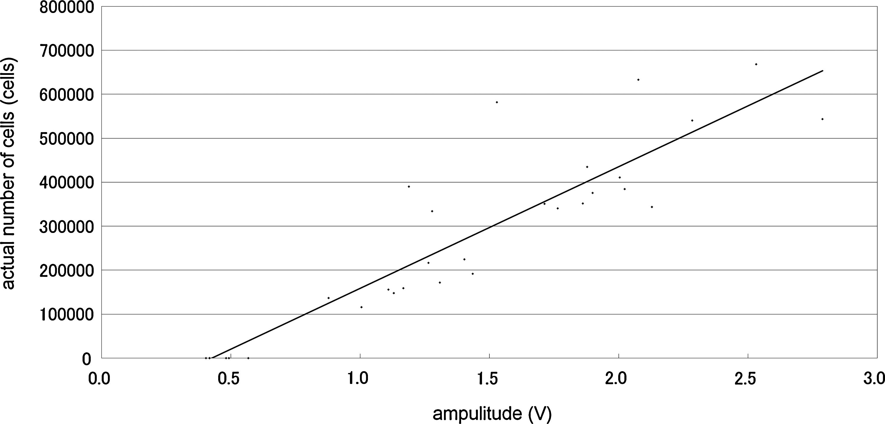
The correlation between ultrasound amplitude and the actual number of cells is depicted. Ultrasound amplitude is well correlated to the actual number of cells in the β-TCP block (r = 0.903, n = 30).
Discussion
Autologous bone is the ideal graft material for use in reconstructive orthopedic surgery. However, its harvesting is closely associated with donor-site morbidity, and it is available in only limited amounts.1,2 Composites of BMSCs/β-TCP have been increasingly used as bone substitutes and studied as a bone graft model for bone tissue engineering.10–13 These studies have supposed that the number of seeded BMSCs in the β-TCP depends on cell-seeding concentrations. However, few studies have evaluated the relationship between the actual number of cells in composites and the cell-seeding concentration. 15 In this study, we showed that BMSCs barely existed in β-TCP blocks when the cell-seeding concentration was <1.0 × 10⁵ cells/mL. Our results indicated that even when one regular cell-seeding concentration was applied to the β-TCP, the actual number of cells in the individual BMSC/β-TCP composites varied considerably. In addition, the cell-seeding concentration was not correlated to the actual number of cells contained in the BMSC/β-TCP composites (Fig. 4). Thus, the cell-seeding concentration cannot be considered as a reliable indicator of the actual number of cells contained in the β-TCP. These results suggested the importance of developing methods that can evaluate the actual number of cells in individual BMSC/β-TCP composites after cell seeding and allow the use of BMSC/β-TCP composites that contain a favorable number of cells for the future tissue engineering application.
We successfully demonstrated for the first time that our developed ultrasound device could nondestructively evaluate the actual number of cells contained in the individual BMSC/β-TCP 24 h after cell seeding (Fig. 5). Our results showed that ultrasound amplitude effectively responded to the quantity of BMSC/β-TCP composites and was well correlated to the actual number of cells contained (r = 0.903). Thus, by using our ultrasound device it is possible to nondestructively choose BMSC/β-TCP composites that contain an appropriate number of BMSCs. However, our results also indicate the existence of some outlier values that could result from defects in the β-TCP surface. When the contact area between β-TCP and probe is obviously decreased due to the large defect, our developed ultrasound device underestimates the number of BMSCs. Since ultrasound devices can be sterilized with gas, they can potentially be used in clinical situations to evaluate and monitor the number of BMSCs in the individual BMSC/β-TCP composite before implantation.
One potential flaw in our study is the difficulty of distinguishing between living and dead BMSCs using ultrasound. There is a possibility that ultrasound may also respond to dead BMSCs. To test this possibility, cell viability inside the composites was evaluated by the trypan blue dye (Gibco) exclusion technique after the trypsin treatment. We were able to show that the cell viability was >99% by the trypan blue dye in BMSC/β-TCP composites, suggesting that there were no dead cells inside the composites in the current study. Therefore, our ultrasound device did not respond to the dead BMSCs in the β-TCP.
We chose the β-TCP block in this study because we are familiar with it due to its clinical use at our institution. Moreover, β-TCP block is one of the most common biodegradable scaffolds with good osteoconductive properties,26,27 and is widely used in many researches and clinical settings.28–30 The Food and Drug Administration is reclassifying TCP granules for dental bone repair from class III to class II. 31 In future studies, we will use our method in other scaffolds such as hydroxyapatite. In the assessment of our proposed system, the cell-seeding concentration ranged from 1.0 × 10⁶ to 1.0 × 10⁷ cells/mL.22–25 Although our proposed system focused on this range, it could be adapted for larger ranges by changing the device parameters such as the voltage resolution of the oscilloscope, the transmitting voltage of the ultrasound, or the center frequency of the probe.
In summary, our results indicated that even if one regular cell-seeding concentration was applied in the β-TCP, the actual number of cells in the individual BMSC/β-TCP composites varied extensively. At cell-seeding densities <1.0 × 10⁵ cells/mL, BMSCs barely existed in β-TCP blocks after 24-h culture. Our developed ultrasound device could quantify the actual number of cells in BMSC/β-TCP composites nondestructively. Using this ultrasound device, orthopedic surgeons can choose composites that contain favorable number of cells before implantation, which would provide a great benefit to an early treatment. Our device could be a valuable, convenient, and nondestructive tool for tissue engineering, especially in the era of artificial bone tissue generation.
Footnotes
Acknowledgments
We would like to express our gratitude to Mr. Takayuki Kobayashi, Mr. Yoshinori Nagai, and Mr. Satoshi Yamaguchi (Graduate School of Engineering, University of Hyogo, Himeji, Japan) for ultrasound technical assistance and advice. We also wish to express our gratitude to Ms. Yoshiko Masuda (Olympus) for her excellent technical assistance in cell count analysis; Ms. Kyoko Tanaka and Ms. Minako Nagata (Department of Orthopaedic Surgery, Kobe University Graduate School of Medicine) for their technical assistance; Mr. Tomoyuki Sawayama (Japan Medical Instruments Co., Ltd.) for developing the ultrasound device; and Janina Tubby for English rewriting.
Disclosure Statement
No competing financial interests exist.