
Abstract
The objective of the study was to describe a novel small-animal model of tissue-engineered aortic valve conduits and to investigate biological processes in an accelerated and inexpensive fashion. An isogenic Lewis-to-Lewis rat model was used to exclude immunological factors of graft deterioration. U-shaped aortic valvular grafts were decellularized and characterized morphologically. Acellular conduits were repopulated with labeled isogenic cells in a bioreactor under flow conditions. Grafts were anastomosed to the recipient's abdominal aorta in an end-to-side manner (n = 7). Native rat aortas were implanted as a control group (n = 7). Grafts were explanted after 28 days and characterized. After treatment with trypsin–ethylenediaminetetraacetic acid, no residual cells were visualized in the scaffold. Mean DNA content decreased from 0.347 to 0 μg/mg of DNA/tissue, and the content of collagenous connective tissue and proteoglycans appeared slightly reduced. Isolated aortic rat endothelial cells and myofibroblasts were repopulated on the acellularized scaffold, and florescent-labeled myofibroblasts were identified in the meshwork. Endothelial cells formed a monolayer on the luminal surface. Reseeded cells were viable as ascertained using a 3-(4,5-dimethylthiazol-2-yl)-5-(3-carboxymethoxyphenyl)-2-(4-sulfophenyl)-2H-tetrazolium assay. After implantation, Doppler and M-mode echography proved pulsatile cusp movement. All conduits were patent after 28 days. Examination of tissue-engineered explants revealed thickened aortic walls and incompetent valve function. Microscopically, aortic intima and media appeared normal, whereas the adventitia showed hyperproliferation of fibroblasts. Our new model leads to accelerated and reproducible results, suited to investigation of biological patterns of tissue engineering. The observed adventitial fibrosis emphasized the importance of careful selection of optimal cell types for repopulation in tissue-engineered constructs.
Introduction
The concept of tissue engineering may overcome the limitations of available valvular and vascular prostheses. Biodegradable matrices or decellularized tissue scaffolds of allo- or xenogenic origin are repopulated with autologous cells in vitro. These constructs are free of immunogenic cells and are supposed to be suitable for surgical implantation. The low-pressure system has been investigated in detail for more than 10 years, and tissue-regenerated pulmonary prostheses have been introduced into clinical practice.6–8 Because acellularized scaffolds used for tissue regeneration are unsuitable for the high-pressure circuit because of its weak matrix composition, development of tissue-engineered (TE) aortic valved conduits requires use of in vitro reconstitution. Currently, the ideal decellularization protocol for aortic tissue, selection of proper cell types for recellularization, and optimal experimental in vitro setting have to be clarified. Several large-animal models are available to solve this puzzle,9,10 but because these models are expensive and require long-term follow-up, specific biological details are difficult to analyze. Here we describe a novel small-animal model of TE aortic valved conduits suited to address various unanswered biological questions quickly and inexpensively.
Materials and Methods
Experimental design and surgical procedure
An isogenic Lewis-to-Lewis rat model was used to exclude immunological factors of graft deterioration. Male Lewis rats (LEW/CrlBR) weighing 250 to 350 g were obtained from Charles River Laboratories (Boston, MA). After explantation of an aortic valve conduit of donor animals, grafts were decellularized and characterized. Then, grafts were recellularized in vitro with isogenic cells using a bioreactor, followed by morphological characterization. Thereafter, seven recellularized-valve aortas were implanted into the infrarenal aortas of recipients.
For explantation of donor valvular aortic conduits, donor rats were anesthetized using an intraperitoneal injection of a cocktail of Ketamine 10% and Rompun 2%. After a large bilateral thoracotomy, the heart, together with the ascending aorta, aortic arch, and descending aorta, were explanted. Using a microscope (Leica, Wetzlar, Germany), the branches were ligated, and the graft was stored in Earle's medium 199 enriched with 100 IU/mL of penicillin-streptomycin and 10 IU/mL of heparin (all PAA Laboratories, Pasching, Austria). The grafts were prepared for implantation in controls or transported to the laboratory for further process of tissue engineering.
For implantation, the heart was dissected from the U-shaped aortic conduit under a microscope (Leica), leaving a myocardial rim below the valvular annulus. After intraperitoneal anesthesia, the abdomen of the recipient was opened using a midline laparotomy. A segment of the abdominal aorta was dissected approximately 1 cm below the origin of the renal arteries and approximately 1 cm above the aortic bifurcation. This segment was gently cross-clamped, and the graft was anastomosed in an end-to-side manner using 8-0 Prolene (Ethicon, Hamburg, Germany) for the proximal and 10-0 Prolene for the distal anastomosis. The native aorta was ligated to ensure bypass flow. After implantation, graft patency was controlled weekly by palpation of the abdomen.
After 28 days, the animals were anesthesized again, and the grafts were explanted. After control of possible emboli, grafts were flushed with saline solution (Sigma-Aldrich, St. Louis, MO) and transferred to the laboratory for further characterization. For control, seven native conduits were transplanted in the same manner.
The committee of animal welfare of the governmental authority approved all experiments. Rats were housed and fed ad libitum in standard conditions and received human care in accordance with the German animal welfare law and European guidelines.
Decellularization of aortic grafts
Valved aortic conduits were flushed with Hank's buffered salt solution (Biochrom, Berlin, Germany) and incubated for 48 h in 0.1% trypsin–ethylenediaminetetraacetic acid (EDTA) –phosphate buffered saline (PBS) solution at 37°C under shaking conditions (Biochrom). The solution was changed after 24 h, and thereafter, the conduits were interposed into a specially designed bioreactor with the left ventricle and the descending aorta connected to in- and outflow tubes, respectively (Fig. 1). The calculated shear rate in this setting was calculated with 0.096 Pa. The bioreactor was filled with PBS (Biochrom) supplemented with penicillin-streptomycin, and the graft was flushed with continuous flow of 1 mL/min at 37°C for 48 h for removal of residual cells. Grafts were stored in Earle's medium 199, enriched with penicillin-streptomycin and heparin, at 4°C until implantation. To control decellularization, semithin 5-μm scaffold sections were obtained from excessive distal aortic graft tissue and stained with hematoxylin before further processing.

Photography of a rat valve aortic conduit connected to a specially designed bioreactor for acellularization. Color images available online at
Isolation and culture of rat aortic endothelial cells and rat myofibroblasts
Isogenic donor Lewis rats were sacrificed. The thoracic cavity was opened, and the aorta was explanted down to the abdominal branches. All major branches were ligated, and intercostal artery branches were closed using diathermic coagulation (Aesculap, Tuttlingen, Germany). The aorta was cannulated, and the complete inner surface was flushed twice with warm PBS without Ca2+ and Mg2+ (Biochrom). Thereafter, the aorta was perfused twice with 1 mL of warm 0.02% collagenase type IV solution (PAA Laboratories) for 1 min. The enzymatic solution carrying isolated cells was collected in Falcon tubes. The aorta was flushed with modified endothelial basal medium (EGM-2) (Clonetics, Baltimore, MD) and collected in the same Falcon tube. This solution, including isolated rat aortic endothelial cells (RAECs), was centrifuged at 250 × g at 4°C for 10 min. Then the pellet was resuspended in 5 mL of EGM-2 and cultivated in 25-cm2 culture flasks. Freedom from infection and typical morphological appearance of endothelial cells (ECs) was confirmed daily using microscopy. When subconfluence of the EC monolayer was archived, cells were split in a ratio of 1:2 using 100 μl/cm2 of Accutase (PAA Laboratories). Two hours later, after ECs adhered to the culture bottle, medium was changed to remove floating myofibroblasts. EC cultures with a typical cobblestone appearance were used for reseeding of matrices in the bioreactor and for preparation of cytospots for consecutive CD31 immunostaining (for technical details, see below). Myofibroblasts were isolated as previous described 11 and cultivated in complete EGM-2.
In vitro cell labeling
For identification of isolated rat cells after the recellularization procedure, RAECs and myofibroblasts were labeled with carboxyfluorescein diaceteate succinimidy ester (CFDA-SE; Vybrandt CFDA SE Cell Tracer Kit, MoBiTec, Goettingen, Germany) as previously described. 6
In vitro recellularization of decellularized aortic valved conduits
After decellularization, conduits remained connected to the bioreactor, which was filled with EGM-2. Myofibroblasts of one 25-cm2 culture flask (subconfluent, ∼8 × 10⁵ to 1.2 ×10⁶) were resuspended in 400 μL of EGM-2. One hundred μL of cell suspension was instilled into the luminal aorta from the distal side and incubated for 3 h; thereafter, the residual 300 μL of cell suspension was introduced from the proximal side into the aortic lumen. The bioreactor was placed on a rolling device in an incubator and was rolled at 0.1 rpm for 24 h. Then the luminal aspect of the system was connected to a roller pump. Perfusion was started at 1 mL/min, and fluid circulation through the bioreactor system was maintained for 3 days. Thereafter, the procedure was repeated with 1.4 × 10⁶ to 1.6 × 10⁶ RAECs. After fluid circulation was continued for 7 days, the graft was proceeded for in vitro characterization or implantation into the recipient animal.
Ex vivo characterization of acellularized scaffolds and TE conduits
Histology
Cryosections (5 μm thick) of graft tissues were generated using a microtome (Leica) and stained with hematoxylin and eosin (H&E) or Movat-Pentachrome staining for differentiated characterization of the extracellular matrix (ECM) components. Sections were visualized in a bright field using an Olympus BX41 microscope. H&E staining was routinely performed on all sections for detection of possible cellular remnants after decellularization. Movat-Pentachrome staining was chosen for detection of matrix components. Samples were stained for proteoglycans in Alcian-blue, followed by nuclear staining with Weigerts-Fe2 + hematoxylin. Elastic fibers were stained with Crocein-acid fuchsin, and collagen was stained yellow in Saffron-du-Gatinais.
DNA assay
To confirm complete removal of cells from the scaffolds after decellularization, DNA was isolated and quantified from seven acellular and seven native aortic tissue samples. Samples were weighed and centrifuged in RiboLyser Tubes (Hybaid, Basingstoke, United Kingdom) in 500 μL of cell culture medium at 1.500 × g for 20 s. After digestion with 750 μL of Tri-Reagent for 10 min on ice, 200 μL of chloroform was added. After centrifugation at 12.300 × g at 4°C for 15 min., the phase containing DNA was isolated and incubated with 300 μL of ethanol for 3 min at room temperature. After centrifugation, the pellet was picked up in 0.1 M sodium citrate and 10% ethanol, and the step was repeated after incubation. Centrifugation with 9.600 × g at 4°C for 15 minutes followed; then the pellet was resuspended in 75% ethanol, incubated, and centrifuged. The pellet was dried and resuspended in 200 μL of distilled water. The DNA photometric extinction was measured at 260 nm using a Spectronic 1201 (Milton Roy Company), and DNA content was calculated with regard to the weight of the tissue sample.
Metabolic activity test for reseeded grafts (3-(4,5-dimethylthiazol-2-yl)-5-(3-carboxymethoxyphenyl)-2-(4-sulfophenyl)-2H-tetrazolium assay)
Viability of reseeded rat cells was ascertained using a 3-(4,5-dimethylthiazol-2-yl)-5-(3-carboxymethoxyphenyl)-2-(4-sulfophenyl)-2H-tetrazolium (MTS) assay (CellTiter 96 AQueous One Solution Proliferation Assay, Promega, Madison, WI), as previously described. 12
Functional and morphological characterization of explanted grafts
Echographic evaluation of implanted aortic valve conduits
Before graft explantation, animals were anesthesized. In vivo valvular graft function was evaluated using a 15-MHz head for transabdominal color Doppler echography (Sonos 5000, Philips, Eindhoren, The Netherlands).
Functional and macroscopic evaluation
Before explantation, the distal aorta of the recipient was checked for pulsatile flow. All explanted valvular grafts were inspected for competence using retrograde injection of saline solution. Leaflet and wall appearance was assessed under magnification after opening the graft lumen.
Histology
Five-μ
Immunohistochemistry and confocal microscopy
For characterization of rat ECs, RAECs and cytospots were stained using mouse anti rat CD-31 (clone TLD-3A12, Serotec, Kidlington, United Kingdom).
Frozen sections of aortic valves and the aortic wall were mounted in Tissue Tec (Sakura, Torrance, CA) and immediately frozen in liquid nitrogen. Sections 10 μm thick were placed on gelatin-coated slides and fixed for 10 min with 4% paraformaldehyde. To block unspecific labeling, the sections were exposed to 0.5% carboxylated bovine serum albumin and 100 mmol/L glycine in PBS for 10 min, followed by incubation with the primary antibodies against alpha smooth muscle actin (α-SMA; 1E4, Sigma, Hamburg, Germany) and collagen type I (Sigma). After repeated washes with PBS, the sections were incubated with the secondary antibodies, including antimouse immunoglobulin (Ig)G conjugated with Cy3 (Chemicon, Billerica, MA) and antirabbit IgG conjugated with fluorescein isothiocyanate (Dianova, Hamburg, Germany). All actins were labeled with phalloidin conjugated with tetramethyl rhodamine isothiocyanate (Sigma), and nuclei were stained with 4',6-diamidino-2-phenylindole (DAPI; Invitrogen, Carlsbad, CA). Omission of the primary antibody served as a negative control. Native rat aortic tissue samples were stained contemporaneously for comparison and as positive controls.
The tissue sections were examined under confocal microscopy (Leica TCS SP2). Series of confocal optical sections were taken at 0.5-μm intervals using a Leica Planapo × 25/0.75 or × 40/1.0 objective lens. Each recorded image was taken using triple-channel scanning and consisted of 1024 × 1024 pixels. To improve image quality and to obtain a high signal to noise ratio, each image from the series was signal averaged. After data acquisition, the images were transferred to a Silicon Graphics workstation for restoration and three-dimensional reconstruction using Imaris 4.5 multichannel image processing software (Bitplane, Zürich, Switzerland). The principles of this method have been previously published. 13
Results
Characterization of the surgical model
Surgical procedure and follow-up
The technique of heterotopic U-shaped aortic valve conduit transplantation into the abdominal aorta of the recipient rat in an end-to-side manner is feasible with low morbidity and mortality. Initially, we tried to sew the proximal aortic root to the abdominal aorta using a running suture but had technical problems with this anastomosis due to bleeding. With the second animal, we left a myocardial cuff below the aortic root and established the anastomosis using single stitches (instead of a running suture) connecting the myocardial cuff to the abdominal aorta. With this technique, we successfully operated on five animals for establishment of the technique without any deaths. After establishment of the technique, all animals survived the implantation procedure. The recipient procedure was performed with a mean operative time of 124 min; aortic cross-clamp time varied between 51 and 65 min. The patency rate of implanted grafts was 100% up to 28 days in the study and the control group. All animals recovered without signs of neurological or ischemic symptoms of the lower limbs.
In vivo characterization of valvular function
The transplanted conduit in the abdominal cavity and the aortic valve of the conduit were clearly identified using two-dimensional echography (Fig. 2A). Flow through the transplanted valve graft was pulsatile, as shown using M-Mode echography (Fig. 2B). Valvular cusps moved in correlation with pulsatile blood flow, and complete closure of the valve was not clearly identified because of the accelerated heart rate of the rat (>300 bpm). Color Doppler echography confirmed physiologically directed blood flow solely through the graft; no perfusion of the ligated abdominal aorta was seen (Fig. 2C). Blood flow was sufficient to avoid any signs of ischemia abdominally or in the lower extremities in any of the animals.
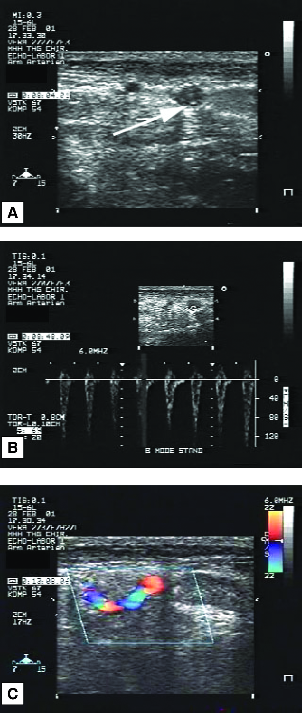
Echographic characterization of aortic valve conduit after transplantation into the abdominal aorta (graft 4). (
Characterization of RAECs
There were few isolated RAECs, but pure samples were isolated using short incubation times, as described in the Methods section. When small cells cluster grew to subconfluency, cells were split in a ratio of 1:2 to avoid overaging of EC cultures. According to this method, we archived a low percentage of contamination with myofibroblasts. If contamination occurred, cultures were discarded. Our culture of RAECs showed a typical cobblestone appearance (Fig. 3A). RAEC cytospots positively stained for CD-31 antibody, demonstrating the endothelial origin of the cells (Fig. 3B). To determine the degree and quality of the reseeding procedure of rat aortas, we labeled RAECs (and myofibroblasts) with fluorescent dye CFDA-SE in vitro. Examination of cells using fluorescent microscopy revealed that 100% of the cells displayed a strong positive signal in the cytoplasm (Fig. 3C). The cellular uptake of CFDA-SE had no influence on cellular viability. Cell labeling was maintained for up to two passages and was inherited by daughter cells after cell division. The ability of cells to maintain the fluorescent dye for a long period of time allowed us to characterize the extent of reseeded cells on the acellular isograft matrix.

Histological characterization of isolated rat aortic endothelial cells (RAECs). (
Characterization of acellularized and TE grafts
Enzymatic treatment with trypsin–EDTA and consecutive washing using the bioreactor led to complete removal of cells from the ECM. Histological examination of acellularized grafts showed no remaining cells in the scaffolds. In the ECM, elastic fibers remain preserved, and the content of collagenous connective tissue and proteoglycans appeared slightly lower. The DNA assay confirmed complete removal of cells, because DNA was not detected in acellularized scaffolds, in contrast to native tissue (mean DNA content 0.347 μg/mg for native tissue versus no detectable DNA in acellularized scaffolds; data not shown).
After recellularization of the acellularized matrix with isogenic RAECs and myofibroblasts, the luminal face was covered with a monolayer of RAECs. Numerous cells were found in the media, but not in the adventitia. H&E staining revealed a morphological aspect similar to that of an arterial vessel (Fig. 4A, B). CD-31 immunohistological staining demonstrated RAECs on the luminal aspect of the graft, forming a monolayer (Fig. 4C). Fluorescence microscopy showed a green signal of cells found in the matrix, demonstrating that reseeded cells were of isogenic origin and not remnants of the acellularization procedure (Fig. 4D).
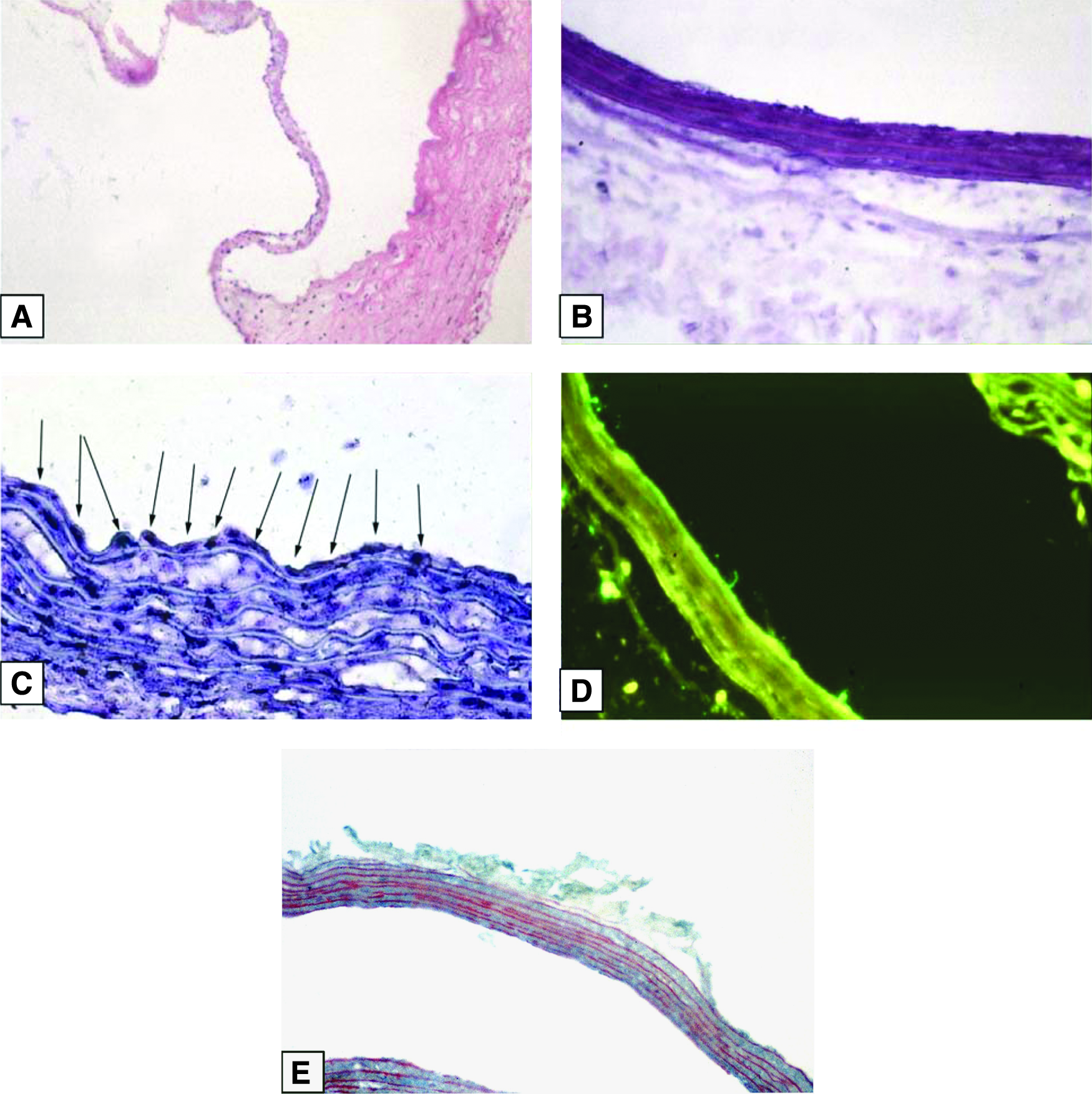
Histological characterization of tissue-engineered aortic valve conduits after recellularization. (
The MTS assay revealed that cells of TE conduits remained viable after recellularization and cultivation in the bioreactor, expressing metabolic activity in all reseeded grafts (n = 7; mean 0.086 (range 0.015–0.185) extinction variance at λ = 490 nm). In contrast, acellularized grafts did not show any metabolic activity (data not shown).
Characterization of TE grafts after explantation
Functional and macroscopic findings
After 28 days, all isogenic native and TE conduits were patent. Macroscopically, native transplanted grafts appeared unchanged in size and shape (Fig. 5A). Retrograde injection of saline solution demonstrated competent aortic valves in the native control group. In contrast, TE grafts appeared thickened, and the shape of the aortic bulb disappeared (Fig. 5D). All aortic valves of TE grafts were insufficient. The sinus of Valsalva was free of thrombotic formation, but the aortic wall and valvular cusps were unphysiologically thickened.
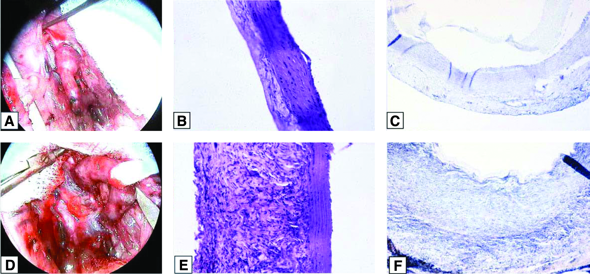
Morphological characterization of explanted grafts 28 days after implantation. (
Morphological characterization
Native conduits of the control group maintained normal morphology without any degenerative or proliferative changes of the tissue (Fig. 5B). The luminal and valvular EC monolayer remained unchanged, without signs of intimal hyperplasia. Size and cell density of the media and adventitia were unaffected. Von Kossa staining revealed no signs of calcification in the tissue (Fig. 5C). In contrast, TE grafts showed marked distension of the aortic wall after 28 days, caused by enormous hyperproliferation of adventitial fibroblasts (Fig. 5E). In comparison, the sizes of the media of TE grafts were unchanged compared to native conduits, and luminal aspect of TE-grafts were covered by ECs. There were no signs of intimal hyperplasia of TE conduits. Von Kossa staining showed few signs of focal calcification in TE grafts (Fig. 5F).
High-power magnification of confocal microscopy of recellularized conduits (aortic wall and leaflet) 28 days after implantation revealed a regular cellularity according to blue-labeled nuclei with DAPI. The monolayer on the intimal aspect appeared normal, without signs of hyperplasia. Abundant collagen all over the conduit and slightly thicker leaflets was shown using immunolabeling for collagen I (green). Alpha-SMA was detected in small amounts, forming a layer at the luminal side of the conduit (red). The merged three-dimensional shadow projection demonstrated the greater quantity of collagen but not SMA in relation to total number of cells (Fig. 6). Under lower magnification of confocal microscopy, the greater thickness of the adventitia is displayed (Fig. 7). No intimal hyperplasia with greater cell numbers was detectable. The tunica media was well preserved, and smooth muscle cells forming the media were compactly arranged. Alpha-SMA (green) and F-actin (red) were detected in greater abundance, and normal cellularity was found using DAPI staining of nuclei. In contrast, the thickened adventitia showed high cellularity but almost no α-SMA (green) or F-actin (red).

Characterization of a representative recellularized conduit (aortic wall and leaflet, graft 4) 28 days after implantation. Representative confocal micrographs showing the cellularity, collagen abundance, and thickness of leaflets (L) immunolabeled for collagen I (green;

Characterization of recellularized conduits (aortic wall, graft 4) 28 days after implantation. Representative confocal micrographs (
Discussion
Tissue engineering represents a multidisciplinary field that combines basic biology and clinical science. The aim of tissue engineering is the construction of viable grafts in vitro to replace deteriorated tissue and organs, repeating the shape, physiological form, and function of native organs. 14 Clinical applications of TE grafts in cardiovascular surgery have been reported,15–17 but before clinical trials, careful examination of biological responses must be conducted in animal experiments, because in vitro–generated bioartificial tissue may behave differently in a complex organism than expected. In the field of valvular and vascular tissue engineering, several large-animal models have been described.10,18 The most commonly used models have been based on swine or sheep. Although close to humans, these models have several disadvantages, for example, high cost, long duration for obtaining relevant results, limited availability of antibodies for histological examination, complex surgical techniques). Therefore, here we report on a novel small-animal model for valvular and vascular tissue engineering based on the rat aimed at overcoming these limitations.
Allo- and xenogenic small-animal rat models with transplantation of an aortic composite graft have been described before,19,20 although these studies were designed to investigate the immunological influence in guided tissue regeneration. Here we first describe tissue engineering in a rat model with reseeding of acellularized composite grafts. We have chosen an isogenic small-animal model of rat to exclude immunological factors that may influence graft deterioration. The biological reaction patterns of the organism to TE grafts in vivo were the sole focus of our study. Our model allows for answering these important questions in an accelerated fashion with lower cost and shorter time than with large-animal models. Although a small-animal model cannot accelerate the time-related process of recellularization, remodeling, and degradation, the same results we observed in this model after 28 days were also seen in a TE large-animal model of sheep after 6 months: normal-like intima with endothelial cell lining, but with abundant adventitial tissue (data not shown). Therefore, we believe that this small-animal model is helpful to address fundamental questions of pathophysiological and pathomorphological mechanisms of remodeling as fundamental studies. The next step would be the translation of these findings into preclinical large-animal models before clinical studies in humans can be considered.
Furthermore, a wider range of available antibodies, especially of rats, allows for addressing questions that cannot be investigated in large-animal models. Various surgical techniques of heterotopic aortic conduit transplantation have been described before. Yankah et al. first reported on infrarenal aortic valve transplantation in an end-to-end manner. 19 The disadvantage of this technique is the obligatory resection of an aortic cusp to prevent sinus thrombosis. This will result in unphysiological flow patterns and a permanent open valve that may affect valvular degeneration. Ross et al., who used the same end-to-end technique, proposed creation of valve insufficiency by perforation of the native aortic valve to create pendeling flow in the distal part of the aorta that should result in near-physiological open-and-closing behavior of the transplanted valve. 21 Disadvantages of this technique are high mortality and cardiac morbidity of the recipient animals. Here we used the “Rotterdam” Model in an end-to-side manner as described by Oei et al. with slight technical variations. 22 The advantage of this model is absence of Valsalva thrombosis of the transplanted aortic valve, although neither the native nor the transplanted valve required direct manipulation. Furthermore, for the first time, we described the echographic evaluation of the transplanted valve in the rat model, demonstrating pulsatile opening and closing movement of the cusps. The highly reproducible model, although technically demanding, can be established without significant mortality or morbidity of the recipient.
As the first step of tissue engineering, target of decellularization techniques is a delicate balance of complete removal of potentially immunogenic cellular components but optimal preservation of the resulted ECM. 23 Acellularization of native aortic conduits with trypsin–EDTA led to acellular grafts with complete removal of viable cells, as confirmed by the DNA assay. ECM components remain conserved if washing after trypsinization is undertaken in a special bioreactor.
Although isolation of rat aortic myofibroblasts can be easily achieved with a standard protocol, 11 isolation and cultivation of RAECs remain difficult. The major problem of isolation of RAECs is the contamination of cultures with fibroblasts that will overgrow RAECs. Several different techniques have been proposed,24–26 but a fast and simple enzymatic isolation technique has been speculated as impossible. 27 Our novel strategy is based on usage of low concentrated collagenase and short incubation time aimed to obtain a small but pure RAEC cluster. Early passaging of RAEC cultures stimulated rapid growth to confluence and prevented over-aging of cells. With this technique, we were able to obtain pure RAEC cultures suitable for the recellularization procedure in a specially designed bioreactor.
Based on our previously described experience with recellularization of human heart valve scaffolds, 6 reseeding of acellularized rat aortic conduits with RAECs and myofibroblasts was performed using pump flow in a bioreactor. Cultivation of cells on acellular scaffolds under shear stress conditions provides maintenance of physiological cell functions and the ability to adhere to the acellular matrix surface. 28
In our series, all grafts were patent, without any thrombus formation. The absence of thrombus even in the sinus of Valsalva indicates the antithrombotic effect of in vitro reseeding with ECs. Coverage of ECM with a monolayer of RAECs, as observed after recellularization, prevents attachment of platelets and erythrocytes on the rough surface of the ECM with unprotected fibers. Consecutively, formation of intimal hyperplasia as a physiological repair process in denuded vessels29,30 did not occur. The cell density of the media in TE grafts was lower, and the integrity and structure appeared similar to those of native control vessels but with changes in the composition of the ECM. After 28 days of follo- up, we did not observe aneurysm formation. This may be related to stabilization of the scaffold with reseeded cells, because reseeded vessels show nearly the same mechanical stability as native control vessels (unpublished data). However, aneurysm formation could occur after longer observation periods. On the other hand, all TE grafts showed marked distension of the aortic wall after 28 days caused by enormous hyperproliferation of adventitial fibroblasts. This thickening involved the cusps of the aortic valve, leading to valvular incompetence. The underlying reason for this phenomenon remains unclear. One explanation for cusp thickening could be the reseeding of the scaffold in vitro by use of culture medium supplemented with fibroblast growth factor. Myofibroblasts have been introduced into the valvular conduit lumen during reseeding in the bioreactor and reached the surface of the cusps by this process. The myofibroblasts may have been stimulated already after seeding in vitro, but the effect, hyperproliferation of myofibroblasts in the adventitia, may have become visible after explantation of grafts. Another explanation for thickening of the arterial wall could be migration of host myofibroblasts into the adventitia stimulated by fibroblast growth factor attached to the ECM. The origin of fibroblasts in the adventitia remains unclear. Therefore, further experiments are mandatory to identify the appropriate cell type and quantity for in vitro recellularization. Progenitor and bone marrow–derived cells may represent an interesting option for further experiments,31,32 although the same finding of adventitial thickening and fibrosis has been observed in a large animal model of TE pulmonary valve transplantation in sheep after 3 and 6 months. 33 Furthermore, in the same large-animal model, results with regard to intima and media were comparable with those in our small-animal model. These comparable results of both animal models demonstrate the value of our small-animal model for investigation of TE graft biology in vivo, because the same results were obtained in a shorter time with lower cost.
Several limitations need to be addressed. First, the physiological function of the transplanted valve remains questionable. Although cusp movement has been documented, complete closure of the valve has not been proven. However, histological examination of the explanted grafts of controls revealed normal morphology of the valve. Second, the origin of fibroblasts in the fibrotic enlarged adventitia remains unclear. CFDA-SE labeling of RAECs and myofibroblasts before seeding on acellularized scaffolds confirmed the attachment of seeded cells to establish the model. However, we did not implant TE grafts with labeled cells. The stability of CFDA-SE dye in transplanted cells during reseeding in vitro and their exposure for 28 days in vivo require further investigation.
In conclusion, our new model leads to accelerated and reproducible results, suited to investigation of biological patterns of tissue engineering. The observed adventitial fibrosis emphasizes the importance of careful selection of the optimal cell types for repopulation in TE constructs.
Footnotes
Acknowledgment
The work was supported by a grant Hochschulinterne Forschungsförderung (HILF) of Hannover Medical School.
Disclosure Statement
The authors have no competing financial interests.