




Abstract
Human adipose-derived stem cells (hASCs) have been shown to be multipotent and could be induced into various cell types, which make them the ideal cell source for cell therapy or tissue engineering. However, differentiation of ASCs into hepatocytes on three-dimensional scaffold, an important part of tissue engineering, has not been reported. In this study, to investigate the hepatogenesis of ASCs on porous poly-lactide-co-glycolide (PLGA) scaffolds, we loaded hASCs on these scaffolds. The cell–scaffold complex was implanted into the peritoneal cavity of 70% hepatectomized rats with or without 14 days of induction in hepatic inducing medium. Our results indicated that hASCs cultured on the PLGA scaffolds in the hepatic inducing medium proliferated more efficiently and could be induced into cells with hepatocyte-like phenotypic and functional properties. In vivo studies showed that induced hASCs on PLGA scaffolds survived and maintained hepatic phenotype and function for at least 14 days after implantation; moreover, noninduced hASCs on PLGA scaffolds expressed human albumin 14 days after transplantation. Collectively, these results suggest that porous PLGA scaffolds are suitable for the hepatogenesis of hASCs. These findings might be helpful in the application of hASC-based tissue engineering for liver disease therapy.
Introduction
Poly-lactide-co-glycolide (PLGA) is a type of highly biocompatible material that has been approved by the U.S. Food and Drug Administration for several biomedical applications in humans and widely used as scaffold materials in tissue engineering.7–9 Additionally, compared with natural materials, PLGA, a synthetic polymer, has good physical properties such as insolubility and slow degradation. Therefore, in this study, we selected porous PLGA scaffolds as the 3D setting to investigate the hepatogenesis of human ASCs (hASCs) in vitro and in vivo, with a long-term goal of developing stem cell-based composite grafts for hepatic tissue repair and regeneration.
Materials and Methods
Isolation and culture of hASCs
Human lipo-aspirates were obtained from patients undergoing selective liposuction at the Plastic and Aesthetic Surgery Center, Peking Union Medical College Hospital. Isolation of hASCs was done using a modified method as previously described.10,11 Briefly, the lipo-aspirates were washed with equal volumes of phosphate-buffered saline (PBS) to remove contaminating blood cells and local anesthetics and then digested with 0.075% collagenase NB 4 (Serva, Heidelberg, Germany)/PBS for 1 h at 37°C with gentle shaking. The collagenase was then neutralized with an equal volume of low-glucose Dulbecco's modification of Eagle's medium (DMEM) supplemented with 10% fetal bovine serum (FBS). After suspension in 160 mM NH4Cl to eliminate erythrocytes and passage through a mesh filter, the cells were resuspended in DMEM/10% FBS and plated at a concentration of 1–5 × 10⁶ cells/75 cm2. The culture medium was changed twice weekly thereafter.
Scaffold preparation
Porous PLGA (PLA:PGA ratio of 85:15; Shandong Institute of Medical Instruments, Jinan, China) film was fabricated using the standard salt leaching procedures with NaCl as the leachable component. 1 Briefly, PLGA was dissolved in 1,4-dioxane and 1-methyl-2-pyrrolidinone solvents (10%, w/v) and 75% (w/w) ratio of varying sizes (30–50, <50, 50–120, 120–200, and >200 μm) of NaCl particles or 50–120 μm size of different weight fraction (50%, 75%, and 90%, w/w) of NaCl particles was added. The PLGA/NaCl mixture was poured into glass Petri dish with a thickness of 2 mm, followed by solvent evaporation and extensive salt leaching in dH2O. For cell differentiation experiments, the scaffolds were cut into 96-well and 24-well constructs. Before cell seeding, they were sterilized in 70% (v/v) ethanol for 1 h, washed three times with PBS, and then soaked in DMEM/10% FBS overnight.
In vitro differentiation
Fourth to seventh passage cells were treated with different inducing media for different lineage differentiation for 14 or 21 days with media changes twice weekly. Unless otherwise stated, we used low-glucose DMEM supplemented with 10% FBS as the basic medium.
Adipogenic induction
DMEM supplemented with 1 μM dexamethasone (Sigma-Aldrich, St. Louis, MO), 1 mM 3-isobutyl-1-methylxanthine, 10 μg/mL insulin, and 60 μM indomethacin was used for adipogenesis. Intracellular lipid deposition was verified by oil-red staining. 12
Osteogenic induction
DMEM supplemented with 100 nM dexamethasone 10 mM beta-glycerophosphate, and 0.05 mM ascorbic acid-2-phosphate was used for osteogenesis. Calcium phosphate depositions were visualized by von Kossa staining. 12
Hepatogenic induction
Cells were seeded on standard culture plates or PLGA scaffolds (unless otherwise stated, scaffolds fabricated using 75% [w/w] ratio of 30–50 μm size of NaCl particles were used for this study) and treated with hepatic inducing medium. Hepatic inducing medium consists of low-glucose DMEM/10% FBS supplemented with 0.1 μL dexamethasone, 1 × insulin–transferrin–selenium (Sigma-Aldrich, St. Louis, MO), 30 ng/mL basic fibroblast growth factor (bFGF), 40 ng/mL hepatocyte growth factor (HGF), 30 ng/mL FGF-4 (all from R&D Systems, Minneapolis, MN), and 20 ng/mL oncostatin M (OSM; Sigma). OSM was added after 7 days of induction. Cells were seeded at the density of 2 × 10⁵ cells/24-well scaffold or 4 × 10⁴ cells/96-well scaffold.
FACS analysis
For cell surface phenotyping, the fourth to seventh passage cells were detached and stained with the following labeled monoclonal antibodies: CD34-fluorescein isothiocyanate (FITC), CD45-FITC, CD44-FITC, CD71-FITC, CD29-phycoerythrin (PE), CD90-PE, CD105-PE, or isotype control immunoglobulin G-PE/FITC (Serotec, Oxford, United Kingdom) and then analyzed with a FACS Calibur cytometer (Becton Dickinson, Franklin Lakes, NJ).
DAPI staining
Scaffolds with hASCs cultured in hepatic inducing medium for 5 days were embedded, sectioned, and stained with a fluorescent DNA-specific stain, DAPI, and viewed under a fluorescence microscope.
Cell counting kit-8 assay
Cell counting kit-8 (CCK8) is a method that allows sensitive colorimetric assay for the determination of the number of viable cells in cell proliferation and cytotoxicity assay by WST-8, which is reduced by dehydrogenases in cells to give a yellow-colored, soluble product, formazan. For cell growth assays, CCK8 (Dojindo, Kumamoto, Japan) was used following the manufacturer's instruction, and absorbance values at 450 nm were determined.
Scanning electron microscopy
Scaffolds without hASCs and with hASCs cultured in hepatic inducing medium for 0, 3, 5, 7, and 14 days were prepared for scanning electron microscopy (SEM) by fixation in 3.0% glutaraldehyde in 0.1 M Na-cacodylate (pH 7.4). SEM images were obtained using a scanning electron microscope (Hitachi S-450, Tokyo, Japan) after coating samples with an ultrathin layer of gold under vacuum.
Transmission electron microscopy
Scaffolds with hASCs differentiating for 14 days were processed for transmission electron microscopy as previously described. 13 Electron micrographs were taken using a transmission electron microscope (Jeol-100CXII, Tokyo, Japan).
Semiquantitative polymerase chain reaction and real-time polymerase chain reaction
Scaffolds collected in 1.5 mL Trizol were vortexed thoroughly to release total RNA completely. RNA was reverse transcribed into first-strand cDNA using oligo (dT) primer for real-time polymerase chain reaction (PCR) and semiquantitative PCR. Real-time PCR was performed using the SYBRs Green PCR Master Mix and the ABI Prism 7300 Sequence Detection System (Applied Biosystems, Foster City, CA). PCR was performed at 95°C for 10 min, followed by 40 cycles of denaturation at 94°C for 30 s and annealing/extension at 60°C for 1 min. After amplification was complete, dissociation analysis was performed to ensure that no primer dimers were formed in the samples and that the reaction was specific. For semiquantitative PCR, cDNA was amplified by 35 cycles (94°C for 30 s, 60°C for 30 s, and 72°C for 30 s) of PCR and products were semiquantified by image analyzer. The primers used are listed in Table 1.
Primer used for real-time polymerase chain reaction.
Alb, albumin; AFP, α-fetoprotein; bp, base pair; CK18, cytokeratin 18; CYP, cytochrome P450.
Immunofluorescence
The porous PLGA scaffolds with hASCs were embedded in optimum cutting temperature (OCT), compound, and frozen sections of 8 μm thickness were prepared with a cryostat (Microm, Heidelberg, Germany). The specimens were fixed in cold acetone for 10 min. After being rinsed with PBS, samples were permeabilized with 0.1% Triton X-100 for 10 min and blocked with 5% bovine serum albumin in PBS for 30 min. Then samples were incubated with anti-human albumin (Alb) monoclonal antibody (Sigma) and anti-human α-fetoprotein (AFP) monoclonal antibody (R&D Systems) diluted 1:200 for 60 min. After washing with PBS, samples were incubated for 30 min with tetramethylrhodamine isothiocyanate (TRITC)-conjugated goat antimouse IgG secondary antibody for 1 h. All staining procedures were performed at room temperature.
Periodic acid-Schiff staining for glycogen
Glycogen depositions were visualized with periodic acid-Schiff (PAS) staining system (Sigma), according to the manufacturer's instructions.
Albumin production
Albumin level in culture media was measured using an ELISA kit specific for human albumin (Bethyl Labs, Montgomery, TX), according to the manufacturer's instructions.
In vivo implantation
Animals
Male Wistar rats (6–8 weeks old) were purchased from Vital River Experimental Animal Center of Beijing in China. The animals were housed in the animal facilities at the Beijing Institute of Pharmacology and Toxicology. Animals received care according to the Division of Laboratory Animal Medicine guidelines, which were approved by the Association for Assessment and Accreditation of Laboratory Animal Care. Before the operation, the rats were fasted for 12 h. The experimental protocol was approved by the Institutional Animal Care and Use Committee (Beijing Institute of Transfusion Medicine, Beijing, China).
Implantation
The rats were anesthetized and received 70% partial hepatectomy. Directly after hepatectomy, 24-well PLGA scaffolds without and with hASCs cultured in basic medium or in hepatic inducing medium for 14 days were implanted into the peritoneal cavity, specifically the space between diaphragm and remnant liver, with cells prelabeled with CFDA-SE for 10 min. All recipients were immunosuppressed by intraperitoneal injection of cyclosporin A (10 mg/[kg · day]) 1 day before implantation. Rats that had received cell-free scaffolds and scaffolds with noninduced hASCs were sacrificed at 14 days postimplantation, and rats that had received scaffolds with preinduced hASCs were sacrificed at 3, 7, or 14 days postimplantation. The implants were harvested, and serum samples were collected. Frozen sections of all implants were immunohistochemically examined for human-specific Alb expression. For implants of scaffolds with preinduced hASCs at 14 days postimplantation, human-specific AFP expression was also examined immunohistochemically. Implants of scaffolds with preinduced hASCs at 14 days postimplantation were frozen to isolate RNA for reverse transcription (RT)-PCR analyses of human liver-specific genes' expression. Serum samples were analyzed for human albumin levels by ELISA.
Statistical analysis
Data were expressed as the mean ± standard deviation. The results were analyzed with analysis of variance using the Prism 4.0 software package (GraphPad Software, San Diego, CA). Bonferroni test was used to make pairwise comparisons between the groups. Statistical significance was established at p < 0.05.
Results
Characterization of hASCs
Before investigating the hepatic differentiation of hASCs on 3D PLGA scaffold, we first characterized the hASCs isolated in our study by their cell surface markers and their abilities to undergo adipogenic/osteogenic differentiation in 2D cultures. The freshly isolated cells were positive for CD29, CD44, CD90, CD71, and CD105, but were negative for CD34 and CD45, as assessed by flow cytometry (Fig. 1A). Cells subjected to adipogenic and osteogenic differentiation conditions accumulated intracellular lipid droplets as revealed by oil-red staining and displayed extracellular calcium phosphate precipitates, which were identified by von Kossa staining (Fig. 1B).
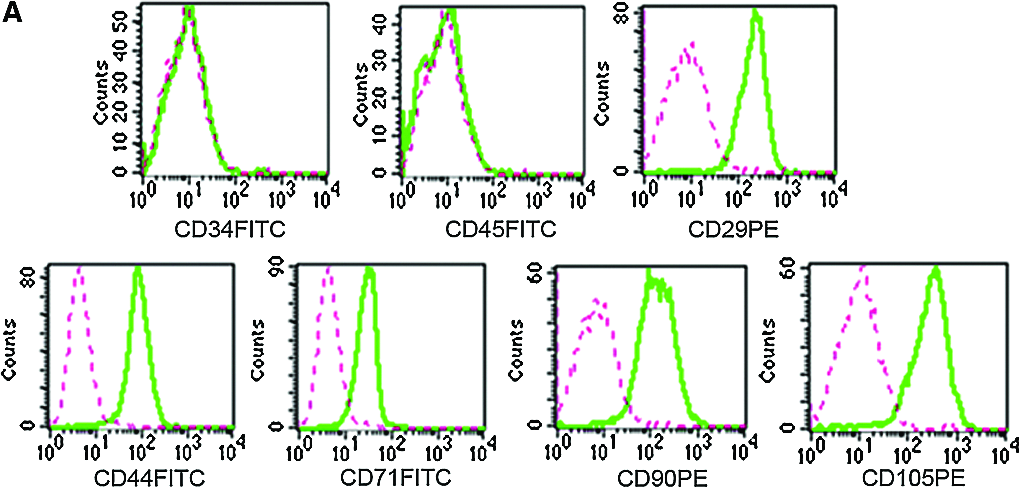
Characterization of hASCs. (
Differentiation of hASCs on scaffolds into hepatocyte-like cells in vitro
We cultured hASCs in monolayer culture system in the presence of HGF, bFGF, FGF-4, and OSM, which are usually used in hepatic differentiation.5,6,14–16 Results showed that the differentiated cells in monolayer culture system displayed several characteristics of hepatocytes, including the round epithelial-like cell morphology, hepatic-specific protein expression, and functionality (PAS stain for glycogen syntheses and indocyanine green (ICG), uptake and secretion) (Supplemental Fig. S1, available online at
Attachment and growth of differentiating hASCs on scaffolds
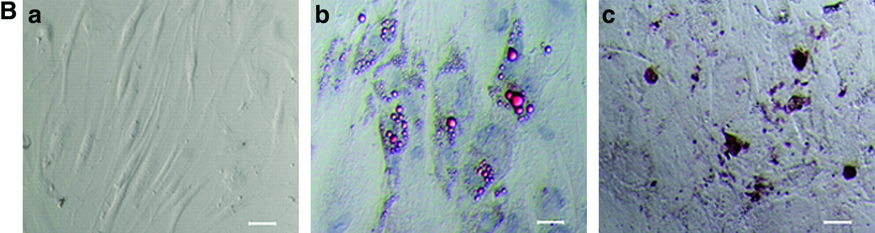
DAPI staining was used to assess hASC distribution throughout the scaffolds. Results showed that hASCs were evenly distributed and integrated into the PLGA scaffolds (Fig. 2A).

Biocompatibility of porous PLGA scaffolds with hASCs in vitro. (
The growth of hASCs was evaluated by the CCK8 assay and indicated by the OD value. When hASCs were seeded in monolayer culture system, the number of hASCs cultured in hepatic inducing medium increased significantly from 3 to 21 days of culture in comparison to hASCs cultured in basic medium (p < 0.001). When cultured in basic medium, no significant difference was observed between hASCs seeded on PLGA scaffold and hASCs seeded in monolayer culture system. Compared with that of hASCs either cultured with basic medium on PLGA scaffold or cultured with hepatic inducing medium in monolayer culture system, the number of hASCs cultured with hepatic inducing medium on PLGA scaffold increased significantly from 3 to 21 days of culture (p < 0.01; Fig. 2B).
SEM was used to examine the morphology of attachment and the growth of hASCs on the PLGA scaffolds (Fig. 2C). Results showed that the fabricated PLGA scaffold is composed of numerous evenly distributed pores and the pores of the scaffold were interconnected without any closed spaces (Fig. 2C-a). hASCs attached to the PLGA scaffold surface appeared somewhat round and penetrated into the pores of the scaffold at 0 day of induction (Fig. 2C-b). Various extracellular matrices were apparently synthesized onto the PLGA scaffolds over induction time. By day 5 (Fig. 2C-d), hASCs apparently synthesized a substantial amount of extracellular matrix components in comparison with day 3 (Fig. 2C-c). HASCs formed a confluent layer on top of the PLGA scaffolds at day 7, and PLGA scaffolds were not visible (Fig. 2C-e). Fourteen days after induction, the cells changed to an elongated shape and displayed numerous cytoplasmic extensions that linked the cells on top of the cell sheet, which eventually formed 3D organoid structures (Fig. 2C-f).
Characterization of hASC-derived hepatocyte-like cells on scaffolds
Transmission electron microscopy of hepatic differentiated hASCs on scaffolds indicate hepatocyte-specific characteristics, including plentiful of mitochondria, endoplasmic reticulum, glycogen, tight junctions, and microvilli on the surface of the cells (Fig. 3A).
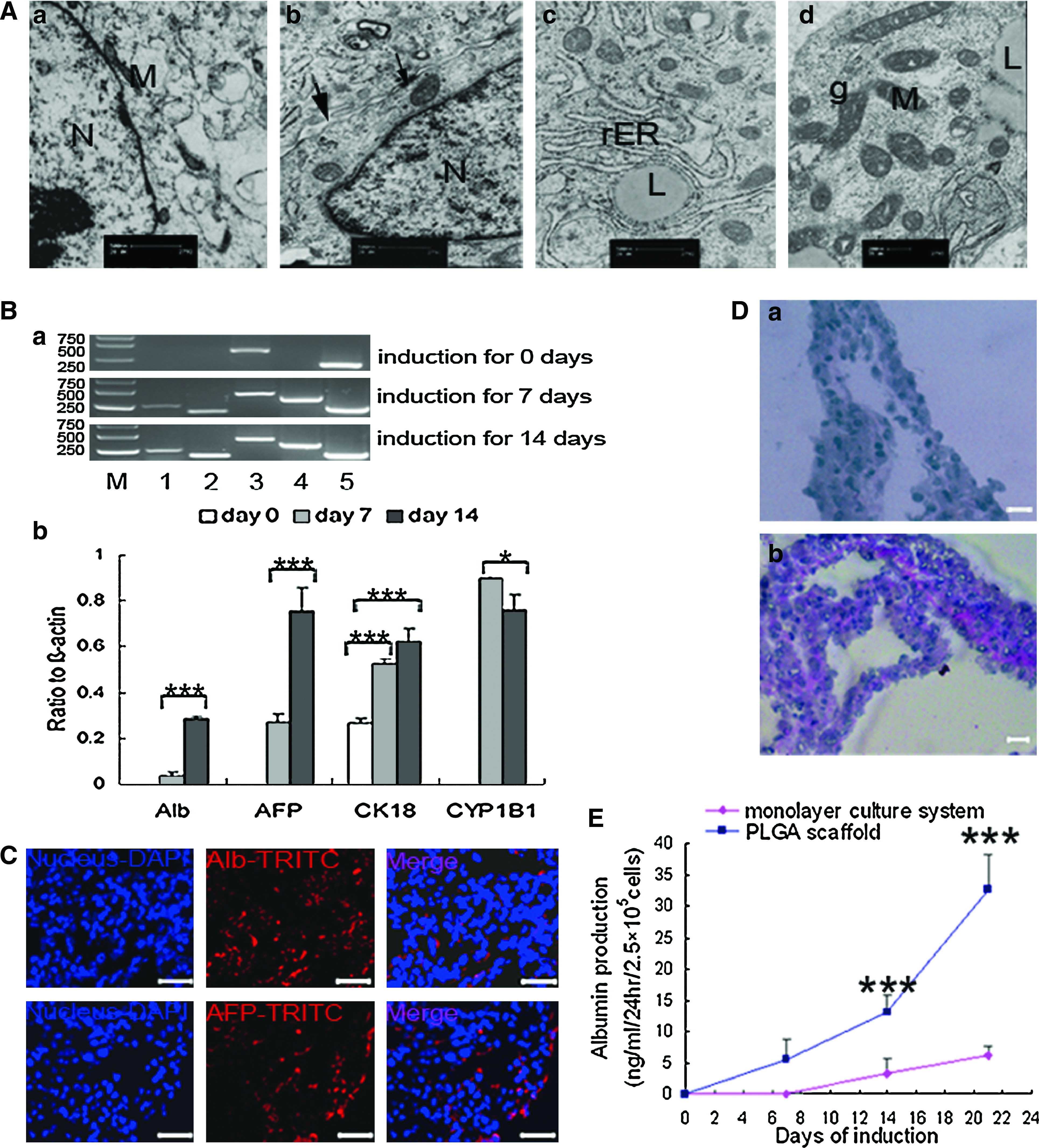
Differentiation of hASCs on scaffolds into hepatocytes in vitro. (
Liver-specific genes analyzed by semiquantitative RT-PCR showed that Alb and AFP mRNA was detectable at day 7 and showed a significant increase at day 14 of induction (p < 0.001, compared with day 7). Cytochrome P450 1B1 mRNA was also detectable at day 7, but was downregulated at day 14 (p < 0.05, compared with day 7). Cytokeratin 18, the marker of epithelia, was expressed earlier by day 0 of induction and showed a significant increase after induction for 7 and 14 days (p < 0.01, both compared with day 0 of induction; Fig. 3B). Immunohistochemical analysis showed positive staining of Alb and AFP after 14 days of induction (Fig. 3C).
PAS is a staining method used in histology and pathology to identify glycogen in tissues. The reaction of periodic acid selectively oxidizes the glucose residues and forms aldehydes that react with the Schiff reagent and create a purple-magenta color. The presence of stored glycogen, as determined by PAS staining, was observed in differentiated cells at 2 weeks postinduction (Fig. 3D). The secretion of albumin from hASCs was examined by ELISA. Albumin production from cells on porous PLGA scaffolds was detected at 7 days of induction and increased in a time-dependent manner, whereas undifferentiated hASCs (day 0) did not produce albumin. In comparison, albumin production from cells cultured in monolayer culture system at the same seeded number was detected at 14 days of induction and was significantly lower than those from cells cultured on porous PLGA scaffolds at 14 and 21 days of induction (p < 0.001; Fig. 3E).
Effect of PLGA scaffolds' physical properties on hepatogenic differentiation of hASCs
To evaluate how scaffolds' physical properties influence hepatogenic differentiation, 10% (w/v) PLGA scaffolds, fabricated with various size and weight fraction of NaCl particles, were seeded with hASCs and cultured in hepatic inducing medium for 14 days, followed by real-time PCR analysis of Alb gene expression. Results showed that hASCs seeded on scaffolds prepared with 120–200 μm size of NaCl particles had the highest Alb gene expression in the four NaCl particle size ranges and showed significant differences when compared with that on scaffolds prepared with <50, 50–120, or >200 μm size of NaCl particles (Fig. 4A). The hASCs seeded on scaffolds prepared with 50% weight fraction of NaCl particles had the highest Alb gene expression, and statistical analysis showed significant differences compared with Alb expression on scaffolds prepared with 75% or 90% weight fraction of NaCl particles (Fig. 4B).
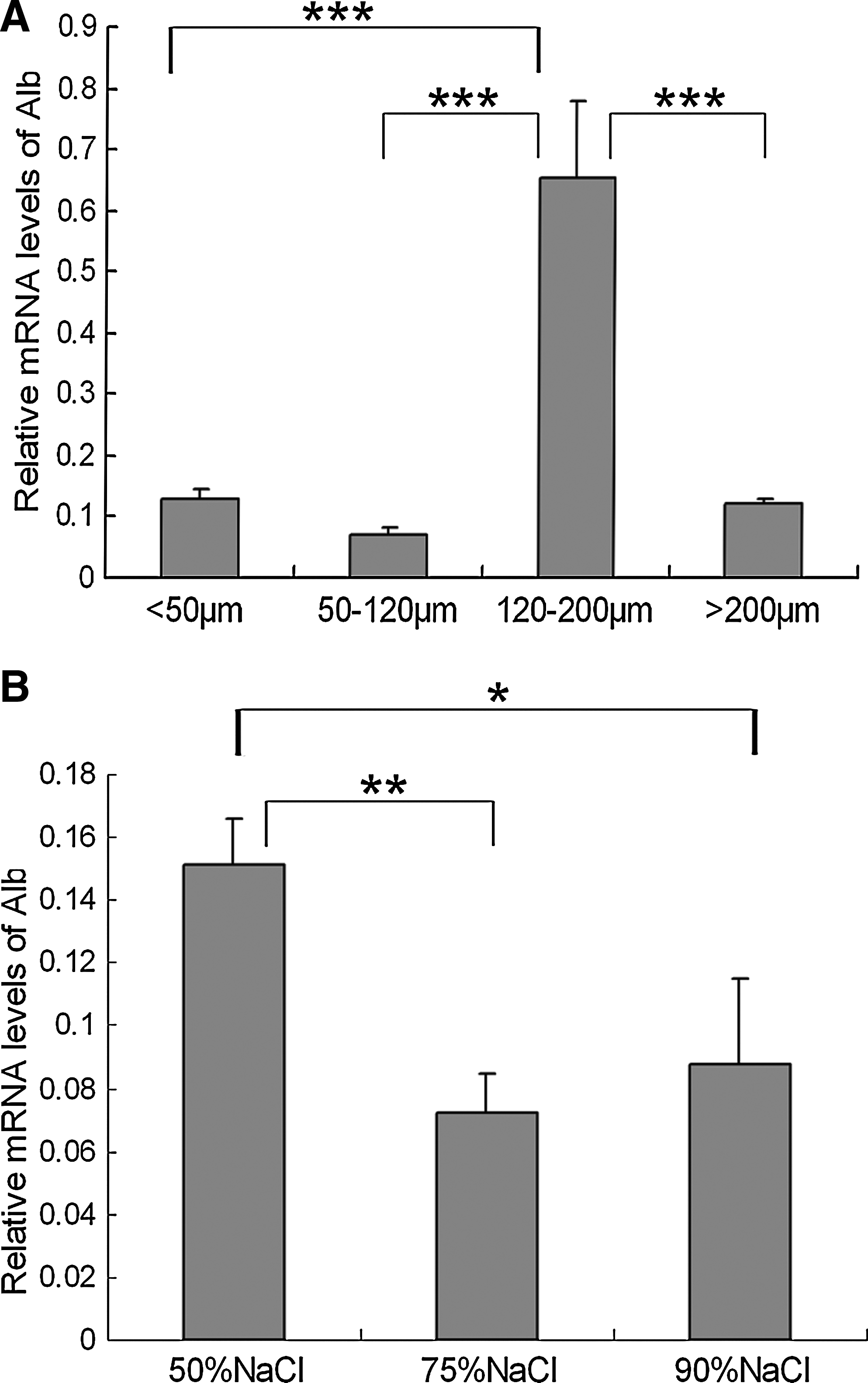
Effect of PLGA scaffolds' physical properties on hepatogenic differentiation. Alb expression in hASCs cultured on porous PLGA scaffolds prepared with various size (
Survival and differentiation of hASCs following implantation
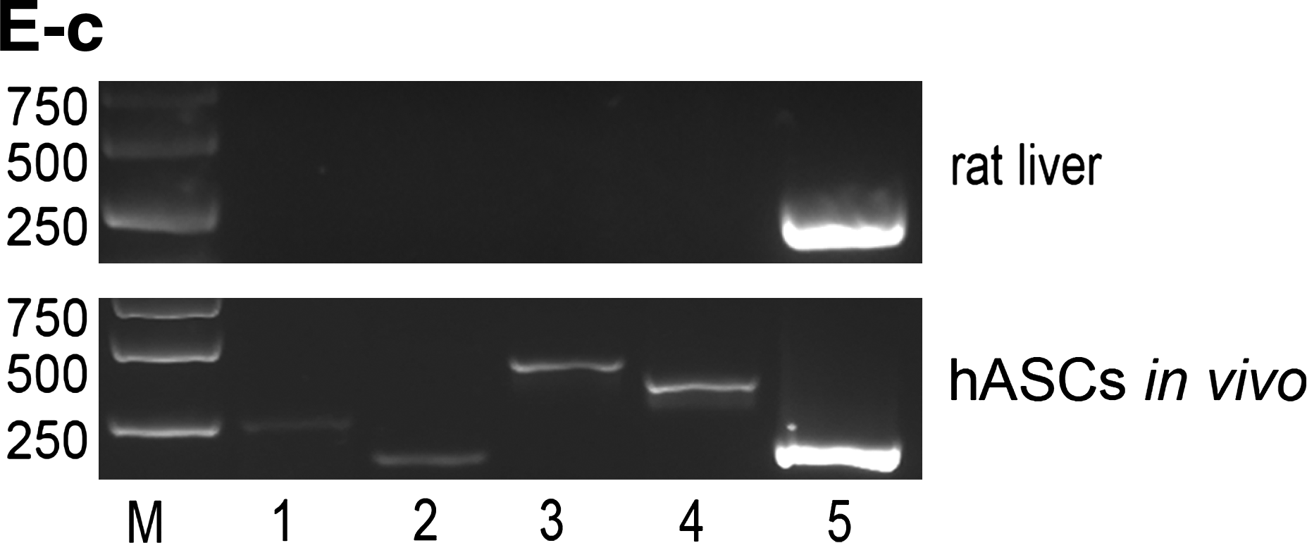
To distinguish hASCs from host cells, hASCs on scaffolds were prelabeled with CFDA-SE (green) before transplantation. Frozen sections of all implants were analyzed for human albumin expression by immunofluorescence, with intention to evaluate the hepatic differentiation of hASCs in vivo. The invasion of host cells was observed in implants of cell-free scaffolds, but no hASCs existed (Fig. 5A). Except for the invasion of host cells, the existence of some hASCs (green) was easily found, and most human Alb-positive cells were observed among those hASCs (red) in all cell-loaded scaffolds (Fig. 5B–D, E-a). Compared with scaffolds with preinduced hASCs, the existing hASCs were relatively few in scaffolds with noninduced hASCs at 14 days after transplantation (Fig. 5B, E-a). The existing hASCs in scaffolds with preinduced hASCs did not decrease significantly with increasing in vivo implant period (Fig. 5C, D, E-a). For scaffolds loaded with induced hASCs at 14 days postimplantation, many human AFP-positive cells could be found (Fig. 5E-b), and RT-PCR experiments demonstrated the expression of human liver-specific genes that were not expressed in rat livers (Fig. 5E-c). However, in this study, human albumin was undetectable in the serum of all rats after implantation of the hASC–scaffold complex.
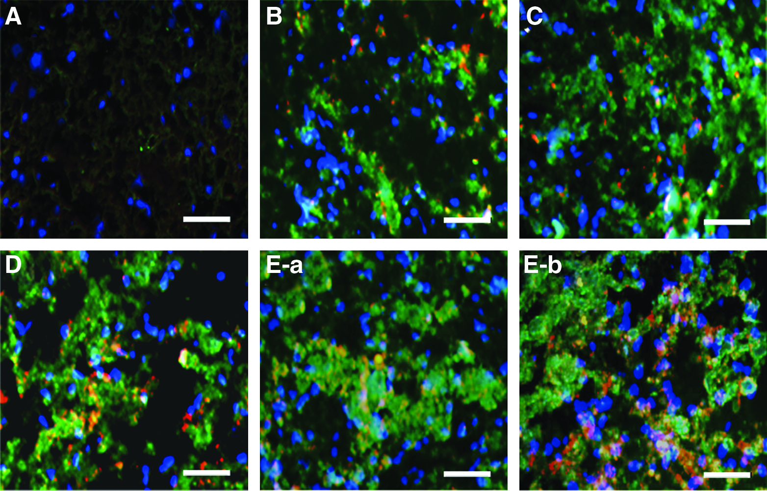
In vivo survival of hASCs. Immunofluorescence staining of human albumin protein for PLGA scaffolds without cells at 14 days postimplantation (
Discussion
Previous studies have shown that hASCs are able to differentiate into hepatocytes in conventional monolayer culture systems; however, the formation of a transplantable organoid graft with an architecture resembling that of natural liver could not be achieved in such culture system. The goal of this study was to explore appropriate 3D scaffold for hepatogenesis of hASCs. To our knowledge, this is the first study demonstrating the ability of PLGA scaffold to support differentiation of hASCs to a liver cell lineage both in vitro and in vivo.
Biocompatibility of biomaterials with stem cells is the foundation of stem cell survival and differentiation on scaffolds. Although many groups focusing on tissue engineering have used the combination of PLGA with hASCs in vivo,17–19 limited characterizations on the biocompatibility of porous PLGA scaffold with hASCs in vitro have been performed. Our studies using DAPI staining, CCK8 assay, and SEM thoroughly demonstrate that porous PLGA scaffolds are biocompatible with hASCs.
The growths of stem cells on scaffolds with and without differentiation are two different processes, and most studies are focused on the latter. So, we examined hASC growth on PLGA scaffold during hepatic differentiation process in this study. Consistent with previous findings that HGF and FGF cytokines promote cell proliferation as well as differentiation,20,21 our results demonstrate that the hepatic inducing medium consisting of HGF, bFGF, and FGF-4 promotes significant simultaneous proliferation and differentiation of hASCs both in monolayer culture system and on the PLGA scaffolds. Compared with 2D system, 3D scaffolds have more surface area for the cells' attachment, expansion, and differentiation. Consistent with this, hASCs cultured on 3D PLGA scaffold proliferated more efficiently than in monolayer culture system, especially in hepatic inducing medium.
One of the factors that affect cell morphology is the type of scaffolds.22,23 Present SEM analysis represented an original morphological investigation of continuously differentiating hASCs on PLGA scaffolds. In this study, cells seeded onto scaffolds appeared somewhat rounded at 0 day of induction, in contrast with spindle-like cells seeded onto monolayer culture plate. This might be due to the effects of the specific substrata used for the cells. Unlike the cell–cell aggregates of hepatocytes found on nonwoven hyaluronic acid esters (HYAFF) fabrics, 23 differentiating hASCs spread along the surface of the porous scaffolds and gradually formed a cell sheet. Remarkably, at day 14 of induction, cytoplasm extension was seen linking the cells on top of the cell sheet, and cells were reorganized into 3D structures resembling those in vivo.
Previous studies have demonstrated that hASCs cultured on PLGA scaffolds can successfully differentiate into osteogenic and chondrogenic lineages.24,25 In this study, we investigate the ability of PLGA scaffolds to support the hepatic differentiation of hASCs. Data in vitro showed that hASCs on PLGA scaffolds in hepatic inducing medium could differentiate into cells with phenotypic characteristics of hepatocytes with functional properties of glycogen deposits and albumin secretion. Interestingly, we also observed that hASCs cultured on porous PLGA scaffolds (3D) produced more albumin than those cultured in monolayer (2D) culture system, suggesting that the 3D PLGA scaffolds that mimic the in vivo environment more closely might be more inductive for hepatic differentiation than the monolayer culture system.
Studies by Taqvi and Roy 26 demonstrated that the physical property (e.g., porosity and pore size) of scaffolds, which mainly depend on weight fraction and size of the NaCl particles, also affected cell differentiation. We varied the size and weight fraction of NaCl particles and explored their effect on hepatic differentiation, and results showed that sizes of 120–200 μm and a weight fraction of 50% would be the most beneficial condition for hepatic differentiation of hASCs.
To investigate the in vivo influence of PLGA scaffolds on hASCs, we implanted the scaffolds with noninduced hASCs or preinduced hASCs into hepatectomized rats. The partially hepatectomized host microenvironment has been demonstrated to be beneficial for hepatic differentiation of stem cells by many studies.27–30 In this study, preinduced hASCs seeded on PLGA scaffolds survived and maintained the hepatic phenotype for at least 14 days, and noninduced hASCs seeded on PLGA scaffolds could differentiate into hepatocytes and express human albumin at 14 days after transplantation under this microenvironment. However, compared with preinduced hASCs, the number of noninduced hASCs both before and after implantation was significantly fewer when the cells were seeded at the same number. These results suggested that preinduced hASCs might be better than noninduced hASCs when applied in vivo, though much work needs to be done to test this speculation. Many studies have demonstrated that the ASCs have the ability to suppress immunological reactions.31–33 Here we showed that preinduced hASCs in scaffolds did not decrease significantly from 3 to 14 days postimplantation, which might be related to the ASCs-mediated immunosuppression. However, injection of small dosages of immunosuppressant cyclosporin A might also contribute to this. Collectively, all these results suggest that PLGA scaffolds do not exert undesirable side effects on hASCs in vivo.
Recent studies by Ong et al. 30 have demonstrated that human albumin was detectable in serum at 14 days after transplantation of pellets containing differentiated mesenchymal stem cells (MSC) into rat livers. However, in this study, human albumin was undetectable in serum of all rats after implantation of the hASC–scaffold complex. This could be due to the specific sites of implantation or an insufficient number of transplanted cells.
In conclusion, we have demonstrated that porous PLGA scaffolds supported hASC attachment, proliferation, and differentiation into hepatocytes in vitro, as well as the survival and hepatic differentiation in vivo. These results suggest that porous PLGA films have the potential to be used as scaffolds for hASC-based hepatogenic tissue engineering for end-stage liver disease therapy.
Footnotes
Acknowledgments
The authors thank Ms. Xue Nan and Shuangshuang Shi for technical support. The authors also thank Dr. Lola M. Reid in the Department of Cell and Molecular Physiology, University of North Carolina School of Medicine, Chapel Hill, for critical review of the manuscript. This work was supported by the National High Technology Research and Development Program of China (no. 2006AA02A107 to X.T.P. and Y.F.W.), the Major State Basic Research Program of China (no. 2005CB522702 to X.T.P. and Y.F.W.), National Nature Science Foundation of China (no. 30671098 to Y.F.W.), and National Nature Science Foundation of China (no. 30901441 to H.Y.P.).
Disclosure Statement
No competing financial interests exist.
References
Supplementary Material
Please find the following supplemental material available below.
For Open Access articles published under a Creative Commons License, all supplemental material carries the same license as the article it is associated with.
For non-Open Access articles published, all supplemental material carries a non-exclusive license, and permission requests for re-use of supplemental material or any part of supplemental material shall be sent directly to the copyright owner as specified in the copyright notice associated with the article.
