




Abstract
Bioactive and degradable scaffolds made from bioactive glass–polycaprolactone with a mineralized surface and a well-defined three-dimensional (3D) pore configuration were produced using a robotic dispensing technique. Human adipose–derived stem cells (hASCs) were cultured on the 3D scaffolds, and the osteogenic development of cells within the scaffolds was addressed under a dynamic flow perfusion system for bone tissue engineering. The bioactive glass component introduced within the composite assisted in the surface mineralization of the 3D scaffolds. The hASCs initially adhered well and grew actively over the mineralized surface, and migrated deep into the channels of the 3D scaffold. In particular, dynamic perfusion culturing helped the cells to proliferate better on the 3D structure compared to that under static culturing condition. After 4 weeks of culturing by dynamic perfusion, the cells not only covered the scaffold surface completely but also filled the pore channels bridging the stems. The osteogenic differentiation of the hASCs with the input of osteogenic factors was stimulated significantly by the dynamic perfusion flow, as determined by alkaline phosphate expression. Overall, the culturing of hASCs upon the currently developed 3D scaffold in conjunction with the dynamic perfusion method may be useful for tissue engineering of bone.
Introduction
A range of materials with bioactive compositions have been developed as bone tissue engineering scaffolds, which include calcium phosphate and glass inorganics, degradable polymers, and their composites.5–8 The porous structure and bioactive composition should be considered to achieve optimal performance for bone tissue engineering.
Recent progress in technology has allowed the production of scaffolds with a custom-tuned geometry and pore configuration.9–12 A robotic dispensing (or robocasting) methodology, which is a type of rapid prototyping technique, is also considered to be an effective processing route for providing scaffolds with a pore configuration in a controllable manner, that is, defined porosity and pore size, which is in direct contrast to the conventional scaffold fabrication methods, such as foaming, negative molding, and salt-leaching.9–13
Previous studies on robotic dispensed scaffolds generally used degradable polymers, such as poly(lactic acid), poly(ɛ-caprolactone) (PCL), chitosan, and collagen.14,15 On the other hand, bioactive inorganics were obtained by the high-temperature consolidation of the robotic dispensed preforms.16,17 While inorganics have good cell responses and bone bioactivity, polymeric scaffolds have the merit of low temperature processing, which can be used to combine biofunctional molecules. Therefore, robotic dispensing of composites using a polymer with a bioactive inorganic phase is considered a suitable way of reaping the benefits of the individual components.18–20
Along with the scaffolds, the cell source is another important parameter for determining the success of bone tissue engineering. Recent advances in cell biology have spurred the widespread use of stem cells in tissue engineering. Compared to an embryonic origin, adult stem cells are easily accessible, directly applicable to autologous surgery, and without ethics problems. 21 Among autologous adult stem cells, bone marrow–derived stem cells have been studied as a cell source for tissue engineering with multipotent differentiation potential for the repair of cartilage, bone, muscle, and neural tissue. 22 As an alternative autologous adult stem cell source, adipose-derived stem cells (ASCs) have attracted considerable interest.23–26 Human adipose tissue can be easily obtained by a suction-assisted lipectomy in relatively large quantities and with minimal discomfort.23,24 Recent studies have shown that ASCs retain their multipotent trait to differentiate into adipogenic, chondrogenic, osteogenic, myogenic, and neurogenic lines with appropriate biochemical cues.24–26
For ASC-based tissue engineering of bone, the appropriate choice of material, which is to serve as a three-dimensional (3D) substrate for cells to grow and differentiate and further to deposit bone mineral, is of special importance. Herein, we use a bioactive and degradable composition, the composite of PCL and bioactive glass (BG), and the surface of the 3D scaffold is functionalized with calcium phosphate mineral to provide a bone-bioactive and tissue-compatible biointerface. Moreover, the 3D structure of the scaffold is constructed by means of the robotic dispensing method. In particular, the ASCs are cultured upon the 3D scaffold under dynamic perfusion flow, which is expected to allow favorable microenvironment for cell multiplication and osteogenic development. The potential of the scaffold and culturing condition to populate and differentiate the ASCs is addressed.
Materials and Methods
Robotic dispensing of composite scaffolds
For the inorganic component of the composite scaffold, sol-gel–derived BG was prepared using the method described in a previous study with slight modifications. 27 Briefly, tetraethyl orthosilicate, calcium nitrate, and triethyl phosphate were mixed in ethanol-distilled water using HCl as a catalyst and stirred vigorously for 6 h. The sol was aged at 50°C for 3 days to allow gelation. The gel was dried and then heat treated at 650°C for 3 h. The powder was crushed, and sieved to a fine powder using a 10 μm sieve. The powder was dispersed in acetone at 50°C. PCL (molecular weight = 80,000; Sigma-Aldrich, St. Louis, MO) was dissolved in a glass–acetone solution at varying ratios (PCL:glass = 3:1, 1:1, and 1:3 by weight) and mixed at 50°C to prepare the mixture solution for robotic dispensing.
The mixture solution was loaded into a syringe and injected through a needle (520 μm diameter) with the assistance of a force-controlled plunger to regulate the mass flow rate of 3 μL/s. A positional control unit linked to a personal computer was operated to guide the dispensing path along the xy-plane and z-direction. The xy-movement speed was fixed at 10 mm/s to produce uniform fiber deposits and well-developed pore configuration. The working temperature of the solution was kept at 50°C using a thermostatically controlled heating jacket, and the ejected fiber was placed in contact with a cooled ethanol bath. The dispensing program was set to produce a BG-PCL porous scaffold with dimensions of either 5 × 5 × 3 mm or 10 × 10 × 3 mm, consisting of six successive layer-by-layer building units with an intended pore size of approximately 500 × 500 μm.
Surface mineralization of scaffolds and characterization
To mineralize the surface with crystalline apatite, the robotic dispensed BG-PCL scaffold was further treated with simulated body fluid (SBF; Na+ 142 mM, K+ 5.0 mM, Mg2+ 1.5 mM, Ca2+ 2.5 mM, Cl− 147.8 mM, HCO3− 4.2 mM, HPO42− 1.0 mM, and SO42− 0.5 mM), which was prepared according to the procedure described elsewhere. 28 Each sample was placed in a plastic tube containing 10 mL of SBF and incubated at 37°C with gentle agitation for different times. From a pilot study, the SBF immersion for 7 days was observed to be the optimal to cover the scaffold surface almost completely with an apatite mineral phase while no surface cracks or delaminations being observed, and thus the scaffolds mineralized for 7 days were used for further cell-culturing tests.
To examine the morphology, the scaffolds were vacuum-dried and coated with Pt. Scanning electron microscopy (SEM; 3000; Hitachi, Ibaraki, Japan) was carried out under operation conditions of an accelerating voltage 15 kV and magnifications ∼20 × to 1000 ×. The pore configuration was assessed from the SEM images. The morphological changes during immersion in SBF were examined by SEM. Energy dispersive spectroscopy was employed to determine the composition of the scaffolds before and after the SBF immersion test.
Isolation and 2D culturing of human ASCs
Human adipose tissues were obtained from the inside of the thigh of a patient who gave informed consent (40-year-old woman) at The Yeonsei Ellefine Clinic (Cheonan, South Korea). The lipoaspirate was processed through a series of sterile phosphate buffered saline washes and digested with 0.075% collagenase I (type I; Sigma-Aldrich) at 37°C for 30 min. The collagenase was then neutralized with an equal volume of the growth medium, α-minimum essential medium containing 10% fetal bovine serum and antibiotics (100 U/mL penicillin G and 100 μg/mL streptomycin). The solution was centrifuged at 1200 g for 10 min. The cell pellet was resuspended in 160 mM NH4Cl and incubated at room temperature for 10 min. The cellular remains were removed through a 100 μm Cell Strainer (Falcon, France). The cells were then incubated at 37°C in a humidified, CO2-controlled (5%) incubator. The adherent cells were allowed to reach 80% confluence (12–17 days for the first passage and 6 days for later passages). The cells were trypsinized and replated every 6–8 days until they reached 80% confluence.
To confirm the osteogenic development of the human ASCs (hASCs) on 2D well plate, the cells were seeded on a 12-well plate at a density of 5 × 10⁴ cells/well and allowed to grow in Dulbecco's modified Eagle's medium containing 10% fetal bovine serum, coupled with osteogenic factors (100 nM dexamethasone, 50 mM ascorbic acid, and 10 mM β-glycerophosphate). Each medium was changed three times per week. Osteogenic development was confirmed by alizarin red S staining (Sigma, St. Louis, MO), which stains calcium red.
Static and dynamic perfusion culturing on 3D scaffolds
Before seeding cells onto the prepared 3D scaffolds, the scaffolds were sterilized by immersion in 70% ethanol for 1 h and washed twice with phosphate-buffered saline. An aliquot of 5 × 10⁴ cells were plated onto each scaffold and cultured overnight to allow the cell adherence and spreading. The cell culturing was continued for up to 28 days (referred to “static culturing”), and the medium was exchanged every 2 days.
On the other hand, after the cell adherence, the cell–scaffold construct was transferred to a perfusion chamber (referred to “dynamic culturing”). The perfusion system consists of one gas exchanger, two perfusion containers, and one medium reservoir. The perfusion container is designed to hold a specimen (10 × 10 × 2 mm), where the scaffold z-axis was parallel to the perfusion flow. In particular, the xz- and yz-face of scaffold were tightly held onto a groove (1 mm depth × 3 mm width) made in the inner wall of the container. The medium was supplied to the perfusion containers through nozzles operated by a peristaltic pump (Masterflex, Barrington, IL). The flow rate was fixed to 0.6 mL/h. The constructs were cultured for up to 28 days under osteogenic culturing medium. Each construct received a total of ∼100 mL of fresh medium each week.
Proliferation assay and cell morphology observation
An MTT (a tetrazolium salt [3-(4,5-dimethyldiazol-2-yl)-2,5-diphenyl tetrazolium bromide]) assay and direct count were carried out to measure the proliferation of ASCs on the bioactive scaffold. At each culturing time, one milliliter of MTT solution was added to the scaffolds, which was incubated for 3 h. The MTT solution was aspirated, and an equal volume of a 10% v/v dimethyl sulfoxide (DMSO)–isopropanol lysis solution was added to each scaffold. The results were read on a spectrophotometer (Kontron Instruments, Helsinki, Finland) at A540 nm.
The cell growth morphology was observed by SEM at an accelerating voltage of 15 kV. The cell–scaffold constructs were fixed with glutaraldehyde (2.5%) for 10 min, dehydrated with a graded series of ethanol (75%, 90%, 95%, and 100%), treated with hexamethyldisilazane, and sputter-coated with Pt.
Confocal laser scanning microscopy (CLSM: LSM 510; Carl Zeiss, Jena GmbH, Germany) was used to observe the cells grown on the samples. The cells grown on each sample were fixed with 4% paraformaldehyde, treated with 0.2% Triton X-100, and then blocked with 1% bovine serum albumin to prevent nonspecific protein binding. The ProLong® Gold antifade reagent with DAPI (P36935; Invitrogen, Carlsbad, CA) was used to stain the nucleus. A fluorescence image was obtained by CLSM.
Osteogenic differentiation and alkaline phosphatase activity determination
The osteogenic differentiation of cells on the 3D scaffolds was carried out using the same medium as that used in the 2D culture well. The cells seeded at a density of 5 × 10⁴ cells on each scaffold were cultured for up to 21 days, either by static or dynamic perfusion culturing methods. The alkaline phosphatase (ALP) activity of the cells was assessed as an index of osteogenic differentiation. At each culturing time, the cell layers were harvested from the scaffolds by disruption with Triton X-100 and cyclic freezing–thawing processes. The ALP activity was assessed colorimetrically by applying a p-nitrophenyl phosphate substrate to each sample. The enzymatic product p-nitrophenol was detected by measuring the optical density at A410 nm, which was converted to the level of ALP activity. The total protein content was quantified using a DC protein assay kit (BioRad, Hercules, CA), and the ALP level was normalized to the total protein content. A protein standard curve was obtained using bovine serum albumin. Three replicate samples were tested under each condition.
Statistical analysis
Data are presented as mean ± one standard deviation from three sets of measurements (n = 3). Statistical analysis was carried out using Student's t-test, and significance was considered at p < 0.05 between groups.
Results and Discussion
Robotic dispensing and bioactive scaffolds
Figure 1 shows a schematic diagram of the robotic dispensing apparatus and the experimental procedures to obtain the 3D bioactive scaffold. As a first step, the preparation of a slurry with the appropriate rheological properties is of particular importance for allowing the fiber injection through the nozzle. The BG and PCL mixture in acetone was homogenized within a high-temperature ball-miller, which helps disperse the BG particles well within a viscous PCL slurry. Immediately after dispensing, the slurry was allowed to solidify into a cylindrical fiber using a cooled ethanol bath. 20 The dispensing process was programmed to move along the xy-plane to construct the 2D alignment of a fibrous structure (as shown in Fig. 1A, B); 3D scaffolding was facilitated by layer-by-layer assembly of the 2D structure using a computer-aided program. Figure 1C shows a typical image of the robotic dispensed 3D scaffold. In particular, the BG component added had a high level of bone bioactivity, as showed by the rapid formation of a surface mineral phase within an SBF. At 1 day of immersion, mineral aggregates formed on some parts of the BG granule surface, and the mineral phase covered the granule surface completely at 3 days of immersion, forming a thick mineral layer (Fig. 1D).
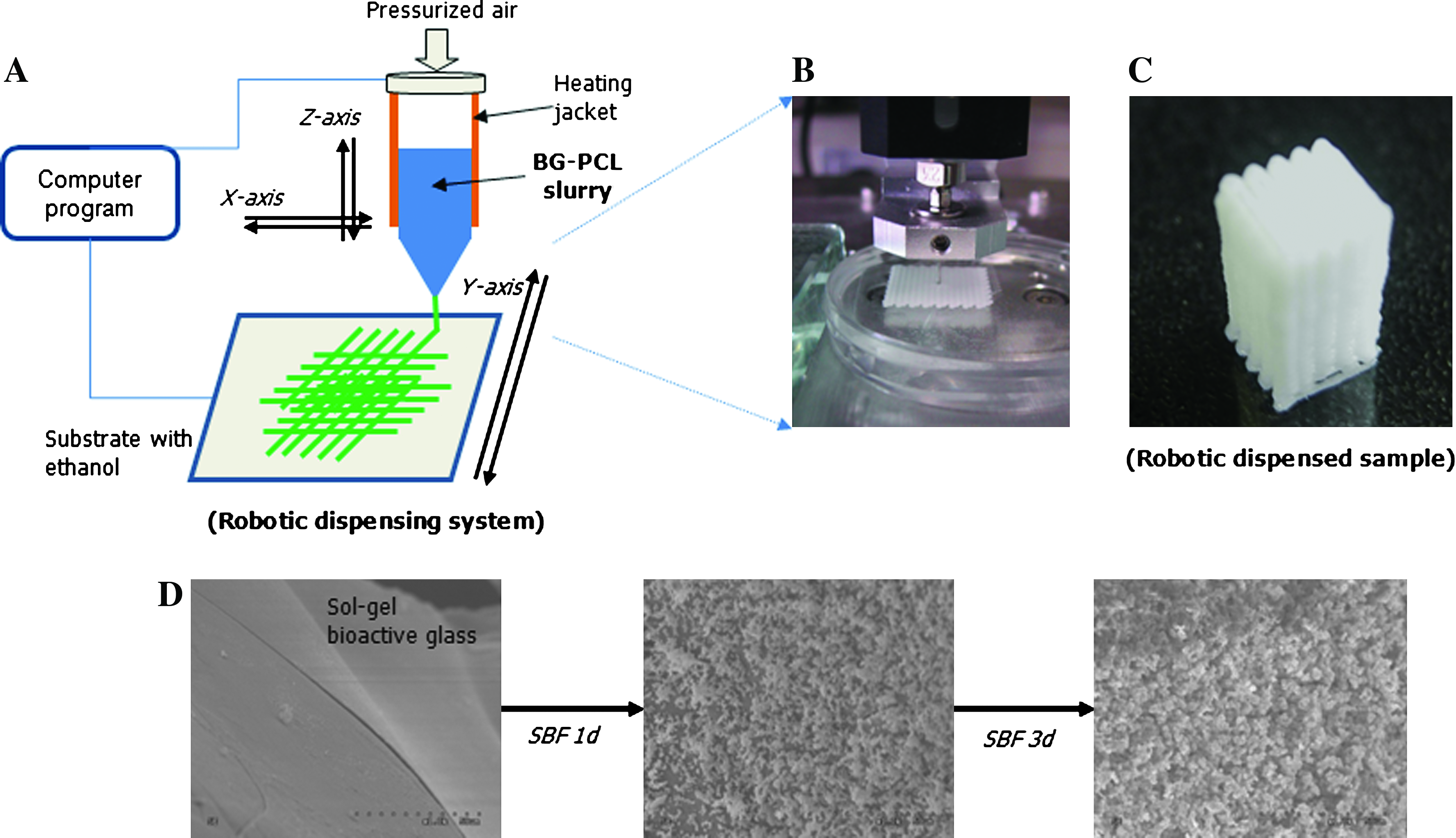
(
Therefore, the introduction of BG powder within the PCL biopolymer enhances the bone bioactivity of the composite scaffolds. The surface bone bioactivity in SBF varied according to the amount of BG added to the PCL. When 25% BG was added to the composite, mineral induction began at ∼7 days, and became significant at ∼14 days (Fig. 2A). As the BG content was increased to 75%, mineral induction began in as little as ∼3 days, and was significant at 7 days (Fig. 2B). Considering the potential of the composite scaffold in the bone regeneration area, a composition with a higher BG content (BG:PCL = 3:1) was used in further study.
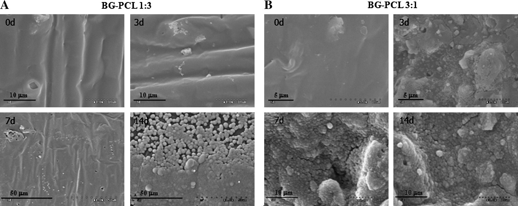
Surface mineralization pilot study of the BG-PCL composite scaffolds, showing the surface morphological change in the scaffolds with two different compositions: the BG-to-PCL ratio was (
The 3D morphology and pore configuration of the robotic dispensed bioactive composite scaffold was examined, as shown in Figure 3. A tilted image of the BG-PCL (3:1) scaffold showed the generation of a well-constructed 3D geometry with interwoven cylindrical fiber stems and square-shaped interspacing pore channels (Fig. 3A). An enlarged image of the scaffold surface revealed the existence of BG particles embedded uniformly in a PCL matrix (Fig. 3B). The 3D pore configuration was evaluated from the SEM images. The obtained fiber thickness was ∼301 μm (±22.7) and the channel dimension was ∼352 μm (±24.6). The geometrical features that support the 3D scaffold are believed to present stable substrate conditions for the cells to adhere to and populate on, as well as to provide a sufficiently large pore size and space for blood circulation and cellular ingrowth. 20

Three-dimensional morphology of the BG-PCL 3:1 composite scaffold: (
The scaffold surface was further tailored with a calcium phosphate mineral phase. The BG-PCL composite scaffolds were found to induce a mineral phase rapidly and profoundly within the SBF under in vitro conditions. Hence, this mineralization behavior was used to coat the surface of the scaffold. Figure 4 shows the robotic dispensed BG-PCL scaffold after the SBF treatment for 7 days. Throughout the scaffold surface calcium phosphate precipitation occurred and the 3D pore configuration was not changed significantly (Fig. 4A). A higher magnification of the surface showed a uniform thick layer of nanocrystallites (Fig. 4B). The atomic composition of the mineral phase was detected by energy dispersive spectroscopy (Fig. 4C), which shows a significant increase in the Ca and P peaks, and the disappearance of the Si peak. The Ca/P ratio was 1.45, which was slightly lower than that for stoichiometric hydroxyapatite (Ca/P = 1.67) but similar to the characteristics of a biological carbonated apatite found in bone and teeth.29,30 Other studies using BG–polymeric substrates have reported the evolution of a mineral phase with a similar morphology and a Ca/P ratio to those in this study.31,32 The added BG component contributed to surface mineral formation via an ionic release and precipitation process. After immersion in SBF, ionic exchange, that is, Ca release from the materials and H3O+ from the SBF, occurred at the surface. This was followed by the formation of Si-OH groups, which later induced the nucleation of apatite mineral. The ions released from the material supersaturated the SBF with respect to apatite, accelerating mineral nucleation and growth on the surface.
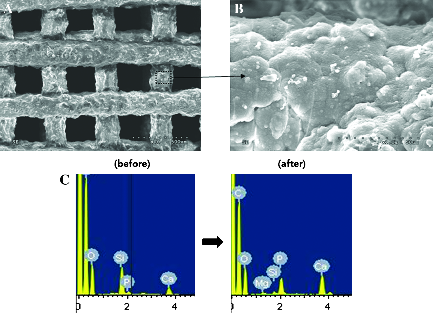
Morphology of the BG-PCL 3:1 composite scaffold with surface mineralization for 7 days: (
The aim of this treatment of the scaffold with apatite mineral was to produce a scaffold surface that mimics the bone extracellular matrix and further combines specific biofunctional molecules, such as growth factors and osteogenic proteins. 33 The latter is convincing considering that the calcium phosphate mineral, particularly, hydroxyapatite nanocrystalline phase, has high biological affinity to a range of bone-associated proteins, such as osteocalcin, bone sialoprotein, osteonectin, and many growth factors, forming specific bonds, which are also dependent on the surface properties of the mineral, including the chemical composition, surface charge, and crystallography.34–38 In vivo, biological molecules play their roles in bone formation by binding to the hydroxyapatite mineral phase.34–37 In this manner, a further study will be needed to introduce growth factors/osteogenic proteins, such as fibroblast growth factor and bone morphogenetic proteins, to better utilize the surface-tailored 3D scaffold. The performance of the 3D scaffold was addressed on the population and osteogenic differentiation of hASCs under a dynamic perfusion culturing system.
Dynamic perfusion culturing and hASC responses to scaffolds
Compared to other types of adult stem cells, such as those derived from bone marrow, the hASCs can be accessed in large quantities through liposuction with minimal discomfort,23,24 which highlights the potential of the hASCs in tissue engineering including bone. Moreover, in response to the appropriate cues, the hASCs undergo osteogenic development, finding promising usefulness in skeletal regeneration.25,26
When the hASCs were cultured in the osteogenic medium for 2 weeks, the cells were covered with a layer of visible deposits (Fig. 5A), which is indicative of cell differentiation. This was not observed in the control cells without an osteogenic treatment (Fig. 5B). Positive staining with alizarin red S showed that these deposits were indeed calcified extracellular matrix (Fig. 5C), which was not observed in the control cells (Fig. 5D).
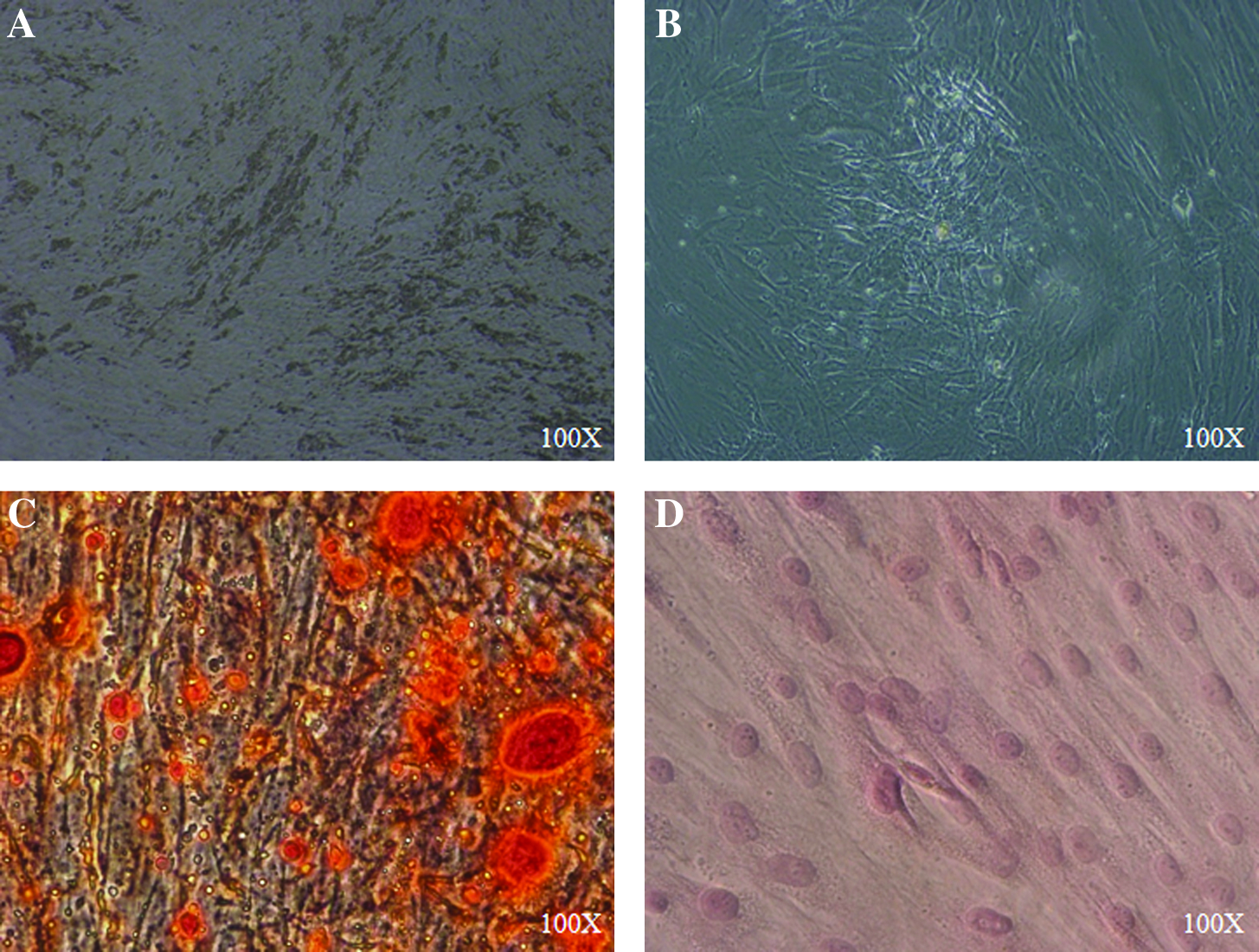
Human adipose–derived stem cells (hASCs) cultured on a 2D culture plate either (
After confirming the osteogenic development of the hASCs under the proper cues within a 2D culture well, the robotic dispensed 3D scaffolds were used to cultivate the cells for bone tissue engineering. A dynamic perfusion system was used to provide continuous medium flow conditions to the 3D scaffold. After seeding the hASCs, the scaffolds were placed in a chamber through which the culturing medium was flowed in and out at a constant rate (herein 0.6 mL/h). During perfusion culturing, a larger quantity of flow was designed to be supplied to the cells and at the same time to remove the waste medium quickly. 39 At the flow rate used in this study, a total 100 mL was consumed each week, which was almost 3 times more than that used in static culturing (medium refreshed every 2 days).
Figure 6 shows confocal laser scanning microscopy images of the populated cells on surface-mineralized 3D scaffolds by either static or dynamic perfusion culturing for 3 and 7 days. The cells stained in red were viable and grew well on the fiber stems of the scaffolds. There appeared to be more cells at both periods by the perfusion culturing (Fig. 6B, D) than by the static culturing (Fig. 6A, C). The confocal cell growth image by the perfusion culturing was recorded over a longer period (4 weeks), as shown in Figure 7. In particular, a construction along the z-line was made to clarify the presence of cells inside the porous scaffold. Along with the significant staining of cells in the stems, the porous channels were covered almost completely with cells. A higher magnification of the pore channel showed that the cells penetrated well into the spacing with a highly networked structure. This image of the cell–material constructs suggests the potential use of the biological hybrid in tissue engineering when cultured dynamically in a perfusion system for an appropriate period. The cell image throughout the composite scaffold after 14 days of culturing was further observed by SEM, as shown in Figure 8 under different magnifications. Actively proliferating cells, their secreted extracellular matrix, and cell–scaffold construction were more noticeable in the electron images. The scaffold stems were covered almost completely with a number of cells that formed a thick cell layer (Fig. 8A, B). The cell layer appeared to secrete a fibrous extracellular matrix (Fig. 8C), and bridged some parts of the pore channels (Fig. 8D).
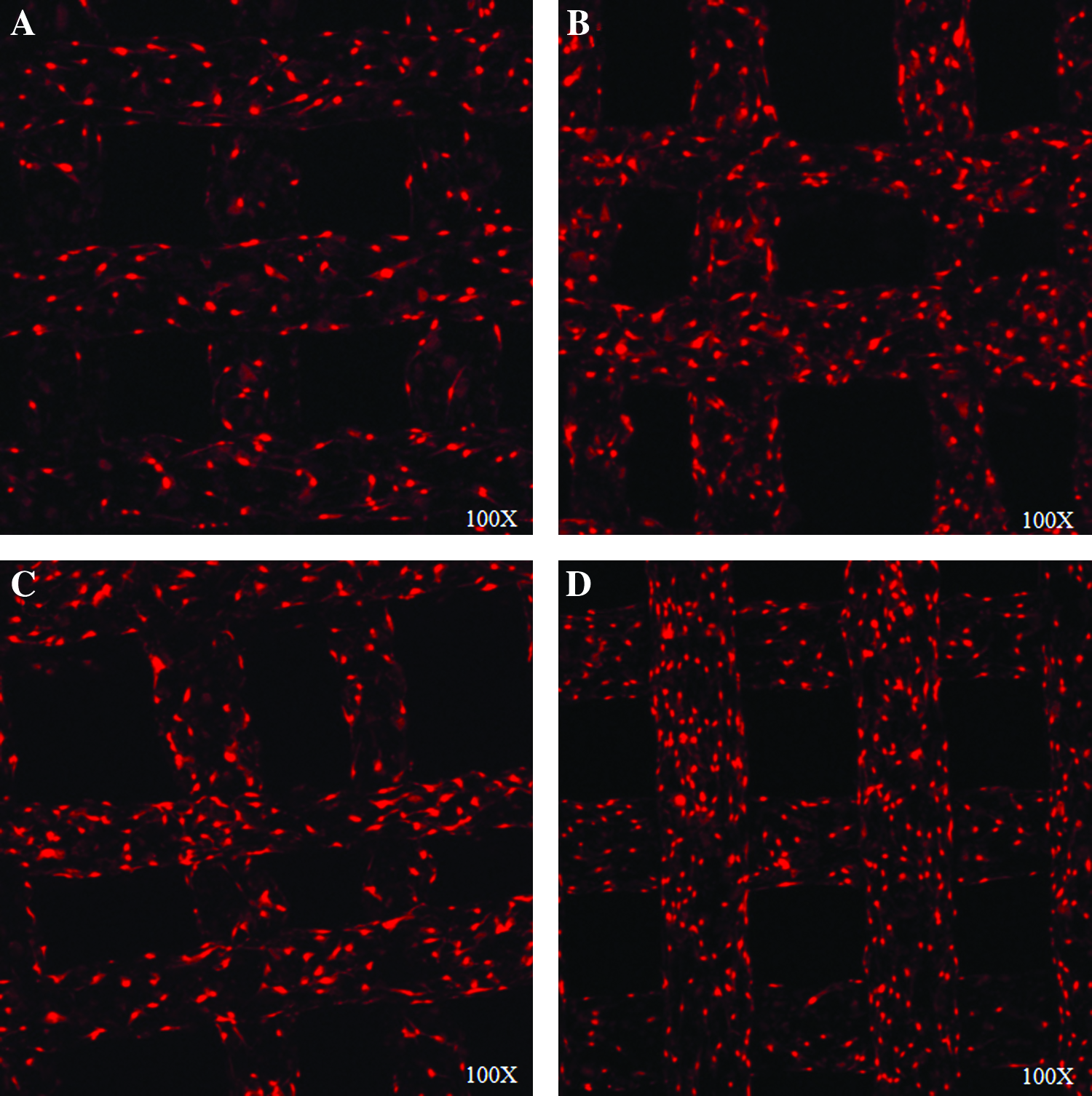
Confocal scanning electron micrographs with propidium iodide (PI) obtained at 3 days (
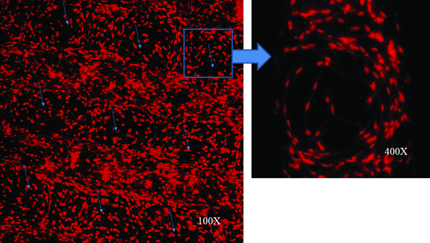
Confocal scanning electron micrographs with propidium iodide (PI) obtained 4 weeks after perfusion culturing of hASCs within the mineralized BG-PCL scaffold. z-line reconstructed image, showing the porous channels (arrowed) of the scaffolds covered with the bridging of proliferated cells, which in direct contrast to the cell–scaffold construct image at the short period shown in Figure 6. Color images available online at
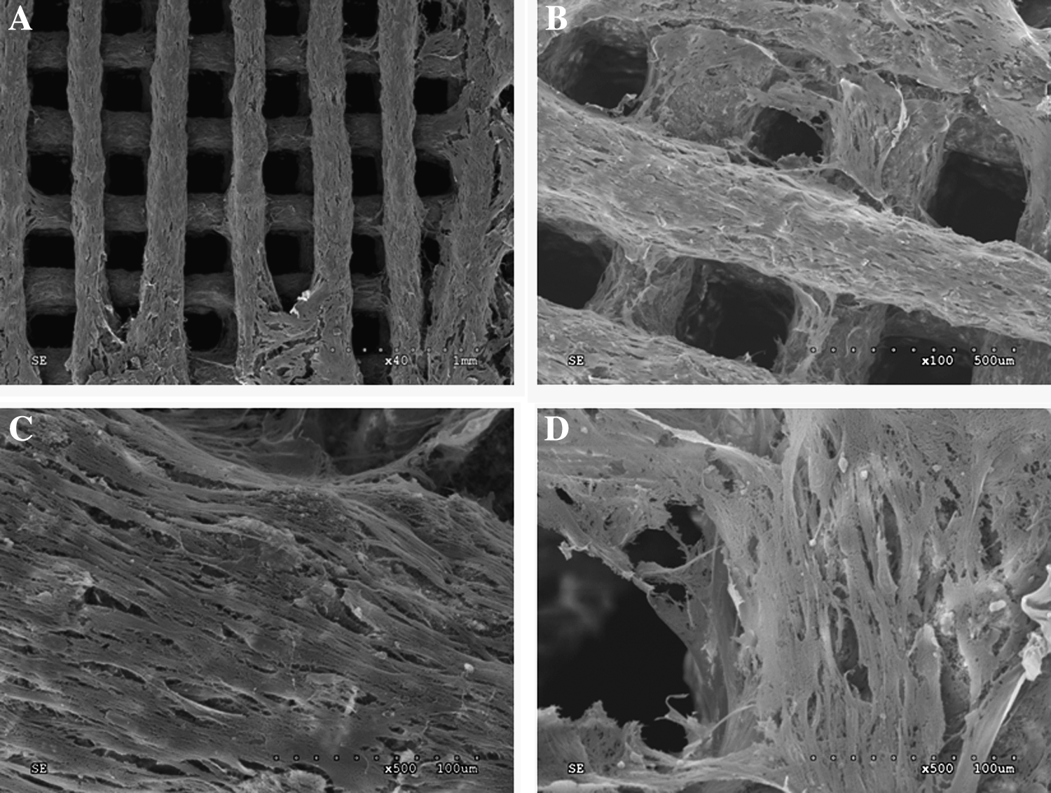
Scanning electron micrographs of the cells populated on the mineralized BG-PCL scaffold with culturing for 2 weeks by the perfusion system, taken at different magnifications. Scaffold stems were completely covered with a thick layer of cells (
The proliferative potential of the hASCs was measured from the cell count using a hemocytometer (A) and from the MTT cell viability (B), as shown in Figure 9. A similar trend was observed for both proliferation results. At all culturing times, perfusion culturing produced a higher level of cell proliferation than static culturing. Moreover, the difference was more significant (approximately twice) at the prolonged stage of culturing (28 days).
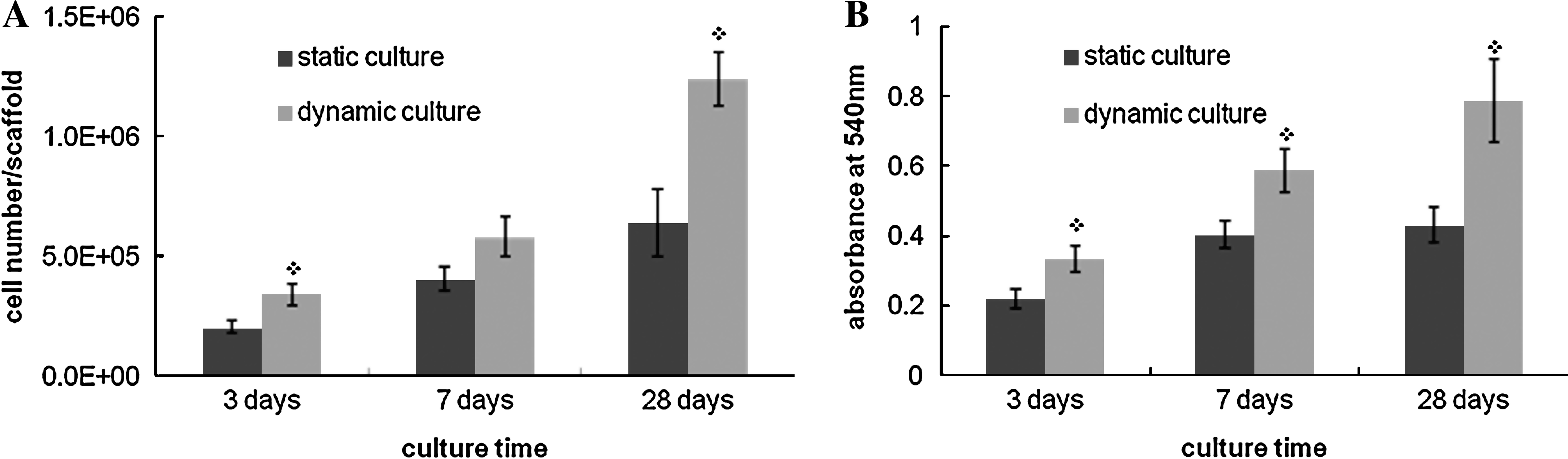
Cell population on the mineralized BG-PCL scaffolds during dynamic culturing for up to 28 days (3, 7, and 28 days), in comparison to static culturing: (
The osteogenic differentiation of the hASCs that proliferated on the scaffold was examined by the production of ALP (an important osteogenic marker of progenitor/stem cells). Figure 10 shows the ALP production by the cells populated on the mineralized scaffold for up to 21 days, either by dynamic perfusion or by static culturing. The total ALP activity produced on each scaffold was significantly higher in the dynamic perfusion system than in the static culturing system at both culturing times (Fig. 10A). When the level was normalized to the total protein content of the cells, the difference was still significant at all periods (Fig. 10B). This suggests that the accumulative ALP produced on the scaffold and ALP secretion from the individual cell component were improved by the dynamic perfusion culturing.
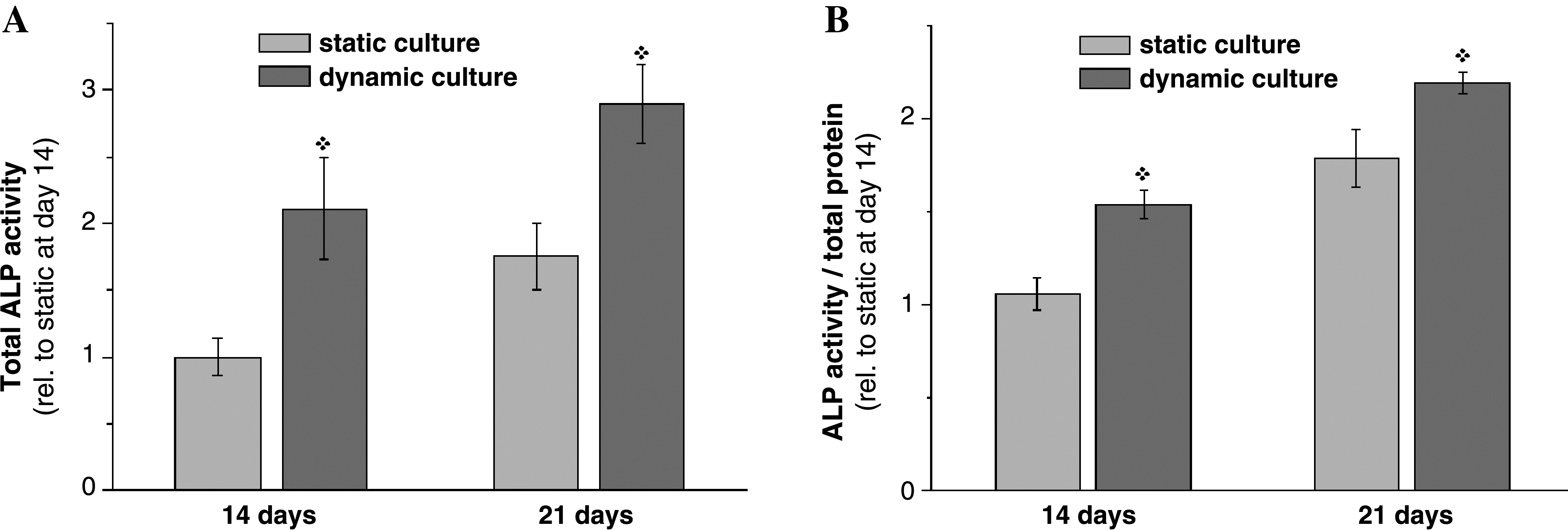
Alkaline phosphatase (ALP) production of the hASCs proliferated on the mineralized scaffold after 14 and 21 days of dynamic perfusion or static culturing: (
Based on the proliferation and osteogenic development of hASCs, perfusion culturing is believed to provide better microenvironmental conditions for the stem cells to populate on and differentiate into an osteogenic pathway. It is believed that the continuous and larger supply of culture medium by the peristaltic pump allows a more rapid and efficient refresh of exhausted cellular metabolites during hASCs mitosis and differentiation. Some studies also examined the effect of perfusion culturing on cell growth and differentiation into specific tissues, including bone and cartilage,40–43 where the design of the container, flow dynamics, and perfusion rate were raised as some important issues. Regarding the perfusion rate, a certain flow rate was observed to be needed to gain optimal cellular behaviors.42,43
In this bioactive and degradable scaffold system, the flow rate is considered to be of particular importance because the products released from the scaffold also vary according to the medium flow rate, which ultimately controls the cellular behavior. The degradation products from the polymeric part should be washed out as quickly as possible. Some ionic elutes from the BG can have a favorable effect, particularly those related to osteogenic development.44,45 Although herein the flow rate was fixed to 0.6 mL/h, which is an approximately three times faster refresh than under a static medium change, there was significant improvement in the cell proliferation and ALP production. More study will be needed to determine the optimal flow rate condition.
Specifically as to the geometry of the macrochanneled scaffold produced by the robotic dispensing (RD) technique, some points also need to be discussed. The channel size is relatively large and the connectivity and flow permeability are far better than the pore structure of other types of scaffolds containing complex pore connections that were made using conventional methods. As such, the cells seeded by a static gravity generally penetrate the scaffold channels directly resulting in a low cell adhesion density. 20 Therefore, a dynamic adhesion technique, such as using a spinner flask, is expected to achieve a better cell population for bone tissue engineering. 46 Another aspect is the benefit of perfusion flow in the cultivation of cells within the macrochanneled scaffold designed in this study. With its pore structure to allow high permeability, the flow can easily penetrate and refresh without significantly altering the structure of the scaffold or breaking down the cell–scaffold construct.
Overall, the results suggest that current bioactive and degradable scaffold with a surface-mineralized and macrochanneled pore structure has potential as a supporting 3D matrix to populate hASCs and induce their osteogenic differentiation. This scaffold–hASC construct is expected to find applications in tissue engineering targeting bone in the near future.
Conclusion
A macrochanneled bioactive and degradable scaffold of BG-PCL was produced from a 3D construction using a robotic dispensing machine. Moreover, the surface was tailored with in situ mineralization to improve the surface functionality. Human adipose–derived stem cells (hASCs) were cultivated within the 3D scaffolds under perfusion dynamic culturing. The cells were quite viable and grew actively on the scaffold assisted by the perfusion flow to a significantly higher population level than that achieved by conventional static culturing. Osteogenic development of the populated hASCs under the appropriate cues was significantly upregulated by perfusion culturing with respect to static culturing, which suggests that the newly developed scaffold and its construction with hASCs under dynamic perfusion culturing condition may be potentially used for the tissue engineering of bone, which remains in the near future.