
Abstract
Polymers that are used in clinical practice as bone-defect-filling materials possess many essential qualities, such as moldability, mechanical strength and biodegradability, but they are neither osteoconductive nor osteoinductive. Osteoconductivity can be conferred by coating the material with a layer of calcium phosphate, which can be rendered osteoinductive by functionalizing it with an osteogenic agent. We wished to ascertain whether the morphological and physicochemical characteristics of unfunctionalized and bovine-serum-albumin (BSA)–functionalized calcium-phosphate coatings were influenced by the surface properties of polymeric carriers. The release kinetics of the protein were also investigated. Two sponge-like materials (Helistat® and Polyactive®) and two fibrous ones (Ethisorb™ and poly[lactic-co-glycolic acid]) were tested. The coating characteristics were evaluated using state-of-the-art methodologies. The release kinetics of BSA were monitored spectrophotometrically. The characteristics of the amorphous and the crystalline phases of the coatings were not influenced by either the surface chemistry or the surface geometry of the underlying polymer. The mechanism whereby BSA was incorporated into the crystalline layer and the rate of release of the truly incorporated depot were likewise unaffected by the nature of the polymeric carrier. Our biomimetic coating technique could be applied to either spongy or fibrous bone-defect-filling organic polymers, with a view to rendering them osteoconductive and osteoinductive.
Introduction
The technology whereby calcium-phosphate layers are deposited upon a substratum has been so greatly improved during recent years as to render the process possible at physiological9,10 rather than at grossly unphysiological temperatures (>1000°C).11–13 Moreover, the structure of the crystals formed (carbonated apatite) is more akin to that of bone mineral than is the hydroxyapatite or tri-/tetracalcium phosphate 11 that is produced at exceedingly high temperatures. One of the main assets of the so-called biomimetic coating technique is that it renders possible the coprecipitation of an osteogenic agent and, consequently, its incorporation into the crystalline latticework of the mineral layer.14–16 Formerly, such agents, being proteinaceous and thus inactivated at high temperatures, could be only superficially adsorbed onto a preformed mineral layer. 17 In a biological milieu, a depot of an osteogenic agent that has been merely adsorbed onto a preformed calcium-phosphate coating is released so rapidly as to be exhausted within a few hours, and the transiently high local concentration of the drug that is thereby generated is not conducive to sustained bone-formation activity. 18 On the other hand, a coating-incorporated depot of such an agent is liberated gradually and in a cell-mediated manner for some weeks. 19 Under these conditions, bone-formation activity can be efficaciously induced and sustained at both ectopic and orthotopic sites in animal models.18,20
The aim of the present study was to ascertain whether the morphological and physicochemical characteristics of unfunctionalized and bovine-serum-albumin (BSA)–functionalized calcium-phosphate coatings, as well as the release kinetics of the protein, were influenced by the surface properties of the underlying polymeric material. For this purpose, we tested four polymers: two with a sponge-like structure (Helistat® [collagen] and Polyactive® [a copolymer of ethyleneoxide and butylene terephthalate]) and two with a fibrous one (Ethisorb™ [a copolymer of glactin and ρ-dioxanone] and poly[lactic-co-glycolic acid] [PLGA]).
Materials and Methods
Experimental design
In this study, four polymers (Helistat, Polyactive, Ethisorb and PLGA) with different surface geometries and chemistries were coated according to a two-step biomimetic procedure with a biphasic layer of calcium phosphate, which was either unfunctionalized or coprecipitated with the model protein BSA. The polymers and the coatings were characterized by scanning electron microscopy, histomorphometry (polymers only), Fourier-transform infrared spectroscopy and X-ray diffractionometry. The pattern of distribution of the amorphous seeding layer of the coating (labeled with Rhodamine), and of the BSA (conjugated with fluorescein isothiocyanate [FITC]) that was coprecipitated with the crystalline one, was mapped by confocal laser-scanning, dual-channel fluorescence microscopy. The release kinetics of coating-incorporated and of adsorbed (control) FITC-BSA were monitored spectrophotometrically in vitro.
Polymeric materials
Of the four polymers tested, three are synthetic (Polyactive, Ethisorb and PLGA) and one is natural (Helistat). Helistat (Integra) is a sponge-like material that is manufactured from natural collagen. Polyactive (IsoTis B.V.) is likewise sponge-like in appearance, but is a synthetic copolymer of ethyleneoxide terephthalate and butylene terephthalate. Ethisorb (Johnson & Johnson) and PLGA (undergoing clinical trials by, and received as a gift from, Smith and Nephew, United Kingdom) are both fibrous in nature, the former being a copolymer of glactin and ρ-dioxanon and the latter one of lactic and glycolic acids.
Characterization of the surface geometry and chemistry of the four polymers
To reveal the surface geometries of the four polymers, these were examined in a scanning electron microscope (XL 30; Philips). For this purpose, 1-cm-diameter discs of the material were mounted on aluminium stubs and sputtered with gold particles to a thickness of 10–15 nm.
The surface-area density and the porosity of each polymer type were estimated histomorphometrically. One-centimeter-diameter discs of the four different materials were dehydrated in ethanol and embedded in methylmethacrylate according to a standard protocol. 21 By applying a systematic random-sampling strategy, 22 ten 600-μm-thick vertical sections, 1 mm apart, were prepared from each disc using a diamond saw (Leica). The slices were mounted on Plexiglass holders, polished down to a thickness of 80 μm, and surface stained with McNeal's Tetrachrome, basic Fuchsine and Toluidine Blue O. 21 The sections were examined in a Nikon-Eclipse E-1000 light microscope. By applying a systematic random-sampling strategy, 23–30 fields were selected and photographed in color at final magnifications of approximately ×400 (Helistat), × 200 (Polyactive) and ×300 (Ethisorb and PLGA). By applying stereological principles, these prints were used to estimate the surface-area density and the porosity (volume density of the internal space) of each polymer. The former parameter was estimated using a cycloid test system 23 and the latter by point counting. 24
The chemical groups of which each material is composed were revealed by Fourier-transform infrared spectroscopy (Spectrum 1000; Perkin-Elmer). For this analysis, 1-cm-diameter discs of the material were frozen in liquid nitrogen for 10 min, pulverized, and then mixed with powdered potassium bromide for compression into pellets. The phase composition of each material was analyzed by X-ray diffractionometry (X'Pert PRO; PANalytical) using a scanning range 20 of 2.00–60.00°, a scanning speed of 2.00° per minute and a scanning interval of 0.02°.
Coating procedure
One-centimeter-diameter discs of each polymer (with thicknesses of 0.6 mm [Ethisorb], 1 mm [Helistat] and 2 mm [Polyactive and PLGA]) were immersed in 20 mL of fivefold-concentrated simulated body fluid (684 mM NaCl, 12.5 mM CaCl2 · 2H2O, 5 mM Na2HPO4 · 2H2O and 21 mM NaHCO3) under high-nucleation conditions, namely, in the presence of 7.5 mM MgCl2 · 6H2O, to inhibit crystal growth, for 24 h at 37°C. The fine, dense layer of amorphous calcium phosphate thereby formed serves as a seeding substratum for the deposition of a more substantial crystalline layer. This was produced by immersing the samples in 10 mL of a supersaturated solution of calcium phosphate (40 mM HCl, 4 mM CaCl2 · 2H2O, 136 mM NaCl and 2 mM Na2HPO4 · 2H2O), which was buffered to pH 7.4 with 50 mM Tris, for 48 h at 37°C.15,25 If the crystalline layer was to be functionalized by the incorporation of BSA, then this protein was introduced into the latter medium (0.1 mg/mL).
A portion of the samples that bore only an amorphous layer of calcium phosphate were reserved for analytical purposes. These samples, as well as those that bore either an unfunctionalized or a BSA-functionalized bilayer of calcium phosphate, were freeze-dried for at least 24 h before their characterization.
Characterization of the coatings
Analogous to the native polymeric materials, the amorphous seeding layer of calcium phosphate, as well as the unfunctionalized and the BSA-functionalized bilayers with which they were coated, were analyzed by scanning electron microscopy, Fourier-transform infrared spectroscopy and X-ray diffractionometry. Scanning electron microscopy was conducted in conjunction with an energy-dispersive X-ray analysis, which revealed the relative densities of calcium and phosphorus that were present, from which data the calcium-to-phosphorus ratios were calculated.
Distribution of the coating material and of coating-incorporated BSA
By appropriate labeling of the amorphous seeding layer of calcium phosphate and of the depot of BSA that was coprecipitated with the crystalline one, the distribution of the coating material and of the incorporated protein can be mapped by confocal laser-scanning, dual-channel fluorescence microscopy. To this end, Rhodamine B (0.1 mg/mL) was introduced into the fivefold-concentrated simulated body fluid (see section Coating procedure), and FITC-conjugated BSA (Sigma) into the supersaturated solution of calcium phosphate (see section Coating procedure).
For the analysis in a confocal laser-scanning microscope that was equipped for fluorescence imaging (Zeiss LSM 510 META with LSM 510 acquisitions software and images 3D software), freeze-dried samples (see section Coating procedure) were embedded in methylmethacrylate. Six-hundred-micrometer-thick sections were prepared from the embedded material, affixed to Plexiglass holders and ground down to a thickness of 80 μm. The distribution of the amorphous layer of calcium phosphate was revealed by a red fluorescence signal; that of the BSA and, by implication, that of the crystalline layer, were disclosed by a green one.
Release kinetics of BSA in vitro
The release kinetics of coating-incorporated depots of protein were spectrophotometrically monitored for 35 days in vitro using samples that had been functionalized with FITC-BSA. For the purpose of comparison, FITC-BSA was directly adsorbed onto a portion of samples that had been coated in the absence of the protein. This was achieved by applying a 10-μL drop of a stock solution of FITC-BSA (5 μg/mL) to each side of the 1-cm-diameter freeze-dried discs of the coated polymeric materials in turn, allowing for complete evaporation at ambient temperature between the applications.
Each sample (n = 6 for each polymer type) was introduced into a 15-mL centrifuge tube containing 10 mL of 0.9% saline, which was buffered to pH 7.4 with 50 mM Tris. The tubes were incubated for up to 35 days in a shaking waterbath (60 agitations/min), which was maintained at 37°C. Triplicate 200-μL aliquots of the medium (containing released FITC-BSA) were withdrawn for analysis after 3 h, 6 h, 9 h, 1 day, 2 days, 3 days, 5 days, 7 days, 10 days, 14 days, 18 days, 23 days, 28 days and 35 days. Fluorescence density was measured in a spectrophotometer (excitation wavelength: 485 nm; emission wavelength: 519 nm).
To estimate the total amount of FITC-BSA that had been incorporated into the crystalline layer of calcium phosphate during the coating procedure, six samples of each polymer type were immersed in 2 mL of 0.5M ethylenediamine tetra-acetic acid (pH 8.0) and vortexed twice for 5 min. The final volume was made up to 10 mL with Tris-buffered 0.9% saline, and triplicate 200-μL aliquots of this medium were withdrawn for its spectrophotometric analysis.
Fluorescence readings were converted to amounts of protein using a standard curve, which was generated by preparing a dilution-series of FITC-BSA in 10 mL of Tris-buffered 0.9% saline. The temporal release of FITC-BSA was expressed as a percentage of the total amount that had been incorporated into the crystalline layer of the calcium phosphate coating.
Statistical analysis
The surface-area density and the porosity of each polymer type, as well as the kinetic data, are presented as mean values together with the standard deviation. Data pertaining to each group were compared using a one-way analysis of variance. The level of significance was set at p < 0.05. SPSS statistical software (version 15.0 for Windows) was used for this evaluation. Post hoc comparisons were made using Bonferroni corrections.
Results
Scanning electron microscopy
Scanning electron microscopy revealed Helistat and Polyactive to be sponge-like materials with a labyrinthine system of interconnecting pores (Fig. 1). Ethisorb and PLGA consisted of entangled fibers, which were distributed compactly in the former case and loosely in the latter (Fig. 1). At high resolution, the surface relief of continuous portions of Helistat was flat, whereas that of Polyactive was undulated (Fig. 1). The surface profiles of individual fibers of Ethisorb and PLGA were flat on a broad scale, but not smooth: in each case, the surface was scattered with approximately 0.5-μm-diameter particles (Fig. 1).
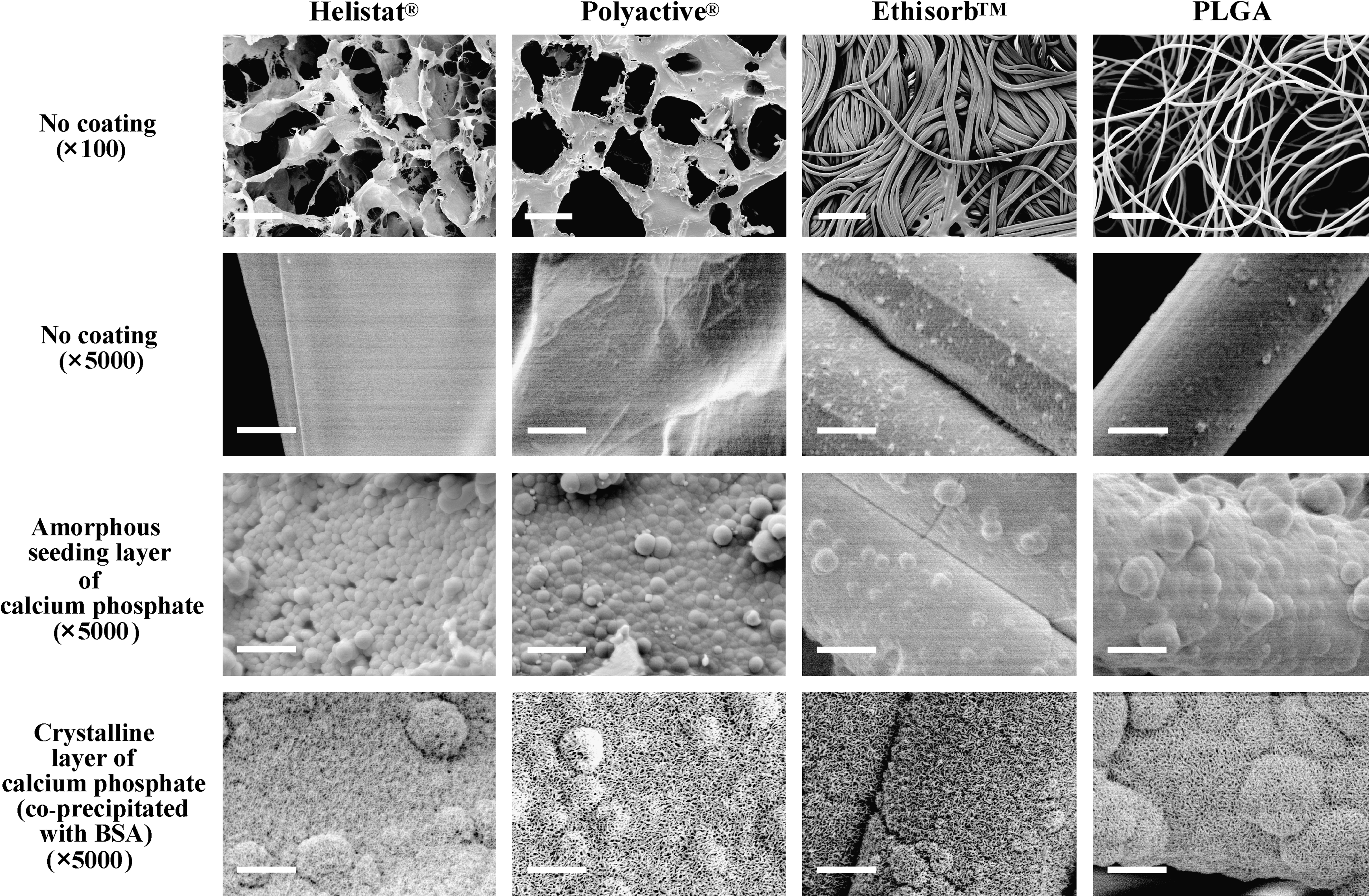
Scanning electron micrographs of the surface of each polymer type (left to right) in the native condition at low (first horizontal panel) and high (second horizontal panel) magnifications, and after coating first with an amorphous seeding layer of calcium phosphate (third horizontal panel) and then with a crystalline one, which was coprecipitated with bovine serum albumin (BSA; fourth horizontal panel). Scale bars = 200 μm (first horizontal panel) and 5 μm (second, third, and fourth horizontal panels).
After the polymers had been coated with an amorphous seeding layer of calcium phosphate, the surfaces of the different materials appeared similar: they were covered with dense, noncrystalline material, which was deposited in the form of spherical particles (Fig. 1). Upon this amorphous seeding layer of calcium phosphate, a thicker crystalline one was deposited, either in the absence or in the presence of BSA. Irrespective of the absence or presence of the coprecipitated protein, or of the nature of the underlying polymer, the crystalline layers had a similar surface appearance in the scanning electron microscope: they were composed of densely packed, small needle-like crystals (Fig. 1).
Surface-area density and porosity of the native polymers
The surface-area density of each native polymer (Helistat: 43.2 ± 3.0 mm−1; Polyactive: 9.2 ± 0.3 mm−1; Ethisorb: 100.2 ±9.4 mm−1; PLGA: 12.6 ± 1.5 mm−1) differed significantly (p <0.001) from that of the other three, except in the comparison between Polyactive and PLGA (p > 0.05). The great difference between the surface-area densities of the two fibrous materials (Ethisorb and PLGA) reflects the compact (Ethisorb: high value) versus loose (PLGA: low value) general organization and surface relief of the fibrous meshworks (Fig. 1). The porosity—namely, the volume density of the internal space—of each polymer (Helistat: 88.2% ± 1.2%; Polyactive: 70.7% ± 5.5%; Ethisorb: 42.2% ± 3.8%; PLGA: 96.8% ± 0.3%) differed significantly from that of the other three in each case (p < 0.05). Once again, the great difference between the values for the two fibrous materials reflects the organization of the fibers, the volume density of the internal surface being much smaller for compactly (Ethisorb) than for loosely (PLGA) arranged structures.
Confocal laser-scanning, dual-channel fluorescence microscopy
In the confocal laser-scanning microscope, the Rhodamine-labeled amorphous seeding layer of calcium phosphate was revealed to follow the surface contours of each of the cross-sectioned polymer types and to be of uniform thickness (red signal in Fig. 2). The crystalline layer of calcium phosphate was coprecipitated with FITC-BSA, and the protein phase (green signal in Fig. 2) was presumed to colocate with the mineral one. As was the case with the amorphous layer of calcium phosphate, the BSA-bearing crystalline one followed the surface contours of each of the cross-sectioned polymer types, and was likewise of uniform thickness in each case (green signal in Fig. 2). Indeed, merger of the two images (red and green signals) revealed the BSA-bearing crystalline layer to be deposited upon, and yet to be clearly demarcated from, the amorphous one (Fig. 2). Some of the fibers of Ethisorb that are illustrated in Figure 2 appear to emit a red fluorescence signal (amorphous layer) but not a green one (FITC-BSA: crystalline layer). Although we cannot categorically rule out the possibility that this imaging result reflects some inefficiencies in the biphasic coating, the analysis in the scanning electron microscope indicated otherwise. This evaluation revealed all fibers of Ethisorb to bear a crystalline layer. A more likely explanation of the phenomenon is an imaging artefact. During the dual-channel fluorescence imaging of the specimens, the red and the green signals were sometimes strongly scattered to variable and differential degrees, depending partially on the level at which a polymer unit was sectioned (e.g., cleanly or tangentially cross-sectioned). To minimize this effect, it was often necessary to reduce the intensity of the fluorescence signal of one color in a bright spot, which occasionally led to a quenching or complete elimination of the fluorescence signal of the other color if this was weak.
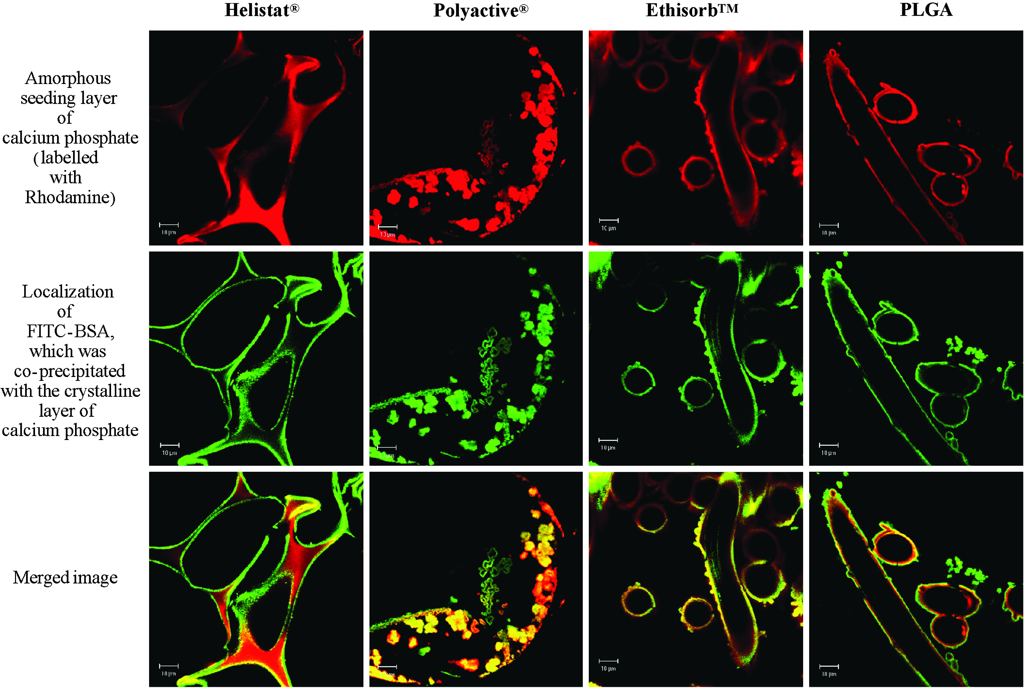
Confocal laser-screening fluorescence micrographs of each polymer type (left to right) after coating first with an amorphous seeding layer of calcium phosphate (labeled with Rhodamine, red signal; first horizontal panel) and then with a crystalline one, which was coprecipitated with BSA that had been conjugated with fluorescein isothiocyanate (FITC, green signal; second horizontal panel). The protein is presumed to colocalize with the crystalline layer. The two sets of images are merged in the third horizontal panel. Scale bars = 10 μm.
Fourier-transform infrared spectroscopy
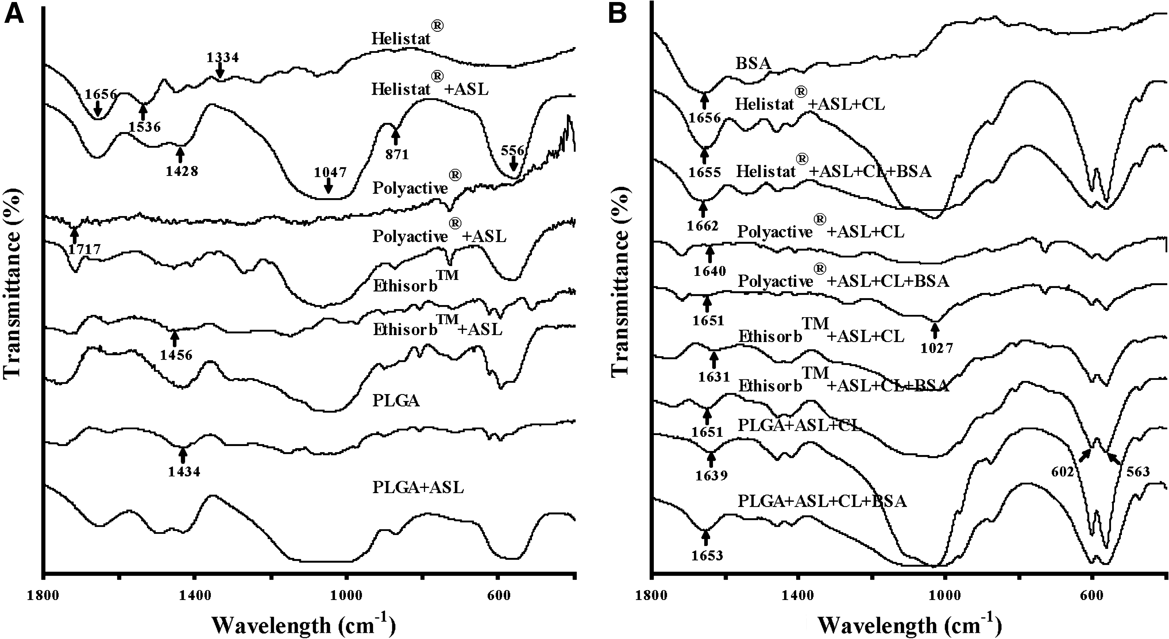
Fourier-transform infrared spectroscopy revealed each of the four polymer types to be characterized by a distinct spectrum (Fig. 3), which reflects its chemical composition. Helistat (collagen) was characterized by bands at wavelengths of 1656 cm−1, 1536 cm−1 and 1334 cm−1, which correspond to amide-I, II and III groups, respectively. Ethisorb and PLGA are characterized by a band at 1456 cm−1 and 1434 cm−1, respectively, which correspond to their CH2 groups, 26 and Polyactive manifests one at 1717 cm−1, which represents its C = O groups. 27
Fourier-transform infrared spectra for each polymer type in a native condition (
After coating the different materials with an amorphous seeding layer of calcium phosphate, several new bands consistently appeared in the spectrum of each polymer type: two at wavelengths of 1047 cm−1 and 556 cm−1, respectively, which correspond to PO43− groups 28 ; one at 871 cm−1, which corresponds to HPO42− groups; and one at 1428 cm−1, which corresponds to CO32−. 29 On the basis of these data, the amorphous seeding layer can be classified as carbonated calcium phosphate.
Additional bands appeared after the crystalline layer of calcium phosphate had been deposited, and these were likewise consistently associated with each polymer type. They included a twin band at 602 cm−1 and 563 cm−1, which corresponds to O-P-O bending, and a single one at 1027 cm−1, which corresponds to P-O stretching.30,31 These bending and stretching modes of the P-O group reflect the crystalline nature of this calcium-phosphate layer. 32 An additional band, the position of which ranged between 1655 cm−1 and 1631 cm−1 according to the nature of the polymer, and which represents molecular water of the crystalline layer, underwent a shift to higher wavelengths when the latter was coprecipitated with BSA (Fig. 3). 33 Also in the spectrum of BSA itself, a band was apparent at this position (1656 cm−1) and, in the case of the protein, corresponds to C = O stretching in amide-I groups (•CO•NH2). The shift to a higher wavelength position that occurred in the spectra of the polymers when the crystalline layer of calcium phosphate was deposited in the presence of BSA indicates that the protein was incorporated into the crystalline lattice work via an interaction involving amide-I groups in the former case and molecular water in the latter.
X-ray diffractionometry and energy-dispersive X-radiography
X-ray diffractionometry revealed each of the four polymer types to be characterized by a unique spectrum (Fig. 4), with distinctive peaks at 2θ = 21.35° and 23.74° for Helistat, at 2θ = 23.54° for Polyactive, at 2θ = 22.08° and 28.31° for Ethisorb, and at 2θ = 21.29°, 21.88° and 28.44° for PLGA.
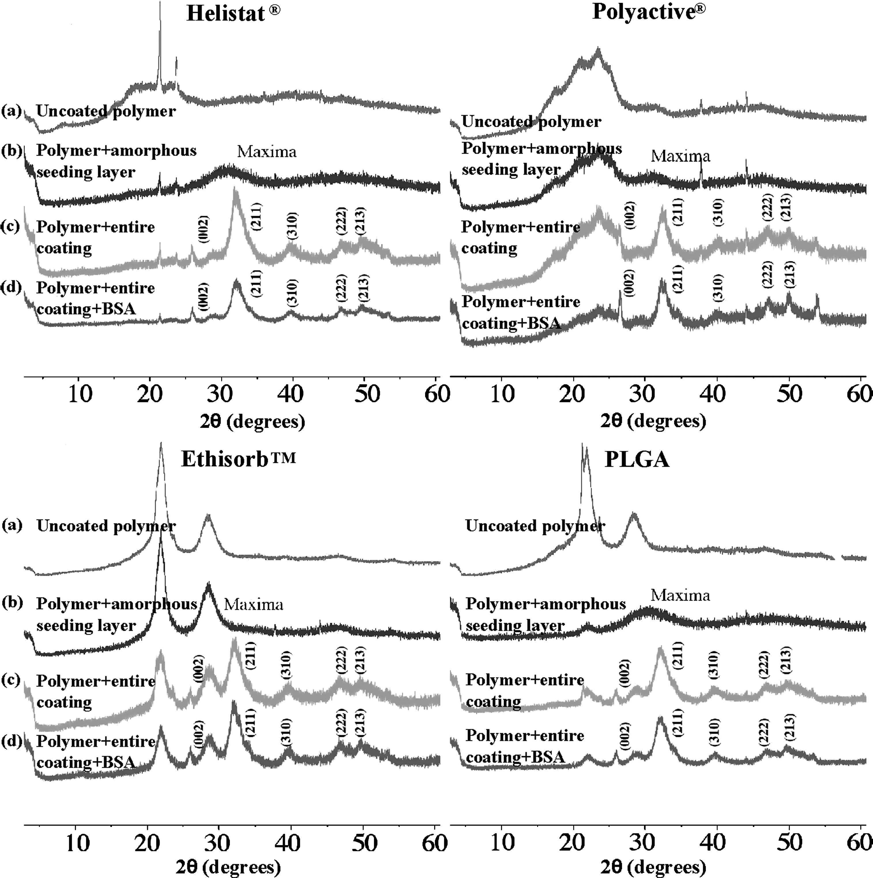
X-ray diffraction spectra for each polymer type in a native condition (a), and after coating first with an amorphous seeding layer of calcium phosphate (b) and then with a crystalline one in the absence (c) or presence of BSA (d).
Coating of the different polymer types with an amorphous seeding layer of calcium phosphate led to the appearance of a new broad peak at 2θ = 28°–32° in the case of Helistat, and to its superimposition upon an existing one in the cases of Polyactive, Ethisorb and PLGA. The position corresponds to that of apatite, and the broadening of the diffraction peak reflects the amorphous nature of the material.
After the crystalline layer of calcium phosphate had been deposited, two major new peaks were introduced into the diffraction spectrum of each polymer type: one narrow band close to 2θ = 26° and a broader one at 2θ = 32°. According to information published by the International Centre for Diffraction, these bands are characteristic of calcium-deficient apatite with a low crystallinity. The incorporation of BSA into the crystalline layer of calcium phosphate elicited no profound change in the diffraction spectrum.
The energy-dispersive X-radiographic analysis (which was conducted in conjunction with scanning electron microscopy) revealed the calcium-to-phosphorus ratios of the amorphous seeding layer of the four polymer types to range from 1.53 to 1.64. After the crystalline layer had been deposited, the calcium-to-phosphorus ratio was lower in each case, and fell within the range of 1.37–1.45. This finding confirms that of the X-ray diffraction analysis, namely, that the crystalline layer was poor in calcium.
Release kinetics of a coating-incorporated depot of BSA in vitro
The release kinetics of a depot of FITC-BSA that had been incorporated into the crystalline layer of calcium phosphate coatings are represented in Figure 5 for each polymer type. For the purpose of comparison, the release kinetics of an adsorbed depot of FITC-BSA are also depicted. As expected, the adsorbed depot of BSA was released rapidly from the coatings of each polymer type when the specimens were immersed in buffered physiological saline. The initial depot was depleted by more than 80% after 6 h, and was completely exhausted after 3 days in all cases.
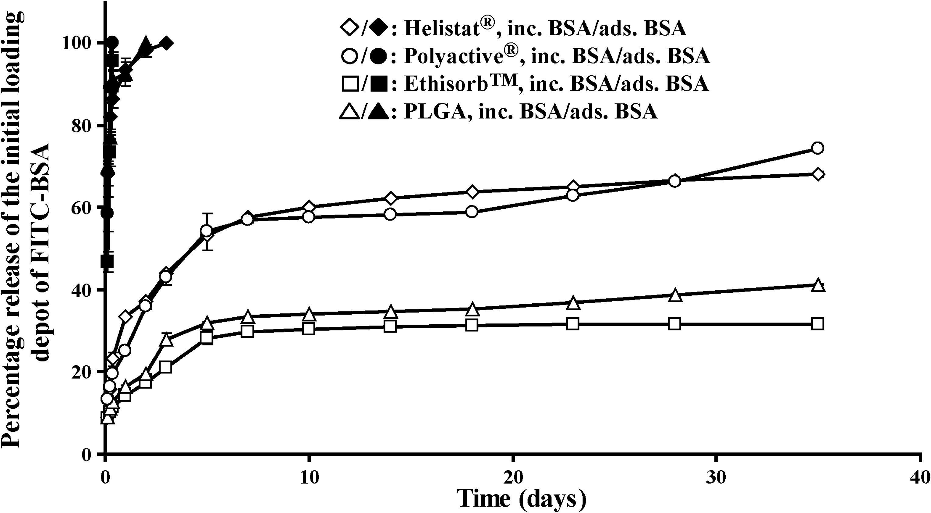
Graph depicting the temporal release profiles of a coating-incorporated (inc.) depot of BSA (open symbols) and of an adsorbed one (ads.; closed symbols) from each polymer type in vitro. Mean values (n = 6 for each polymer type) are presented together with the standard deviation.
The release kinetics of a coating-incorporated depot of BSA followed a biphasic course in the case of each polymer type: an initial rapid, and a subsequent slower one. However, the rate of protein release during the initial rapid phase differed between the sponge-like and the fibrous polymer types. In the case of Helistat and Polyactive, both of which have a labyrinthine sponge-like structure (Fig. 1), 54% of the coating-incorporated depot of BSA had been released by day 5 at a rate of nearly 11% per day in each instance. But in the case of Ethisorb and PLGA, both of which have a fibrous structure (Fig. 1), only 28% (Ethisorb) to 32% (PLGA) of the coating-incorporated depot of BSA had been liberated by the same juncture, at rates of 6% (Ethisorb) and 8% (PLGA) per day.
During the subsequent slower phase (days 5–35), the rates of protein release were similar for each polymer type. By the end of the monitoring period (day 35), the initial coating-incorporated depot of BSA had been depleted by 68% and 74% for Helistat and Polyactive, respectively, and by 31% and 41% for Ethisorb and PLGA, respectively.
Our data indicate that the surface geometry of the underlying polymeric material influences only the initial rapid rate of release of a coating-incorporated depot of protein, not the subsequent slower one.
Discussion
The concept of biomimetic mineralization was introduced by Kokubo et al. in 1990. 9 The layer of calcium phosphate that is thereby produced is more conducive than conventionally prepared ones to the differentiation of bone-marrow stromal cells into osteoblasts, 34 as well as to the ingrowth of osseous tissue and its contact with the implanted material,35–37 which is less vigorously encapsulated with fibrous tissue. 38 Various types of material have been thus coated, including bioceramics, 39 metals 40 and organic polymers. 41 Using the original biomimetic technique, the successful coating of polymers depends greatly upon the surface chemistry of the material,42–44 since this surface furnishes the active chemical groups that serve as nucleation sites for mineralization.43,45,46 Monolayers of dihydrogen phosphate or carboxylic acid have been shown to be highly conducive to biomimetic mineralization, and those composed of methyl groups (CH3) to be unpropitious to the process. 46 However, since many of the polymers that are marketed for clinical use are not equipped with active chemical groups on their surfaces, attempts have been made to covalently attach functional anionic ones. 45 Furthernore, a patterning of these functional groups permits a selective topographical localization and orientation of the mineral deposits, which in turn permits a control of cell binding and alignment. 47 However, most of the polymers that are in clinical use are not amenable to such manipulation; if they are, then the modifications carry the risk of compromising the physicochemical and biological properties of the material. Moreover, not only the surface chemistry but also the surface geometry and the three-dimensional structure of a polymer can influence its biomimetic mineralization. 48
Using the biphasic biomimetic coating technique that was refined in our laboratory,15,16 the four tested polymer types could be coated with an amorphous and a crystalline layer of calcium phosphate, the morphological and physicochemical properties of each of which were independent of either the surface chemistry, the surface geometry or the three-dimensional structure of the underlying carrier. The consistency of the result is to be laid to the merits of the amorphous seeding layer. Previously, we had applied this biphasic biomimetic coating technique to titanium-alloy implants, 20 and the adhesion thereto of the amorphous layer of calcium phosphate was attributed to the affinity of Ca2+ and HPO42− ions for titanium.29,49 However, since the data of the present study indicate that such an amorphous layer of calcium phosphate can be precipitated also on polymers with diverse surface chemistries, and without any change in its morphological or physicochemical properties, the deposition process is probably unrelated to the phenomenon of chemical affinity. A more likely explanation is that the tiny particles of calcium phosphate—which are formed under the nucleation inhibitory influence of Mg2+ and HCO32−—are captured and immobilized on the substratum by a process of mechanical gomphosis.29,50 These particles then serve as seeding centers for the subsequent growth of a crystalline latticework of calcium phosphate under conditions that are conducive to nucleation. 28
One might perhaps question whether an amorphous layer of calcium phosphate would not in itself suffice as an osteoconductive surface and, after its functionalization with an osteogenic agent, as an osteoinductive one. This question must be answered in the negative. An amorphous layer of calcium phosphate undergoes rapid dissolution. 51 Although this circumstance may not interfere with the property of osteoconductivity, it renders the layer an unsuitable vehicle for the carriage of an osteogenic agent. To be efficacious at the target site, an osteogenic agent needs to be liberated gradually and steadily at a low pharmacological level, not rapidly in a single high-dose burst. 20 Therein lies the beauty of a crystalline latticework of calcium phosphate and the advantage that is afforded by the biomimetic coprecipitation of this layer with a protein, which is thereby incorporated into its structure. Evidence for the latter phenomenon is furnished elsewhere15,20,52 and was confirmed in the present study by Fourier-transform infrared spectroscopy.
In vivo, the degradation of a crystalline coating of calcium phosphate is not a spontaneous, but a cell-mediated process,18,20 and in this respect it resembles the resorption of bone mineral, which is mediated by osteoclasts. Indeed, when the crystalline layer of calcium phosphate is functionalized by the incorporation of an osteogenic agent,18,20,25,52,53 the analogy to physiological bone resorption is still closer: osteogenic growth factors are trapped within the mineralized matrix of bone and are released when it is resorbed by osteoclasts.45,46 These osteogenic growth factors then stimulate bone-formation activity. Hence, a biomimetic coating of calcium phosphate that has been functionalized by the incorporation of an osteogenic agent can facilitate bone formation along a course that resembles the physiological process of bone remodeling, which involves the resorption of old and the formation of new bone.20,52
The release kinetics of a coating-incorporated depot of BSA (which served as a model protein) were influenced by the surface geometry and the three-dimensional structure of the underlying polymer only during the initial rapid phase (up to 5 days). The protein was liberated at a higher rate from polymers that had a chambered, sponge-like structure (Helistat and Polyactive) than from those that had an open fibrous one (Ethisorb and PLGA). Albeit so, the rate of release during this phase was, in each case, still markedly slower from a coating-incorporated than from an adsorbed depot of BSA (Fig. 5). During the subsequent slower kinetic phase (5–35 days), the rate of protein release was similar for each polymer type.
During the initial rapid kinetic phase, the BSA that was released in the coating-incorporated groups probably originated predominantly from an incidentally adsorbed depot. The labyrinthine system of interconnecting pores that characterizes the sponge-like materials offers a greater potential for the entrapment of fluid-borne protein (due to the formation of poorly diffusible stagnation pockets), and thus to its incidental adsorption during subsequent freeze-drying, than do the open and more freely diffusible spaces that surround the nonporous fibrous ones. The existence of a larger depot of entrapped and incidentally adsorbed BSA in association with the sponge-like (Helistat and Polyactive) than with the fibrous polymers (Ethisorb and PLGA) would account for the higher rates and percentages of protein release from the former during the initial rapid kinetic phase. The BSA that was liberated during the slower kinetic phase (5–35 days) originated exclusively from the coating-incorporated depot, the rate of release of which was similar for each polymer type. This latter observation is consistent with the findings of the Fourier-transform infrared spectroscopy analysis: for each type of polymer, the incorporation of BSA into the crystalline layer of calcium phosphate elicited a comparable change in the spectrum of the latter, which indicates that the integration mechanism was similar in each case.
Clearly, BSA is not biologically active as an osteogenic agent. In this explorative study, it was used as an inexpensive alternative to bone morphogenetic protein-2 (BMP-2). Although BMP-2 is a much smaller molecule than BSA and has a different charge density, it can nevertheless be incorporated into a calcium-phosphate coating without impairing its osteoinductive efficacy, either in vitro 25 or in vivo. 53 BMP-2 that had been incorporated into a calcium-phosphate coating was previously shown not only to induce bone formation at both an ectopic and an orthotopic ossification site in rats20,53 and miniature pigs, 18 respectively, but also to do so potently at a low pharmacological dose (1.7 μg/coating), and at a level that was sustained for some weeks. These data afford indirect evidence that the coating-incorporated depot of BMP-2 was liberated gradually and steadily in vivo, namely, along a temporal course that resembled the elution of a coating-incorporated depot of BSA in the present study.
It is important here to mention that once the functionalized coating has been degraded in vivo, the underlying carrier will be exposed to the host tissues. The biocompatibility and biodegradability of the carrier will undoubtedly influence the osteoinductive activity of the osteogenic agent that was incorporated into the coating. In an ongoing study, which is nearing completion, the four polymeric carriers that were the subject of the present work were coated with a BMP-2-functionalized layer of calcium phosphate and implanted at an ectopic ossification site in rats. Parameters such as coating stability after implantation, the rates of coating and polymer degradation, foreign-body-giant-cell reactivity (a gauge of biocompatibility) and osteoinductive efficacy were histomorphometrically assessed after 5 weeks. These data, which do not fall within the scope of the present study, will be published shortly. Another elegant coating methodology has been investigated for the controlled release of biologically active molecules, namely, the sol-gel technique. 54 However, the methodology is more cumbersome than the biomimetic coprecipitation of a biologically active agent and a layer of calcium phosphate.
In summary, our data indicate that the biphasic biomimetic coating technique can be applied to polymers with diverse surface chemistries, surface geometries and three-dimensional structures without impacting the morphological and physicochemical properties of either the amorphous seeding layer of calcium phosphate or the crystalline one, or affecting the release kinetics of a protein depot that is incorporated into the latter. The technique could thus be applied to spongy or fibrous polymers that are used in clinical practice as bone-defect-filling materials, with a view to improving their osteoconductivity. Moreover, the technique can readily accommodate a process of coating-functionalization with a proteinaceous agent, whose release kinetics would be highly conducive to the efficacious induction and sustentation of bone-formation activity.
Footnotes
Disclosure Statement
No competing financial interests exist.