
Abstract
To optimize the chondrocyte numbers obtained after collagenase digestion for cartilage tissue engineering, we examined the enzyme concentration and incubation time for collagenase digestion. The appropriate cell density in the chondrocyte primary culture was also verified. The collagenase digestion conditions that maximized the viable cell numbers were 24 h in 0.15% and 0.3% collagenase, 6 h in 0.6%, and 4 h in 1.2%, leading to ∼5 × 10⁵ cells from 0.05 g. When seeded at 10,000 cells/cm2, all of these cells became almost confluent within 1 week. Cells digested in 0.3% for 24 h or 0.6% for 6 h and seeded at 3000 cells/cm2 may also be acceptable, and similarly reached confluence within 1 week. Results regarding expression of the p53, tumor necrosis factor-α, and interleukin-1β genes, as well as apoptosis enzyme-linked immunosorbent assay results, show that excessive collagenase exposure may decrease chondrocyte viability or activity. We recommend a 24-h incubation in 0.3% collagenase or 6 h in 0.6% collagenase, and a cell-seeding density of 3000–10,000 cells/cm2. These conditions maximize the harvest of isolated chondrocytes from a small amount of biopsied tissue and significantly aid in obtaining a large quantity of cultured cells in a short period.
Introduction
Dashes indicate lack of data.
The seeding density has a significant influence on cell viability and proliferation efficacy in the primary culture. If the seeding density is too low, the autocrine/paracrine system may barely work, which leads to slow proliferation. On the other hand, an overdose of chondrocytes may interfere with sufficient cell attachment to the substrate in the culture dish or evoke early contact inhibition, both of which lead to a less effective cell culture. However, the optimum seeding density in chondrocyte cultures for tissue engineering has not yet been investigated in detail. In this study, the appropriate cell density for the chondrocyte primary culture was also verified.
Materials and Methods
Chondrocyte isolation
All procedures were approved by the Ethics Committee of the University of Tokyo Hospital (ethics permission number 622). Remnant auricular cartilage from three microtia patients was obtained during surgery in accordance with the Helsinki Principles. The cartilage tissue was thoroughly minced with scissors and tweezers into fragments of 250–1000 μm (Fig. 1A). Approximately 3 mL of collagenase solution (Wako Pure Chemical Industries) at concentrations of 0.15%, 0.3%, 0.6%, or 1.2% was poured into a 5 mL tube (BD Falcon). Four tubes were prepared for each concentration, for a total of 16 tubes. Approximately 0.05 g of cartilage fragments was put into each tube, which was then placed in a 37°C water bath and shaken at 150 cycles/min. For each concentration, the number of total cells and viable cells as well as the cell viability were measured with a NucleoCounter (ChemoMetec) after 2, 4, 6, and 24 h.
Chondrocyte culture
The viable cells were seeded in 6.4-mm plastic culture dishes coated with collagen type 1. Cells were seeded at densities of 30,000; 10,000; 3000; 1000; 300; and 100 cells/cm2 for the evaluation of the optimal cell-seeding density for the primary culture. For the analysis of gene expression, 35-mm plastic culture dishes coated with collagen type 1 were used for the culturing of cells digested under certain conditions. The culture medium was Dulbecco's modified Eagle's medium Nutrient Mixture F-12 HAM (Sigma Chemical Co.) containing 5% human serum (Sigma Chemical Co.), 100 ng/mL fibroblast growth factor-2 (Kaken Pharmaceutical Co., Ltd.), and 5 μg/mL insulin (MP Biomedicals). 32
Real-time reverse transcription (RT)-polymerase chain reaction analysis
Total RNA was isolated from the chondrocytes with ISOGEN (Wako Pure Chemical Industries) following the supplier's protocol. cDNA was synthesized from 1 μg of the total RNA with the PrimeScript® RT-PCR Kit Perfect Real Time (Takara Shuzo). The full-length or partial-length cDNA of the target genes, including the polymerase chain reaction (PCR) amplicon sequences, was amplified by PCR, cloned into pCR-TOPO Zero II or pCR-TOPO II vectors (Invitrogen), and used as standard templates after linearization. Using the QuantiTect SYBR Green PCR Master Mix (Invitrogen) and the ABI 7700 Sequence Detection System, we performed real-time fluorescence detection with the following protocol: initial denaturation for 10 min at 94°C followed by 40 cycles of 15 s at 94°C and 1 min at 60°C. All reactions were run in quadruplicate. The sequences of the primers were 5′-CCAGCCAAAGAAGAAACCAC-3′ and 5′-CTCATTCAGCTCTCGGAAC-3′ for p53, 5′-CCCCAGGGACCTCTCTCTAATC-3′ and 5′-GGTTTGCTACAACATGGGCTACA-3′ for tumor necrosis factor alpha (TNF-α), 5′-CCTGTCCTGCGTGTTGAAAGA-3′ and 5′-GGGAACTGGGCAGACTCAAA-3′ for interleukin 1beta (IL-1β), 5′-AGAACCTTGTGTGACAAATGAGAAC-3′ and 5′-TACCCATTAGACATATCCAGCTTGA-3′ for bcl-2, and 5′-GAAGGTGAAGGTCGGAGTCA-3′ and 5′-GAAGATGGTGATGGGATTTC-3′ for glyceraldehyde-3-phosphate dehydrogenase.33–36
Photometry analysis of ssDNA apoptosis enzyme-linked immunosorbent assay
Five thousand cells were transferred into each well of a 96-well microplate, and the microplate was centrifuged at 200 g for 5 min. The medium was removed, and 200 μL of fixative (80% methanol in phosphate-buffered saline) was added to each microwell. The microplate was incubated at room temperature for 30 min, after which the fixative was removed, and the microplate was kept at room temperature for 1–2 h to allow attachment of the cells to the plate. Cell apoptosis was evaluated according to the manufacturer's protocol of the ssDNA Apoptosis ELISA Kit (Chemicon® International Inc.). The enzyme-linked immunosorbent assay absorbance was measured using a standard microplate reader at 405 nm.
Results
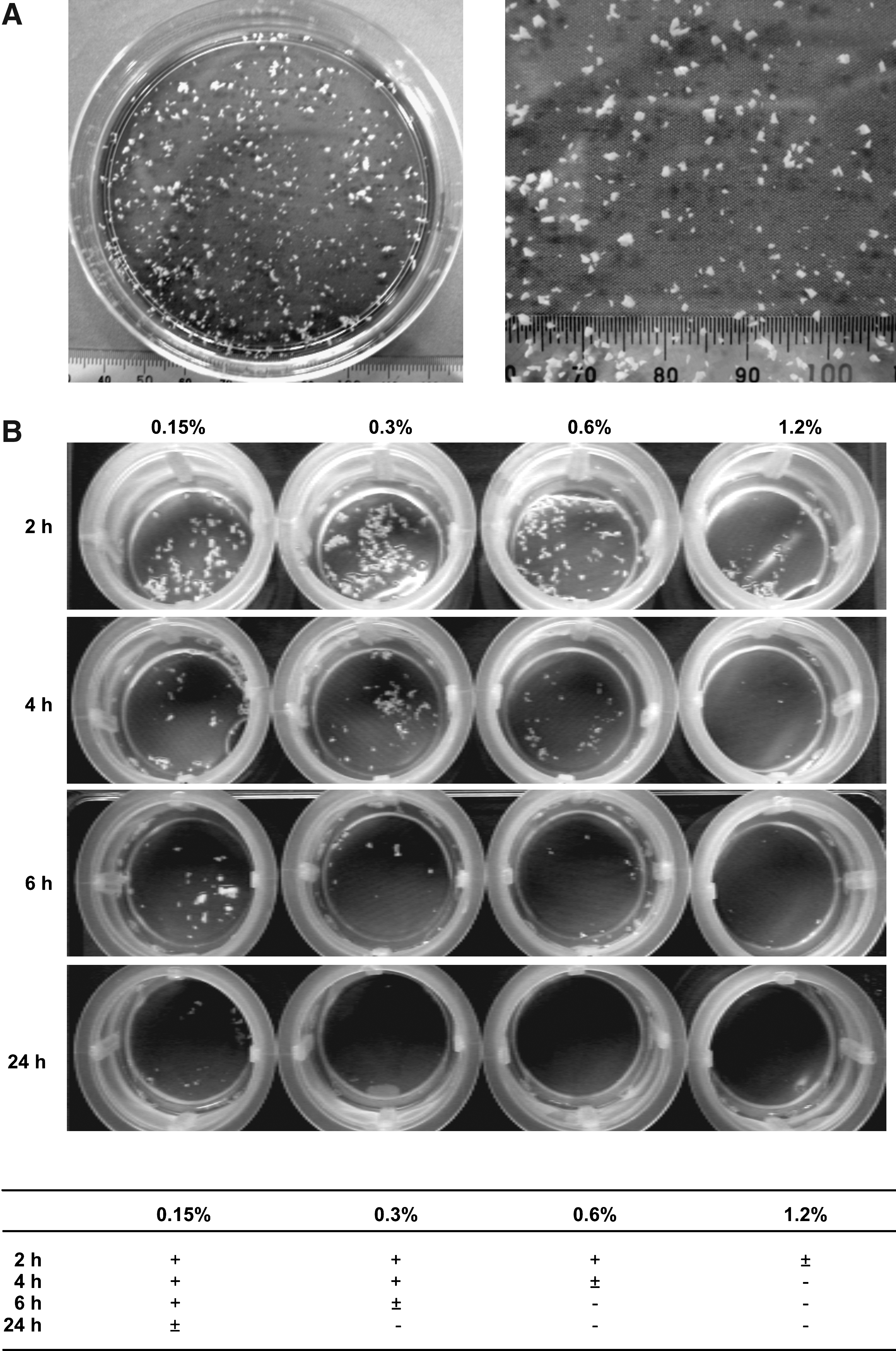
We examined the effects of collagenase concentration and incubation time on cartilage digestion. For all concentrations, the volume of the collagenase digests decreased as the incubation period increased (Fig. 1B). Although the cartilage pieces were somewhat visible after 24 h in the 0.15% collagenase, they had disappeared after the same amount of time in collagenase concentrations >0.3% (Fig. 1B). The amount of time for the cartilage remnants to disappear was shortened to 6 and 4 h in collagenase concentrations of 0.6% and 1.2%, respectively (Fig. 1B).
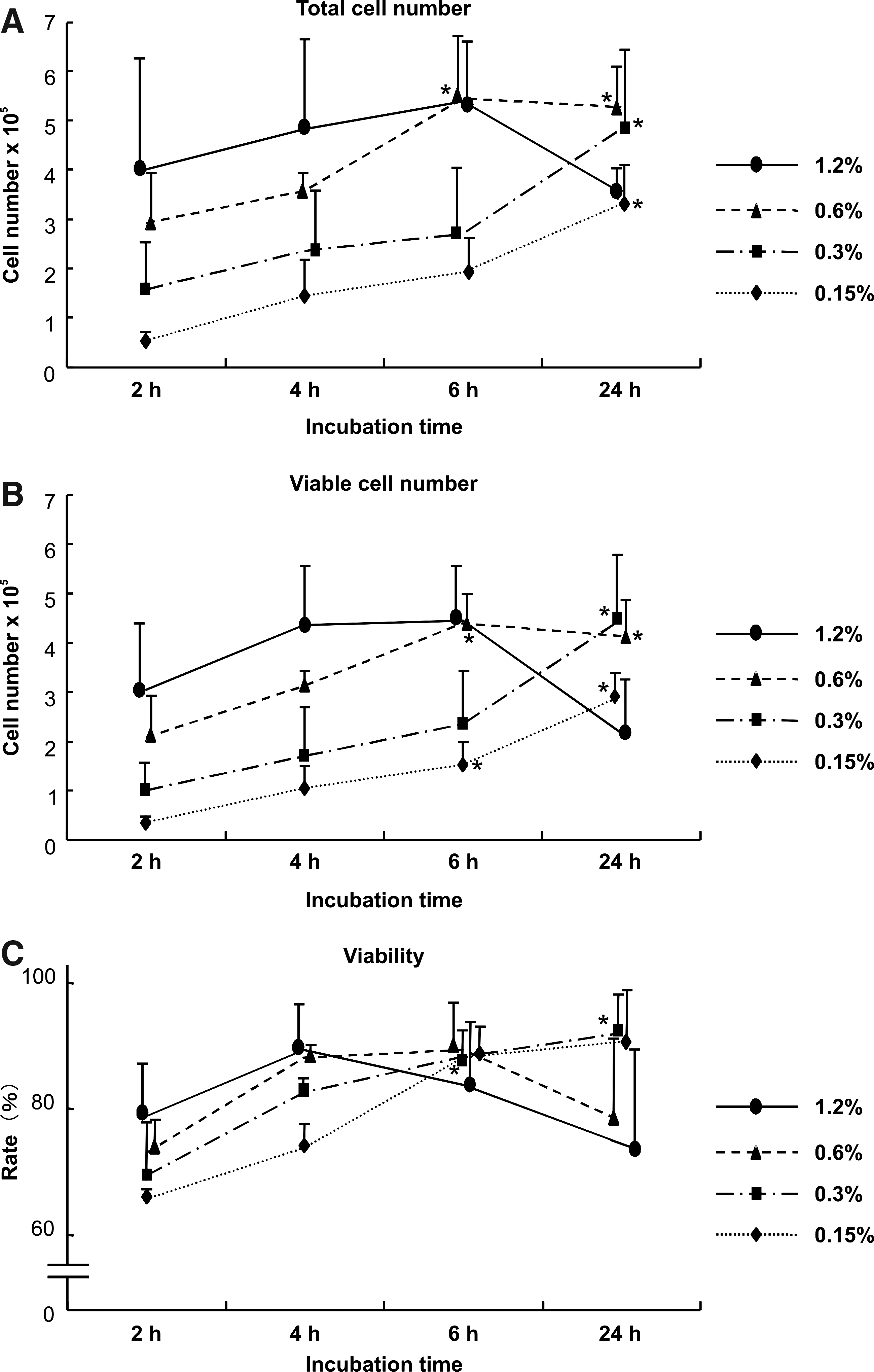
In all concentrations, both the total number of cells and the number of viable cells increased in parallel to the increase in the incubation time, with the exception of the sample with 1.2% collagenase for 24 h (Fig. 2A and B). The maximum number of viable cells, 4–5 × 10⁵ cells obtained from ∼0.05 g tissue, occurred at 4 h in 1.2% collagenase, 6 h in 0.6% collagenase, and 24 h in 0.3% collagenase. However, the sample with 0.15% collagenase provided <3 × 10⁵ cells even after 24 h, possibly because of the abundant remnants of the cartilage digests (Fig. 2A and B). The total cell numbers and viable cell numbers under conditions of 24 h and 0.15% collagenase, 24 h and 0.3% collagenase, and 6 h and 0.6% collagenase were significantly higher than those at 2 h for each dose of collagenase (Fig. 2A and B). The cell numbers had decreased after 24 h in 1.2% collagenase, as the viability deteriorated (Fig. 2C). Therefore, we determined which conditions of collagenase digestion maximized the viable cell numbers and found that the incubation time can be reduced to 24 h in 0.15% collagenase and 0.3% collagenase, 6 h in 0.6% collagenase, and 4 h in 1.2% collagenase.
Cell numbers and viability after collagenase digestion. Both the total
Next, we investigated the effect of cell density during seeding on cell growth (Fig. 3). Chondrocytes that had been digested by collagenase under suitable conditions as determined above were seeded at several cell densities (100–30,000 cells/cm2). All of those seeded at 10,000 cells/cm2 became almost confluent in 1 week. Seeding at 3000 cells/cm2 may be acceptable for the chondrocytes digested with 0.3% collagen for 24 h and 0.6% for 6 h, but the cells digested under other conditions could not sufficiently proliferate at 3000 cells/cm2 (Fig. 3B). The cells were aggregated with each other and formed cell clusters at 30,000 cells/cm2 in the samples digested with 0.6% collagenase and 1.2% collagenase (Fig. 3A and B). When we counted the cell numbers, the cells seeded at a density of 3000–10,000 cells/cm2 after digestion in 0.3% collagenase for 24 h and 0.6% for 6 h reached the maximum cell numbers at 1 week (Fig. 4A), which supported the results shown in Figure 3. However, the cells seeded at <1000 cells/cm2 became confluent as well after >2 weeks (Fig. 4B)
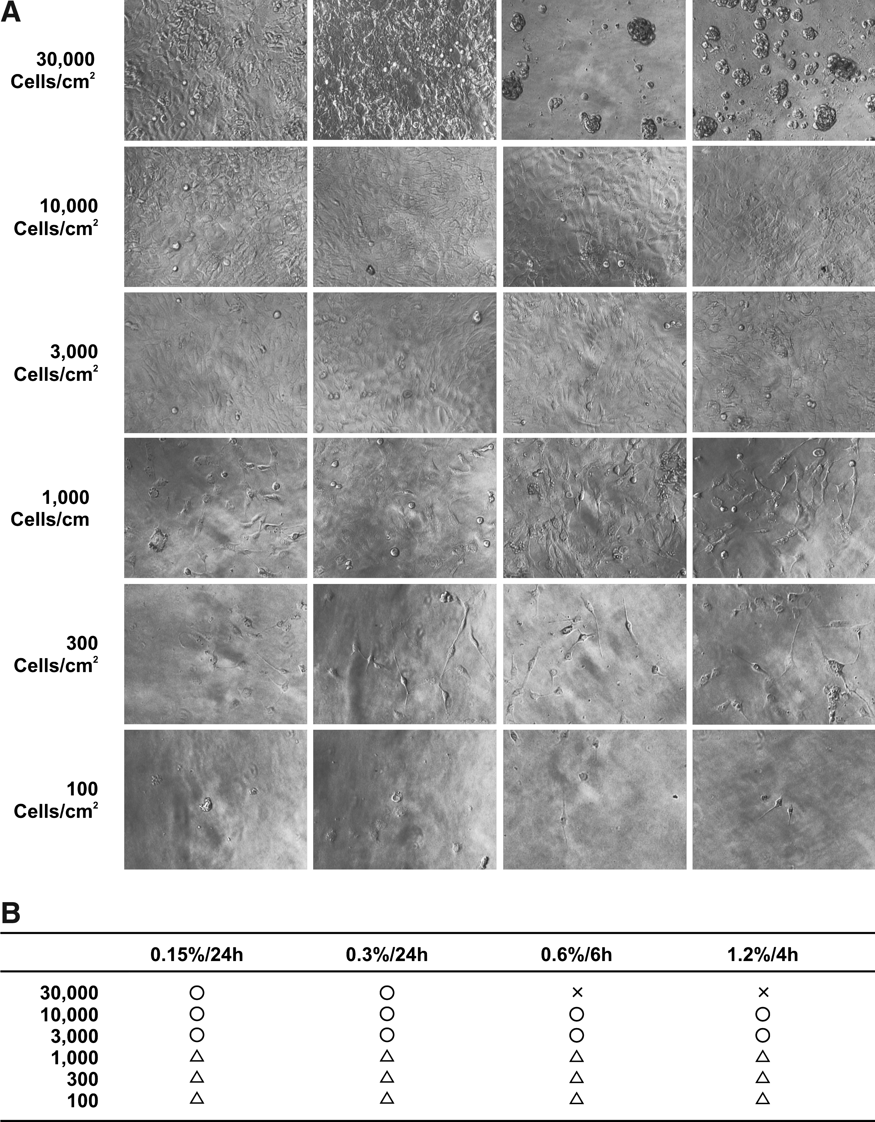
Optimal cell-seeding density to provide favorable cell growth.

Optimal cell-seeding density for obtaining sufficient cell numbers.
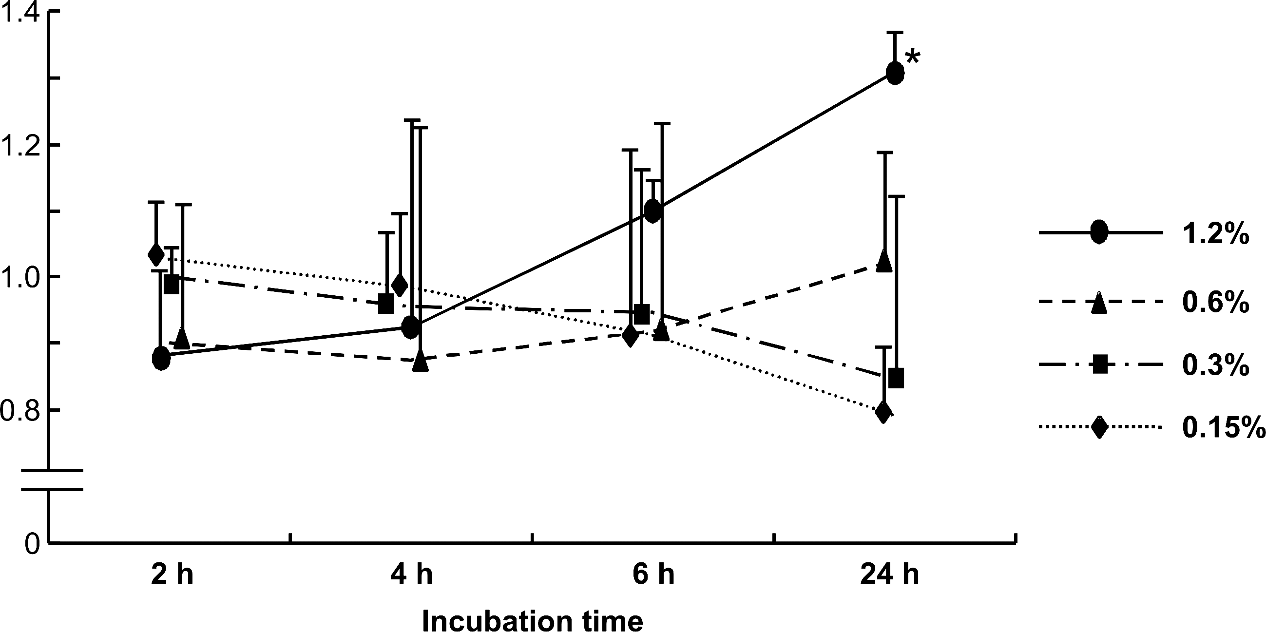
The possibility that the chondrocytes may fall into apoptosis or catabolic situations after the destruction of the native matrices by collagenase digestion could be not ignored. Thus, we examined gene expression of the apoptosis-related molecules and inflammatory cytokines in the chondrocytes for each set of collagenase digestion conditions. As shown in Figure 5, the proapoptotic factor p53 was likely upregulated as collagenase concentration increased, although we could not detect Bcl-2. The inflammatory cytokines TNF-α and IL-1β were scarcely detected in the chondrocytes digested in 0.15% collagenase for 24 h and in 0.3% collagenase for 24 h. Expression of these genes tended to increase in the chondrocytes with the higher collagenase concentrations (Fig. 5), suggesting that a collagenase concentration higher than 0.6% may somewhat affect chondrocyte viability or metabolism. Actually, the results of apoptosis assays also showed that apoptosis tended to be upregulated when the exposure time became longer in the higher concentrations of collagenase (0.6% or 1.2%). In contrast, the chondrocytes in the lower concentrations (0.15% and 0.3%) exhibited minimum apoptosis at 24 h (Fig. 6), which was consistent with the viability of chondrocytes (Fig. 2C) and the results of p53 expression (Fig. 5).
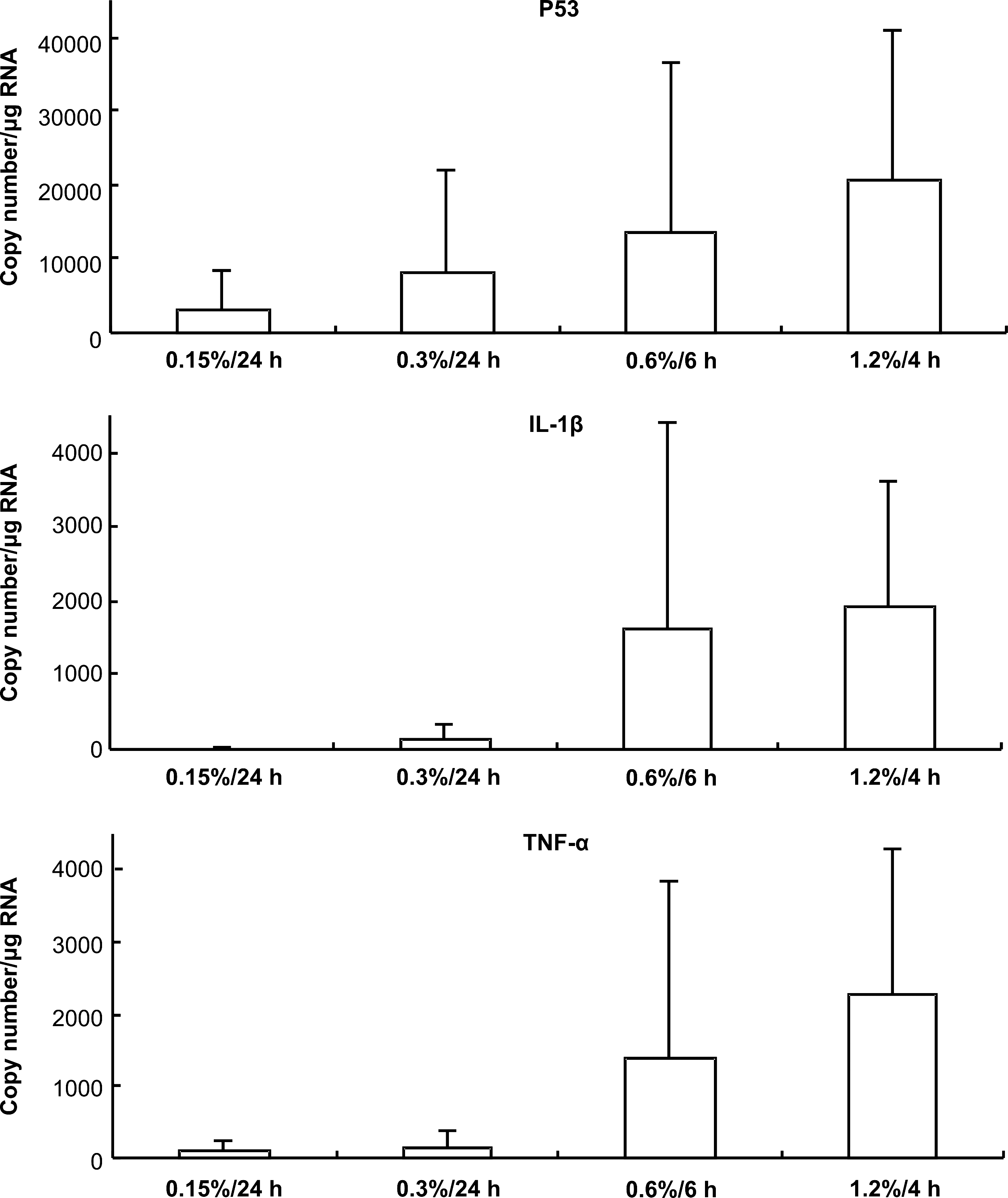
Gene expression of the proapoptotic factor and the inflammatory cytokines. Gene expression of the proapoptotic factor, p53, or the inflammatory cytokines, tumor necrosis factor alpha (TNF-α), and interleukin 1beta (IL-1β) tended to be upregulated in the chondrocytes as the concentration of collagenase increased. All values are presented as mean ± SD of three samples per group.
Apoptosis enzyme-linked immunosorbent assay. Apoptosis appeared to be increased after digestion in 1.2% collagenase, especially at 24 h. All values are presented as mean ± SD of three samples per group. Statistics were assessed using the Dunnett's test (*p < 0.05, vs. 2 h in each concentration of collagenase).
Discussion
Each laboratory engaged in the research and development of cartilage tissue engineering prepares various unique protocols according to their previous findings and experiences. Although they may independently work well, some kinds of standards are needed when we attempt to obtain stable results. However, thus far, no systematic analyses on collagenase doses or incubation times have been found. Although the cartilage includes different tissues and donor ages or diseases affect the cellularity or protein compositions of the tissues, the amount of collagen, which is the major inhibitory factor for chondrocyte isolation, seems almost constant. 37 This implies the possibility of a standard protocol. Thus, we regard the investigation of the optimal conditions for collagenase digestion to be a pivotal task for increasing the steadiness and safety of regenerative medicine.
On the basis of the results of the present study, by the time-course counting of cell numbers in various concentrations of collagenase, the maximum number of viable cells—5 × 10⁵—were harvested from 0.05 g of original tissue at 24 h in 0.15% collagenase, 6 h in 0.6%, and 4 h in 1.2% (Fig. 2B). The incubation time at which the viable cell numbers reached a maximum corresponded to the point at which the collagenase digests completely disappeared (Fig. 1B). The reason why the harvested cell numbers in the 0.15% collagenase did not reach the maximum even at 24 h may be because the remaining cartilage digests in the solution at that time. The viability of the cells harvested at 2 h was lower than that of the later harvests. The cells harvested in this short incubation period had been located at the surface of the cartilage tissues that had been minced to <1 mm3 before the collagenase digest. The mincing damaged the cells located on the surface of the tissues and decreased the viability of those cells. Therefore, the cells harvested after the 2 h incubation period showed a low viability (Fig. 2C). This tendency was also confirmed by the results of apoptosis assay. When the chondrocytes were exposed to lower concentrations of collagenase (0.15% or 0.3%), in which the cytotoxic effects of collagenase may hardly become obvious, those of short-term exposure (2–4 h) showed rather high extent of apoptosis (Fig. 6).
The cytotoxic effects of long-term exposure to collagenase were regarded as the cause of decrease in the viable cell numbers for those cells harvested at 24 h in 1.2% collagenase. In the present study, the maximum number of cells harvested from the native cartilage was ∼1 × 10⁷ cells/g. These cell numbers were lower than those found in human cartilage (1 × 10⁸ cells/g 31 ), but it is a 10-fold increase when compared to previous data (1 × 10⁶ cells/g 26 ).
A cell density of 30,000 cells/cm2 upon seeding was regarded as too high because the cells harvested from the 0.6% to 1.2% collagenase digest tended to be aggregated, as seen in Figure 3. If fragments of the matrices remain around the chondrocytes after collagenase digestion, the chondrocytes may aggregate because cell aggregation is promoted by the matrix fragments that mediate the cell–matrix interaction. 38 Exposure to a higher concentration of collagenase even for a short incubation period could completely release the chondrocytes, but some fragments of the extracellular matrices may remain, which could mediate cell aggregation in a cell density as high as 30,000 cells/cm2. In contrast, the density of 3000–10,000 cells/cm2 showed appropriate adhesion and subsequent proliferation in all concentrations.
In addition, for a cell density lower than 1000 cells/cm2, some viable cells were obtained after more than a week of incubation, as shown in Figure 4B.
It took 2 weeks for cells at initial densities <1000 cells/cm2 to become confluent, and cells seeded at 100 cells/cm2 needed as long as 3 weeks (Fig. 4B). However, if it takes >2 weeks for the chondrocytes to become confluent after seeding, too much time may be spent judging whether or not the chondrocytes have appropriately proliferated. A cell density at which the cells completely reach confluence within 1 week is desirable. Moreover, the paracrine/autocrine signals are needed for adequate cell proliferation. 39 Cells may not sufficiently proliferate at a low cell density because they cannot receive the paracrine/autocrine signals from adjacent cells. On the other hand, when the cell density is higher than 10,000 cells/cm2, the passage would be needed at <1 week of incubation. If the incubation time during one passage is shortened and the passage number increases correspondingly, procedures become more complicated, and the risk of dedifferentiation may increase.
p53 and Bcl-2 are related to apoptosis. p53 induces apoptosis, participating in the maintenance of cell homeostasis. 33 Bcl-2, on the other hand, prohibits apoptosis. 34 TNF-α and IL-1β are inflammatory cytokines that are synthesized by natural immune cells in response to the infection or destruction of tissues.35,36 Analysis of gene expression of these inflammatory cytokines, apoptosis-related molecules and apoptosis assay revealed that damage to the cells is diminished when the collagenase concentration is low. The damage to the cells may result from the cytotoxicity of the collagenase, which is a kind of digestive enzyme.
Therefore, we recommend a 24-h incubation in 0.3% collagenase, or 6 h in 0.6% collagenase, as the optimal condition for chondrocyte isolation from cartilage pieces that are 250–1000 μm in size. Moreover, the cell-seeding density should be in the range of 3000–10,000 cells/cm2. These conditions maximize the harvest of the isolated chondrocytes from a small amount of biopsied tissue and significantly aid in obtaining a large quantity of cultured cells in a short period.
Footnotes
Acknowledgments
We thank Mr. Takashi Nakamoto, Ms. Miki Akizawa, Mr. Tomoaki Sakamoto, Mr. Takayuki Furuichi, and Mr. Makoto Watanabe for their technical support. This work was supported by Grants-in-Aid for Scientific Research from the Ministry of Education, Culture, Sports, Science and Technology of Japan (MEXT, No. 21390532 and 21659462), Research and Development Programs for Three-dimensional Complex Organ Structures from the New Energy and Industrial Technology Development Organization, and Resolving Critical Issues from Special Coordination Funds for Promoting Science and Technology (SCF) commissioned by MEXT.
Disclosure Statement
No competing financial interests exist.