
Abstract
Electrospun scaffolds have been studied extensively for their potential use in bone tissue engineering applications. However, inherent issues with the electrospinning approach limit the thickness of these scaffolds and constrain their use for repair of critical-sized bone defects. One method to increase overall scaffold thickness is to bond multiple electrospun scaffolds together with a biocompatible gel. The objective of this study was to determine whether multiple human adipose-derived stem cell (hASC)–seeded electrospun, nanofibrous scaffolds could be layered via in situ collagen assembly and whether the addition of laser-ablated micron-sized pores within the electrospun scaffold layers was beneficial to the bonding process. Pores were created by a laser ablation technique. We hypothesized that the addition of micron-sized pores within the electrospun scaffolds would encourage collagen integration between scaffold layers, and promote osteogenic differentiation of hASCs seeded within the layered electrospun scaffolds. To evaluate the benefit of assembled scaffolds with and without engineered pores, hASCs were seeded on individual electrospun scaffolds, hASC-seeded scaffolds were bonded with type I collagen, and the assembled ∼3-mm-thick constructs were cultured for 3 weeks to examine their potential as bone tissue engineering scaffolds. Assembled electrospun scaffolds/collagen gel constructs using electrospun scaffolds with pores resulted in enhanced hASC viability, proliferation, and mineralization of the scaffolds after 3 weeks in vitro compared to constructs using electrospun scaffolds without pores. Scanning electron microscopy and histological examination revealed that the assembled constructs that included laser-ablated electrospun scaffolds were able to maintain a contracted structure and were not delaminated, unlike assembled constructs containing nonablated electrospun scaffolds. This is the first study to show that the introduction of engineered pores in electrospun scaffolds assists with multilayered scaffold integration, resulting in thick constructs potentially suitable for use as scaffolds for bone tissue engineering or repair of critical bone defects.
Introduction
To create large structural features within electrospun scaffolds, investigators have recently used a method known as laser ablation. 17 Laser ablation is a technique that removes selective regions of a material and introduces micron-structured features, including pores of a uniform size, specific global porosity, and preferential routes for gel permeation. In laser ablation, pulses of light at a specific wavelength (ranging from below ultraviolet to above the visible light spectrum) are used to remove material in a highly controlled fashion through the absorption of photons by the target material, exciting electrons in the polymer, and resulting in removal of the material in the form of a dust-like plume. 18 One of the benefits of laser ablation is that material is removed with minimal thermal damage. 19 Machining of electrospun tissue engineering scaffolds by laser ablation has previously been reported, and investigators have reported successful creation of channel-like features within an electrospun matrix. 17 However, the successful assembly of multiple laser-ablated electrospun scaffolds via gel permeation has not been investigated. A multilayered construct would be of great benefit for tissue engineering by incorporating the nanoscale architecture of electrospun scaffolds with a facile bonding technique of collagen polymerization for the creation of composite scaffolds for bone defects.
An emerging source of cells for bone tissue engineering applications is adipose-derived stem cells. In recent years, the use of human adipose-derived stem cells (hASCs) for bone tissue engineering has greatly increased because of hASCs' multipotent nature, source tissue availability, and similar performance characteristics to bone-marrow-derived stem cells.20–22 However, no investigations to date have evaluated the use of hASCs on multilayered electrospun scaffolds. We hypothesized that laser-ablated scaffolds would not only support collagen integration but also enhance the proliferation and osteogenic differentiation of hASCs compared to nonablated assembled scaffolds. The objectives of this study were to (1) determine whether laser-ablated pores in assembled electrospun nanofibrous scaffolds would improve the integration of multilayered structures by promoting collagen gel contraction, and (2) evaluate if such structures would promote greater viability, proliferation, and osteogenic differentiation of hASCs. We fabricated assembled collagen gel/electrospun scaffolds by bonding three individual hASC-seeded electrospun scaffolds with type I collagen per assembled constructs and assessed the impact of the assembled constructs with, and without, laser-ablated pores on hASC response.
Methods
hASC isolation and seeding
hASCs were obtained from excess human adipose tissue from liposuction procedures using waste tissue obtained from two Caucasian women (average age 49 ± 3 years) in accordance with an approved IRB protocol (IRB-04-1622) at University of North Carolina–Chapel Hill. hASCs were isolated from tissue based on the original method described by Zuk et al. 23 and described previously by our laboratory.24,25 In brief, 50 g of adipose tissue was washed in phosphate-buffered saline (PBS), minced using scissors, and enzymatically digested at 37°C at 5% CO2 for 30 min in 0.075% type I collagenase (Worthington Biochemical, Lakewood, NJ), supplemented with 100 IU penicillin/100 μg/mL streptomycin (Mediatech, Herndon, VA) in Eagle's minimum essential medium, alpha-modified (Invitrogen, Carlsbad, CA). After digestion, the digested tissue was centrifuged for 10 min at 10,000 g and the supernatant was discarded. The hASC-rich cell pellet was resuspended in a complete growth medium (CGM; Eagle's minimum essential medium, alpha-modified, supplemented with 10% fetal bovine serum, 2 mM L-glutamine, 100 units/mL penicillin, and 100 μg/mL streptomycin), seeded in 75 cm2 flasks, and cultured within a humidified incubator at 37°C at 5% CO2. Cultures were grown till 80% confluency and passaged. For this study second-passage hASCs were grown to 80% confluency, trypsinized, and resuspended in the growth medium.
Electrospun scaffold fabrication
hASC-seeded assembled constructs were created via four main processing steps: (1) electrospinning of scaffolds; (2) laser ablation of scaffolds; (3) seeding of hASCs on scaffolds; (4) bonding of hASC-seeded scaffolds with type I collagen (Fig. 1). Scaffolds were fabricated by dissolving poly (l-lactic acid) (PLA) with a molecular weight of 70,000 Da in chloroform and dimethylformamide at a ratio of 4:1 to yield a 12 wt% solution (both reagents from Sigma, St. Louis, MO). Scaffolds were electrospun by volumetrically dispensing the dissolved polymer solution through a 20-gauge blunt-tip needle at a flow rate of 50 μL/min (New Era Syringe Pump, Wantagh, NY) and electrifying the needle with a positive voltage of 15 kV (Gamma High Voltage Power Supply, Ormond Beach, FL), separated from a 23-cm grounded collector plate by 15 cm, and depositing for approximately 1.5 h (Fig. 1a). Randomly aligned nanofibers were collected onto aluminum foil and maintained in a laminar flow hood for 24 h to ensure solvent evaporation.

Schematic of the creation of ablated and nonablated electrospun scaffolds bonded with type I collagen. PLA, poly(l-lactic acid).
Laser ablation parameters
Micromachining of the pores in the electrospun scaffolds was performed by ablation from a pulsed excimer laser (Fig. 1b). The source light was a Lambda Physik COMPexPro 201 ArF pulsed excimer laser (Coherent, Santa Clara, CA) operating at 193 nm, 25 ns pulse duration, and 90 mJ at the source. The laser was positioned by an Aerotech x-y-z translation stage (Aerotech, Pittsburgh, PA) controlled by NView MMI software (Aerotech). A 6 × 6 array (36 pores) was ablated through the PLA scaffold with a target pore diameter of 300 μm (Fig. 1c). The laser was then used to cut samples with and without the ablated pore pattern into 10 × 10 mm squares. The porosity of the machined area for the ablated scaffolds (4 × 4 mm center region of the 10 × 10 mm scaffold) was 15.9%. Owing to the rectangular profile of the laser beam, rastering was performed to create a more circular pore. Scaffold thickness was measured to be approximately 0.15 ± 0.01 mm with a Mitutoyo absolute micrometer (Aurora, IL), which can precisely determine thicknesses up to 1 μm. Scaffold mass was measured to be 3 ± 1 mg using a Mettler Toledo AB104 scale (Columbus, OH) with an accuracy of 0.1 mg. Scaffolds were sputter-coated with 200 Å Au/Pd and imaged using a JEOL JSM-6400 FE-SEM (Peabody, MA) operating at 5 kV. Images were analyzed using Revolution™ software (4Pi, Durham, NC). At least three different samples for each of the ablated and nonablated scaffolds were imaged, and fiber diameter and ablated pore diameter averages were calculated from at least 30 measurements (minimum 10 measurements per sample).
Construct assembly
After scaffold fabrication, scaffolds were sterilized by soaking in 70% ethanol for 30 min followed by rinsing three times with 1 × PBS. Scaffolds were then placed in nonpyrogenic 24-well plates (Sarstedt, Newton, NC) and soaked in CGM for 12 h before hASC seeding. hASCs (passage 2) were seeded onto the electrospun scaffolds at a density of 20,000 cells/cm2 in a volume of 100 μL CGM and allowed to attach for 30 min (Fig. 1c). After this initial incubation, the wells were flooded with 1 mL of CGM. hASCs were allowed to grow for 24 h after seeding and viability images were captured before scaffold stacking using the Live/Dead Assay Cytotoxicity Kit (Molecular Probes, Eugene, OR) for mammalian cells. hASC-seeded electrospun scaffolds were fit into a custom 10 × 10 × 50 mm polycarbonate mold and stacked three scaffold layers high (Fig. 1d). Type I collagen gel at a concentration of 3 mg/mL (Vitrogen, Angiotech BioMaterials Corporation, Palo Alto, CA) was neutralized to pH 7.0 with 1 N NaOH, and 200 μL collagen was pipetted between the layers for a total of 400 μL collagen per construct (Fig. 1d). The assembled scaffolds were then incubated at 37°C at 5% CO2 for 2 h to allow the collagen gel to polymerize. After polymerization, assembled constructs were transferred to new nonpyrogenic 24-well plates and the wells were flooded with 1 mL CGM. After another 24 h, the medium was changed to either CGM or osteogenic differentiating medium (ODM; CGM supplemented with 50 μM ascorbic acid, 0.1 μM dexamethasone, and 10 mM β-glycerol phosphate) and subsequently changed every 3 days thereafter for up to 21 days.
Construct analysis
Fourier transform infrared spectrometry
Fourier transform infrared (FTIR) spectra of electrospun PLA scaffolds were recorded before and after collagen addition and polymerization using a Thermo Nicolet Nexus 470 (Waltham, MA) fitted with a Continuum microscope and a Mercury Cadmium Telluride detector. A total of 64 scans in reflectance mode were aggregated for each sample spectrum in the range of 800–2000 cm−1 with a 4 cm−1 resolution. Representative spectra are shown.
Scaffold mass
Assembled scaffolds were weighed on days 7, 14, and 21 to quantify any changes in mass due to collagen gel contraction and resulting water loss.
Cellular analysis
hASCs were quantified on assembled scaffolds on days 7, 14, and 21 by carefully separating the assembled scaffolds with forceps into their three separate electrospun PLA layers. DNA content was measured in each electrospun layer using the DNA binding dye Hoechst 33258 in microplate format after an overnight digestion at 60°C in 2.5 units/mL papain in PBS with 5 mM ethylenediaminetetraacetic acid and 5 mM cysteine HCl (all reagents from Sigma). Osteogenic differentiation was assessed by quantifying the amount of mineralized matrix produced by the hASCs by digesting the individual electrospun layers of the assembled constructs in 0.5 N HCl overnight and assaying the supernatant using the Calcium Liquicolor Assay (Stanbio, Boerne, TX).
Histological and scanning electron microscopy analysis
On day 21, samples were fixed with 3.7% formaldehyde for 30 min and rinsed twice in 1 × PBS. Assembled scaffolds were stained for calcium accretion by staining with 40 mM Alizarin Red S, pH 4.2, for 3 min, followed by rinsing with deionized water five times to remove any unbound stain. Assembled scaffolds for histological examination were dehydrated with increasing ethanol series of 50%, 70%, 95%, 100%, and 100% for 15 min in each bath. Histological samples were embedded in paraffin and mounted. Assembled scaffold constructs were cut into 5-μm-thick serial sections using a rotary microtome and stained with hematoxylin and eosin (Fisher Chemical, Waltham, MA) to stain cells and the ECM using standard histochemical techniques. Images were captured with a Leica EZ 4D Digital Dissecting Scope (Leica, Wetzler, Germany). For scanning electron microscopy (SEM) analysis, assembled constructs were cut in half, dehydrated (using the same ethanol series procedure as above), and then critical-point dried with CO2. After critical-point drying, samples were sputter-coated with 200 Å Au/Pd before imaging with a JEOL JSM-6360 scanning electron microscope at 5 kV in secondary electron imaging mode. Images were captured for both cross-sectional and top views of the assembled scaffolds.
Statistical analysis
All experimental sample groups had a sample size of at least n = 3, and experiments were performed in two independent trials using hASCs from two Caucasian women (average age, 49 years). Data are presented as average ± standard error mean. Statistical analysis was performed using JMP 7.0 Software (SAS, Cary, NC) by performing analysis of variance and Tukey honest significance difference (HSD) tests, with a significance accepted at a p-value < 0.05.
Results
Scaffold fabrication and construct assembly
Electrospun PLA scaffolds displayed a random fiber arrangement characteristic of electrospun mats (Fig. 2a). Scaffolds possessed an average fiber diameter of 819 ± 90 nm. Laser ablation was used to create pores with an average diameter of 316 ± 8 μm (Fig. 2b). Highly ordered 6 × 6 pore arrays were ablated into the electrospun PLA scaffolds (Fig. 2c) and were positioned within the central 4 × 4 mm region of the 10 × 10 mm scaffold area. Redeposition of the ablated PLA was minimal around the pore edge, and nanofibers on ablated scaffolds displayed similar morphology to nonablated electrospun scaffold regions (Fig. 2b). Electrospun scaffolds were able to be bonded together using type I collagen as the binding system (Fig. 2d). After polymerization of the collagen, constructs were able to maintain an intact structure approximately 3 mm in thickness (Fig. 2d), compared to a conventional thickness of 150 μm for a single electrospun scaffold. FTIR spectroscopy indicated that the electrospun PLA scaffold had a large absorbance peak at 1757 cm−1 (Fig. 3). Collagen addition and polymerization to the electrospun scaffold resulted in the presence of both amide I and II peaks at 1649 and 1536 cm−1 respectively, verifying the presence of collagen.
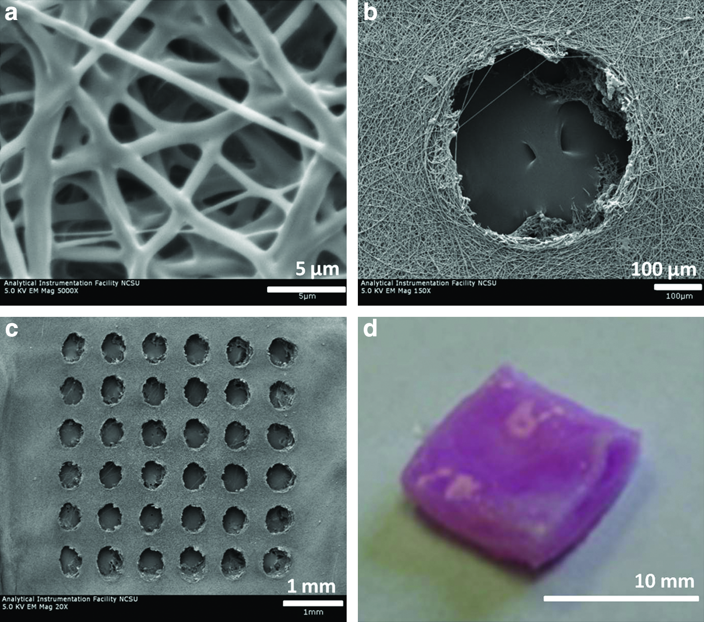
Scanning electron microscopy (SEM) images of electrospun poly(l-lactic acid) (PLA) fibers (

Fourier transform infrared spectra of electrospun PLA scaffolds before and after collagen polymerization and spectra for neat collagen. Fourier transform infrared spectra for the constructs exhibited spectra of both neat electrospun PLA and collagen with peaks at 1649 and 1536 cm−1 for amide I and II, respectively.
Cellular analyses
hASCs were able to adhere on both nonablated (Fig. 4a) and ablated (Fig. 4b) electrospun scaffolds. Cells displayed a normal morphology and adhered around the micromachined pores (Fig. 4b). After layered construct assembly with type I collagen, viability images indicated that hASCs were spreading throughout the collagen layer within 24 h of hASC-seeded electrospun scaffold/collagen gel construct assembly (Fig. 4c). On days 7, 14, and 21 the ablated and nonablated assembled scaffolds cultured in both CGM and osteogenic medium were weighed. The assembled scaffolds continued to lose mass throughout the 3-week experiment (Fig. 5), due to contraction of the collagen I gel and associated fluid extraction. At days 7 and 14 the nonablated scaffolds weighed more than the ablated scaffolds. However, by day 21 there was no significant difference between any experimental groups.
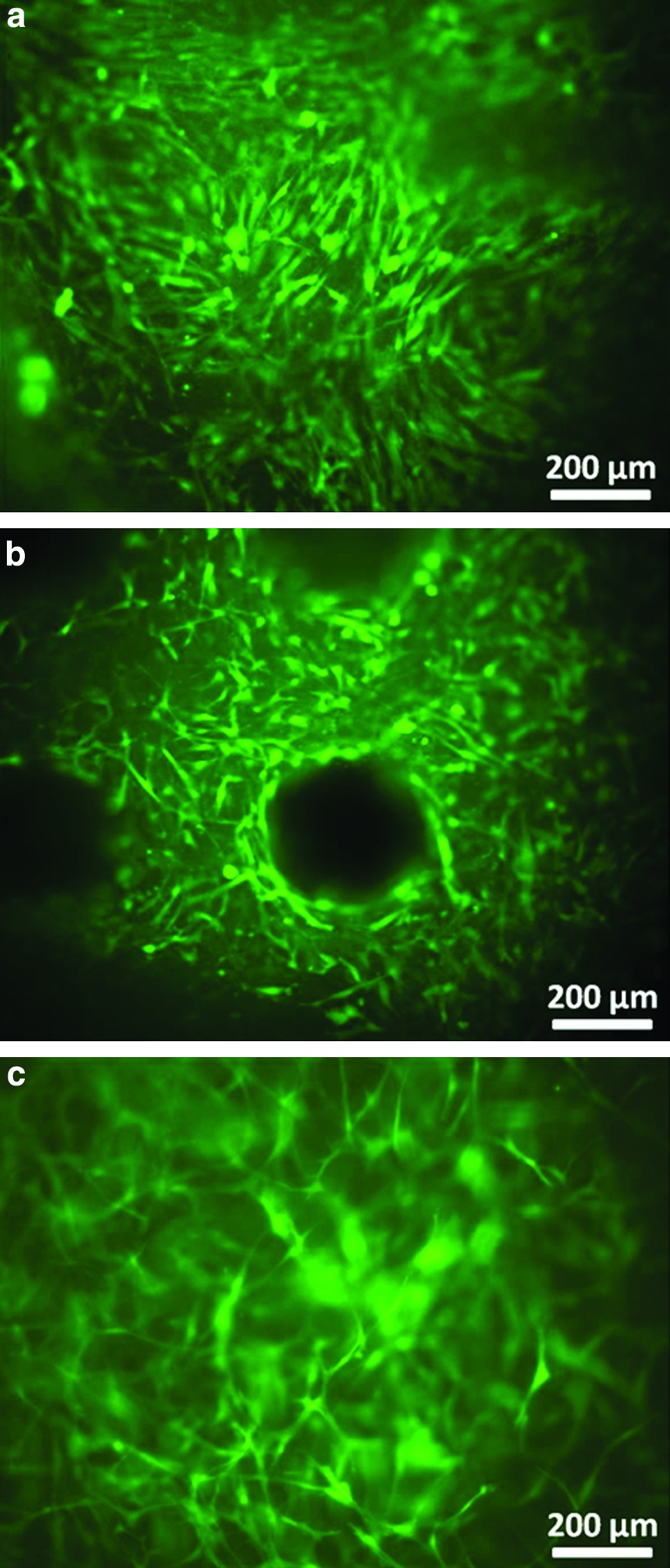
Human adipose-derived stem cell (hASC) viability images on electrospun PLA scaffolds without (
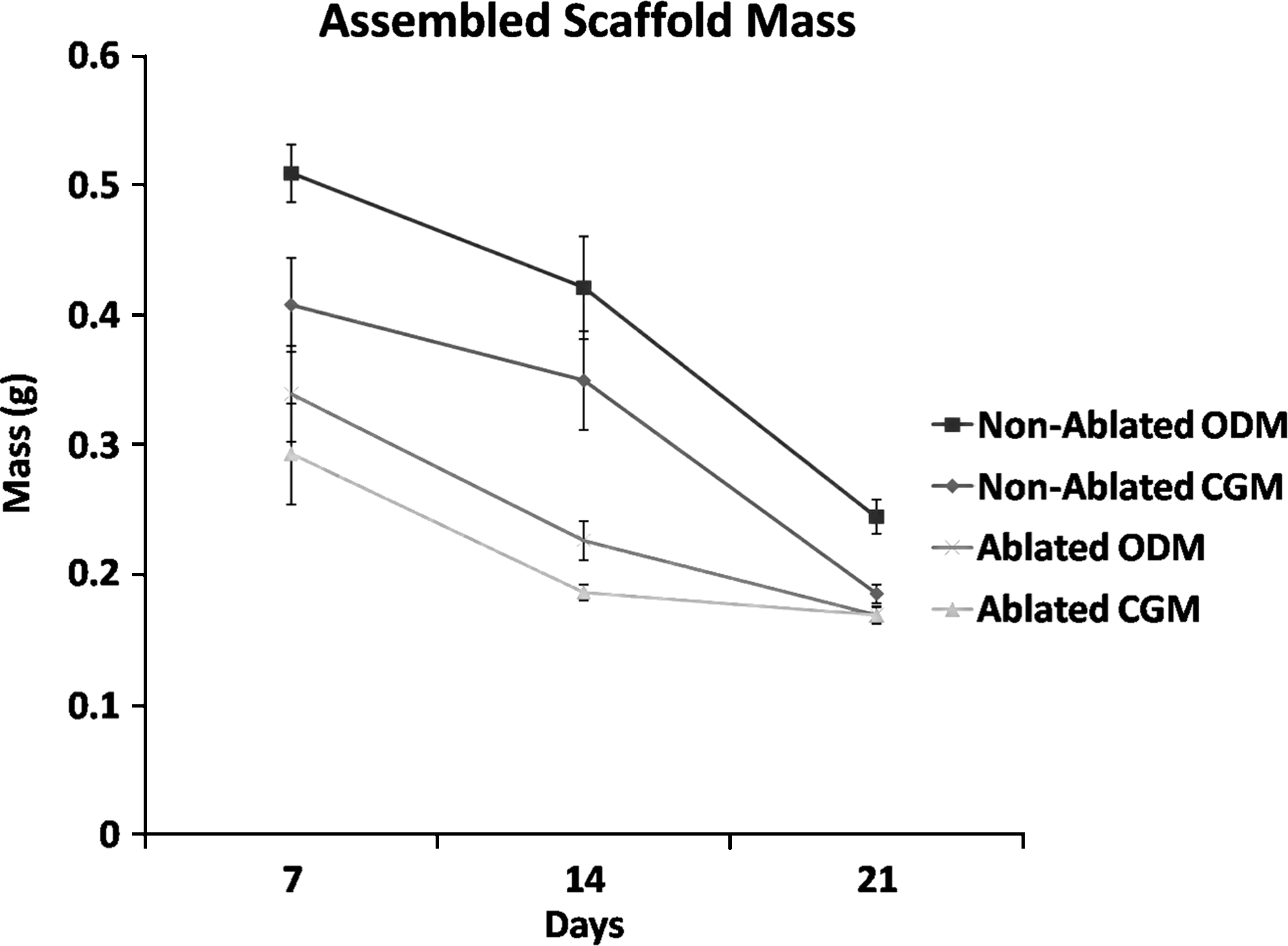
Throughout the experiment the mass of the assembled scaffolds continued to decrease because of collagen gel contraction and associated fluid extraction. ODM, osteogenic differentiating medium; CGM, complete growth medium.
There was no significant difference in hASC proliferation in nonablated and ablated constructs at days 7 and 14 (Fig. 6). By day 21, however, the ablated scaffolds exhibited the highest amount of DNA content, whereas the nonablated constructs cultured in ODM displayed the lowest amount of DNA. To examine the effects of the culture medium and to confirm osteogenic differentiation, assembled scaffolds were stained with Alizarin Red S to identify deposited mineral on day 21. After 21 days of culture, assembled scaffolds cultured in ODM stained positive for deposited calcium, whereas assembled scaffolds cultured in CGM exhibited only slight nonspecific staining (Fig. 7). Mineralized calcium was quantified on days 7, 14, and 21 (Fig. 8). Mineralized calcium content increased in a temporal manner with culture in the osteogenic differentiation medium. By day 21 it was determined that hASCs in the ablated constructs produced significantly more mineral compared to those on nonablated constucts, 233 ± 8 versus 188 ± 6 μg Ca2+/construct for the ablated and nonablated scaffolds, respectively (Fig. 8). As expected, mineralized calcium remained very low at all time points in both scaffold types for hASCs maintained in CGM.
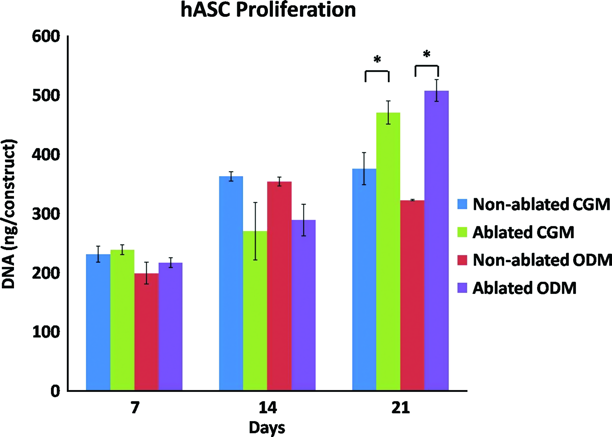
hASC proliferation throughout the duration of the experiment. hASCs were able to proliferate on all assembled constructs throughout the duration of the experiment. By day 21 both of the ablated scaffold groups (CGM and ODM) displayed a significantly higher amount of DNA compared to nonablated scaffolds. *Significant difference (p-value < 0.05). Color images available online at
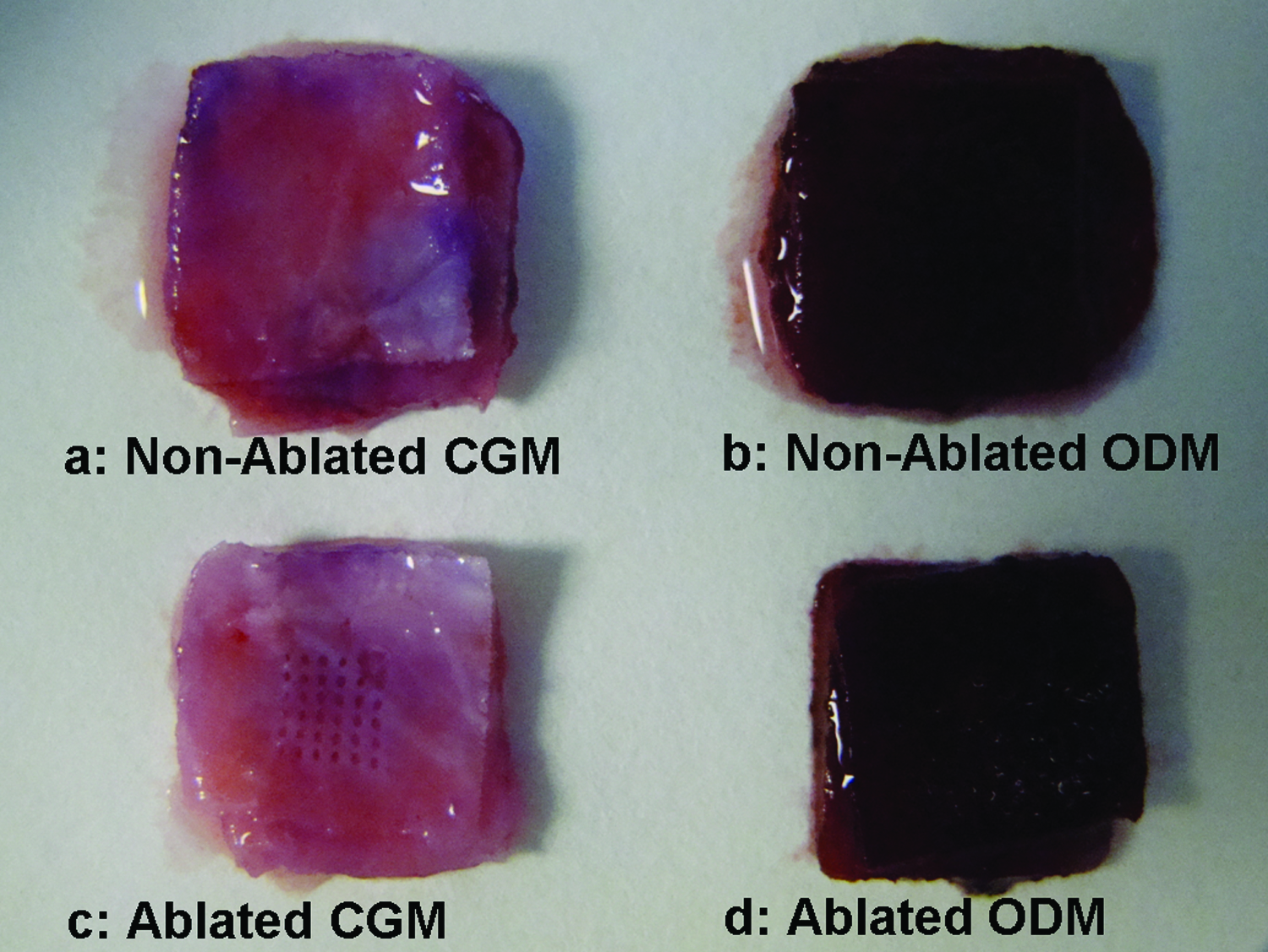
Alizarin Red S staining of assembled constructs with and without laser-ablated pores cultured in the complete growth medium (CGM,

hASC mineralization increased for both ablated and nonablated scaffold constructs cultured in ODM. At day 21, there was a significantly higher amount of calcium in the ablated constructs compared to the nonablated constructs. *Significant difference (p-value < 0.05). Color images available online at
To examine cellular interaction between the three different electrospun layers of the assembled electrospun scaffolds/collagen gel constructs, histological and SEM were performed at day 21. Hematoxylin and eosin staining indicated that hASCs were present within the collagen gel (stained pale pink) and electrospun scaffold matrix (unstained) (Fig. 9a–d). Upon examination, it was seen that extensive delamination occurred between the nonablated electrospun scaffold and collagen gel layers (Fig. 9a, b, arrows). Collagen was present along the interface of the nonablated scaffold maintained in CGM (Fig. 9a, dark pink), but very little was present within the nonablated scaffold maintained in the osteogenic differentiation medium (Fig. 9b). In addition, there were few cells present within the center layer of the construct for the nonablated construct maintained in ODM, indicating low viability (Fig. 9b). The majority of the cells were located on both the top and bottom edges of the construct (Fig. 9b). Partial alignment of the pores for the laser-ablated constructs was achieved for both medium groups (Fig. 9c, d). For the ablated constructs, hASCs were present along the boundary of the electrospun scaffold and also evident along the edges of the pores, noted by the blue staining of cell nuclei (Fig. 9d). Collagen was visible throughout the cross section of the ablated scaffolds and permeated through the individual scaffold layers of the assembled construct (collagen gel stained pink in contrast to unstained electrospun scaffolds, Fig. 9c, d).
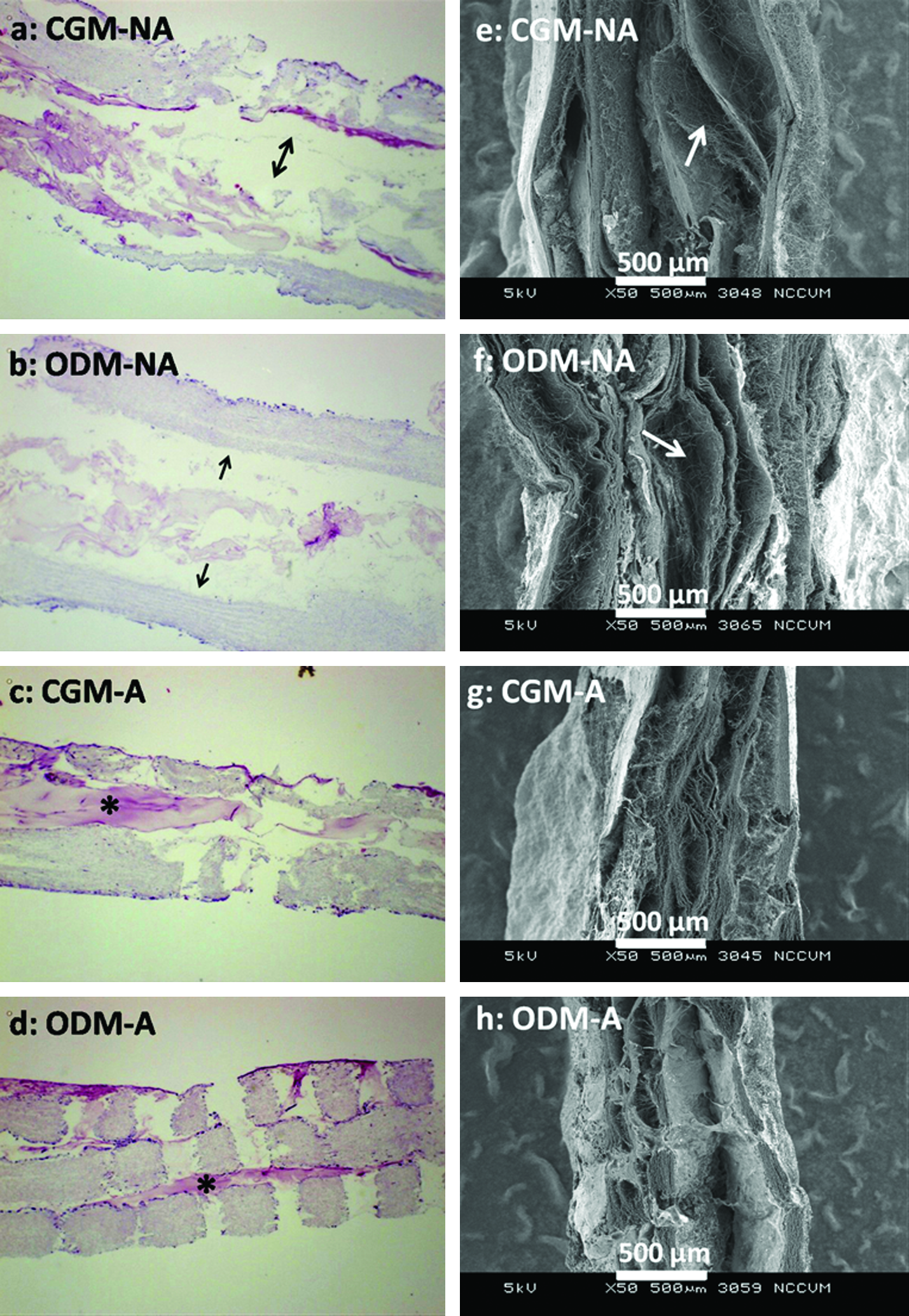
Histological (
SEM images of the assembled electrospun/collagen gel constructs indicated that the individual electrospun scaffold layers of the nonablated scaffold/gel constructs were still largely separate, as open areas were present within the nonablated constructs (Fig. 9e, f, white arrows). SEM indicated that the ablated scaffold/gel constructs, on the other hand, retained a tight, intact structure (Fig. 9g, h). Again, partial alignment of the pores was achieved and noted by the indentations of the scaffold. An aerial view of the assembled electrospun/collagen gel construct with ablated scaffolds indicated that hASCs were able to adhere and display a well-spread morphology around and within the ablated features (pore array indicated by white arrows in Fig. 10a). hASCs were present over the top surface of the construct and were able to adhere and spread around and within the ablated features (hASCs indicated by white arrows in Fig. 10b–d).
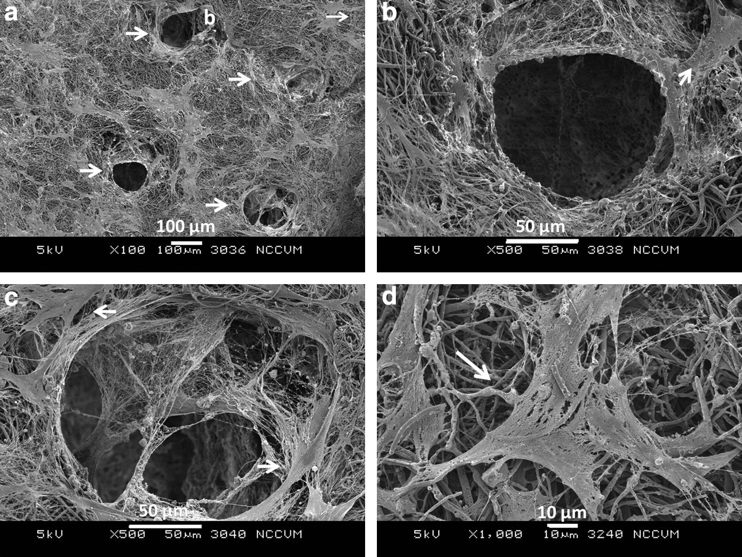
SEM images of ablated scaffold/collagen gel assembled constructs after 21 days in vitro. hASCs were present on the top surface of the constructs and were able to adhere around and in the ablated pores. Pore array is indicated by white arrows in (
Discussion
This study demonstrates that with the addition of engineered pore arrays in electrospun nanofibrous substrates, and subsequent polymerization with type I collagen gel, thick, cell-seeded, electrospun scaffolds/collagen gel constructs can be fabricated. These layered scaffolds maintain hASC viability and support hASC osteogenic differentiation with minimal delamination between the multiple electrospun scaffold layers because of collagen I permeation through the laser-ablated pores. Our findings support previous reports that the combination of gel systems with porous scaffolds leads to enhanced cell distribution, infiltration, and proliferation.
26
For this study, collagen I was used as the binding system and was able to promote rapid contraction and binding of laser-ablated electrospun scaffolds. Collagen I is potentially an ideal binding material to use for bone tissue engineering, given that it is the most predominant protein in bone and it promotes cell migration and proliferation because of the presence of arginine-glycine-aspartic acid sequences (RGD) on collagen.
1
Weinand et al. assessed the ability of several hydrogels to facilitate osteogenesis of porcine bone-marrow-derived stem cells in porous β-tricalcium phosphate scaffolds, and determined that type I collagen led to enhanced radiological density and relative stiffness of cell-seeded constructs compared to both alginate and fibrin glue.
27
FTIR spectroscopy indicated that after collagen polymerization on electrospun PLA scaffolds, the constructs exhibited spectra of both neat electrospun PLA and collagen with specific peaks at 1649 and 1536 cm−1 for amide I and II, respectively. Previous research has indicated that blending of collagen with synthetic polymers such as poly(
As cells grow and spread within a collagen matrix, the matrix is able to undergo cellular remodeling via contraction, resulting in the formation of a dense collagen network. As we have demonstrated previously, when mesenchymal stem cells are seeded within type I collagen gels, rapid contraction occurs, with such contraction detectable after only 1 day of culture. 30 Although we have previously viewed this contraction as a potential limitation in collagen gel systems for some applications, 30 it can be considered beneficial when acting as an integrator between electrospun scaffold layers, as in this work and others. 13 It is well known that cellular remodeling of collagen matrices is a direct effect of the interaction with the α2β1 integrin receptor present on the cell membrane, and this interaction between the α2β1 integrin and collagen has been demonstrated to play a crucial role in osteoblastic differentiation.31,32 The benefit of combining collagen gel and electrospun scaffolds is that the remodeling and degradation time frame is much slower for the synthetic electrospun scaffold compared to type I collagen. Therefore, collagen gel contraction promotes the integration of the separate electrospun scaffolds, whereas the electrospun scaffolds provide a platform to support the morphology, continued growth, and osteogenic differentiation of hASCs and other stem and progenitor cells. The use of nanofibrous scaffolds has been shown to promote osteogenic differentiation and biomineralzation of osteoblasts compared to solid-walled scaffolds. 33 More recently, Woo et al. evaluated the use of nanofibrous PLA scaffolding in vivo with a critical-sized calvarial defect. 34 When compared to solid scaffolds, nanofibrous scaffolds supported substantial increases in bone tissue formation, collagen deposition, and positive staining for both Runx2 and bone sialoprotein. 34 By mimicking the size-scale of the natural ECM, nanofibrous PLA scaffolds are able to provide a superior scaffold for bone regeneration. 34
The addition of large pore arrays in the electrospun scaffolds not only led to cell attachment around these features (Fig. 4) but also provided a means for collagen to permeate through the individual electrospun scaffold layers. The presence of the laser-ablated features also appeared to promote continued proliferation of hASCs in both growth and osteogenic differentiation medium. Nonablated scaffolds exhibited poor hASC viability on the scaffold in the middle of the assembled construct, and overall proliferation was diminished relative to the ablated scaffold constructs. Reduced hASC viability could have been a result of reduced mass transport of nutrients throughout the nonablated scaffold, but that was not investigated in this study. The ablated pores also assisted in maintaining a bound scaffold, as determined by both histology and electron microscopy (Fig. 9).
The addition of channel-like features has previously been evaluated in different scaffold systems. 35 Nazhat et al. created longitudinal channel-like features through the incorporation and subsequent washing of soluble phosphate glass fibers within a dense collagen scaffold. 35 As demonstrated with their technique, the number and size of the pores was based on the periodicity and size of the phosphate glass fibers embedded within a collagen matrix. In our technique, the size and location of the ablated pores can be altered by modifying laser parameters, including laser fluence and stage movement. Although the ablated features were formed on individual scaffolds, alignment of the pores was possible because of the high precision of the laser ablation technique (Fig. 9). Although the pores were precisely engineered to be 300 μm in diameter, slight swelling of the electrospun scaffolds did occur as we noticed a slight decrease in some laser-ablated pores (Fig. 10).
A facile technique for the introduction of micron-scale pores has also been shown by press-fitting a mold into a composite gel composed of type I collagen and β-tricalcium phosphate particles. 36 The addition of pores increased cellular infiltration, whereas the control scaffold with no pores had limited cellularity within the center of the construct. 36 In our study, the addition of micron-scale features in nanofibrous scaffolds allowed for the maintenance of nanofiber morphology to preserve the nanoscale architecture of the natural ECM, while providing prominent micron-sized features for collagen gel permeation.
Conclusions
In this study we have demonstrated that delicate electrospun nanofibrous scaffolds can be laser ablated and imparted with micron-scale features. By utilizing nanofibrous scaffolds, we were able to maintain the unique nanofibrous architecture of electrospun scaffolds that supports cell adhesion and promotes osteogenic differentiation, and to engineer micron-scale features within the scaffold that assisted with collagen gel permeation. The fabrication and assembly of these ablated scaffolds/collagen gel constructs resulted in thick electrospun scaffold/collagen gel constructs that were significantly thicker than traditional electrospun scaffolds but still supported the osteogenic differentiation of hASCs.
This is the first study to create and assess the effects of laser-ablated pores on the layered assembly of electrospun scaffolds via type I collagen gel. We have shown that hASCs are able to proliferate and osteogenically differentiate on ablated scaffolds, while maintaining a normal morphology. We were further able to demonstrate that laser-ablated/collagen gel constructs could be created with multiple electrospun scaffold layers without delamination of the layers, creating thick constructs for potential use in bony critical defects for bone tissue engineering and regenerative medicine applications. These findings indicate that the addition of laser-structured features in electrospun scaffolds assists in collagen gel permeation and helps to reduce delamination as determined via histological and electron microscopy images. SEM revealed that hASCs were well spread and attached around, and within, laser-ablated features of the electrospun scaffolds. In addition, mineralization data indicated a significant increase in mineral content for the ablated electrospun scaffolds/collagen gel construct by day 21. This is the first study to demonstrate the use of ablated assembled electrospun scaffolds with binding by type I collagen gel, and that the addition of structured pore arrays in stacked electrospun scaffolds facilitates collagen gel permeation, preserves layer integration, and promotes the mineralization of hASCs under osteogenic stimulation.
Footnotes
Acknowledgments
The authors would like to acknowledge Dr. Susan Bernacki for her technical assistance and funding by the North Carolina Biotechnology Center (E.G.L.).
Disclosure Statement
No competing financial interests exist.