
Abstract
A new method for encapsulating cells in interpenetrating network (IPN) hydrogels of superior mechanical integrity was developed. In this study, two biocompatible materials—agarose and poly(ethylene glycol) (PEG) diacrylate—were combined to create a new IPN hydrogel with greatly enhanced mechanical performance. Unconfined compression of hydrogel samples revealed that the IPN displayed a fourfold increase in shear modulus relative to a pure PEG-diacrylate network (39.9 vs. 9.9 kPa) and a 4.9-fold increase relative to a pure agarose network (8.2 kPa). PEG and IPN compressive failure strains were found to be 71% ± 17% and 74% ± 17%, respectively, while pure agarose gels failed around 15% strain. Similar mechanical property improvements were seen when IPNs-encapsulated chondrocytes, and LIVE/DEAD cell viability assays demonstrated that cells survived the IPN encapsulation process. The majority of IPN-encapsulated chondrocytes remained viable 1 week postencapsulation, and chondrocytes exhibited glycosaminoglycan synthesis comparable to that of agarose-encapsulated chondrocytes at 3 weeks postencapsulation. The introduction of a new method for encapsulating cells in a hydrogel with enhanced mechanical performance is a promising step toward cartilage defect repair. This method can be applied to fabricate a broad variety of cell-based IPNs by varying monomers and polymers in type and concentration and by adding functional groups such as degradable sequences or cell adhesion groups. Further, this technology may be applicable in other cell-based applications where mechanical integrity of cell-containing hydrogels is of great importance.
Introduction
A biocompatible and biodegradable high-strength material with the ability to encapsulate chondrocytes would be a promising biomaterial for implant-mediated cartilage regeneration, as the synthetic network could perform the structural function of native cartilage while supporting growing cartilage cells. This approach would yield decreased time to recovery, as the synthetic network assumes short-term structural responsibility while encapsulated cells synthesize new cartilage or cartilage-like tissue. Target areas for therapy include the temporomandibular joint, the knee meniscus, and the intervertebral disc, as well as other articular surfaces. From a surgeon's or patient's perspective, the ideal material would exhibit high toughness to avoid implant fracture and subsequent failure. A modulus comparable to native cartilage may also be desirable so that the implant deforms in a similar manner to the surrounding cartilage. A major barrier to this approach is the current lack of an appropriate material with the necessary combination of high mechanical integrity and cytocompatibility.
Hydrogels are a promising class of scaffold materials in tissue engineering.6–11 Any material that contains over 20 wt% water and can maintain a permanent three-dimensional (3D) structure may be considered a hydrogel. 12 However, many hydrogels have been unsuitable for application in joints with high mechanical demands due to unfavorable mechanical properties; hydrogel moduli and toughness are often an order of magnitude too low for cartilage applications. Commonly used hydrogels in cartilage tissue engineering include alginate, agarose, and various modified versions of poly(ethylene glycol) (PEG), and previous studies have explored hydrogels for the purpose of regenerating cartilage tissue, but applications have often been limited by the relatively low mechanical integrity of the hydrogel material. 6 At the high water content necessary for adequate nutrient and waste transport to support encapsulated cells (typically >75%), mechanical integrity is reduced because the polymer is highly diluted by absorbed solution. Aggregate (equilibrium) compressive moduli of native cartilage have been shown to be in the neighborhood of 700 kPa in the human knee. 13 This value provides a reasonable order of magnitude design target for cartilage implant material stiffness.
Hydrogel technology advanced significantly toward achieving high mechanical integrity in 2003, when the research team of Gong and Osada14–22 at Hokkaido University in Japan reported the synthesis of interpenetrating network (IPN) and semi-IPN of various combinations of biological and synthetic polymers with dramatically improved (orders of magnitude) mechanical properties such as fracture strain, fracture stress, Young's modulus, and toughness. The method has two basic steps: first, a hydrogel network is synthesized; then, the gel is soaked in a monomer solution, and once it has equilibrated with the solution, it is photopolymerized to form a second network that interpenetrates the first but is not linked to it. As the two networks are independent of each other while being physically interlocked, this type of network is termed an IPN. A modification of this technique is to begin the synthesis with a polymer of one network dissolved in a solution of another monomer. This monomer can then be polymerized around the first network to form a gel. If the original network is of sufficiently high molecular weight, it will be unable to diffuse out of the second network, and this structure is termed a semi-IPN. The methods for making IPNs and semi-IPNs are well established. In general, IPNs have properties that either retain the characteristics of the single networks or are an average of the two independent networks.23,24 However, Gong and Osada discovered that for a variety of hydrogel IPNs and semi-IPNs, the mechanical properties of the IPNs were far superior to either of the component networks, such that they termed their IPN formulations “dual networks” (DNs) to distinguish them from previously known IPN gel formulations. This technology has been applied in biomedical applications with varying degrees of success, and interest in high-strength IPN gels for tissue engineering has grown rapidly in recent years. For example, Frank and coworkers25–28 reported that DN gels of poly(ethylene oxide) and poly(acrylic acid) had both superior mechanical performance and good biocompatibility in corneal applications. Other investigations with DN gels for tissue engineering include cell encapsulation in semi-IPNs,23,24,29–31 cells seeded onto IPN scaffolds, 32 and in vivo studies with DN gels. 33 Most recently, an article introduced a poly(acrylic acid) and alginate IPN material that incorporated nanosilica to greatly increase compressive strength. 34
Hydrogel scientists may take cues from natural sources in the design of high-strength cell scaffolds. Many body tissues fit the definition of a hydrogel (high water content and a permanent 3D structure), and yet exhibit mechanical properties such as tensile and compressive moduli that are vastly greater than those of synthetic hydrogels, often by an order of magnitude. In contrast to synthetic hydrogels, biological hydrogels are rarely constructed from a single type of molecule and normally have a defined hierarchical structure. It is theorized that the hierarchical structure contributes to the enhanced mechanical properties of biological hydrogels, and we believe that it is not coincidental that extracellular matrix can be considered a semi-IPN of a crosslinked proteinaceous material such as collagen35–41 or elastin interlaced with high molecular weight molecules such as the glycosaminoglycans (GAGs), hyaluronic acid and chondroitin sulfate.42–44 An engineered cartilage construct designed to circumvent the mechanical limitations of common synthetic hydrogels may mimic the microstructure of native cartilage in an effort to maximize its mechanical properties. The key to understanding how biological hydrogels function and how to design human-made hydrogels with improved properties may be in understanding how to select mutually reinforcing components and control their molecular organization. Empirically, improved mechanical performance has been observed in the combination of polymer networks with asymmetric mechanical properties: a rigid, brittle network interpenetrated with a softer, more ductile network, the latter being present in a much higher concentration than the former. This phenomenon was first observed by Gong et al. and has been modified in the current study. 21
The potential value of IPN or semi-IPN scaffolds with improved mechanical integrity that are also capable of encapsulating viable cells has been recognized recently.30,31 However, it has not been demonstrated whether the DN strategy shown to attain dramatically greater mechanical property improvement over previously reported IPNs could be adapted to cell encapsulation. We hypothesized that an IPN hydrogel synthesized as a hierarchical combination of agarose and PEG would yield a new material with enhanced mechanical integrity, while maintaining biocompatibility and the ability to encapsulate living cells. Agarose and PEG were selected as the materials to test this hypothesis based upon their common use in tissue engineering studies, and to approximate the combination of networks of asymmetric mechanical properties—a rigid, brittle network interpenetrated with a softer, more ductile network—theorized to serve as the key to enhanced mechanical performance in DN gels.
Materials and Methods
Materials
2-Hydroxyethyl agarose (Type VII) and PEG diacrylate (DA) (molecular weight = 2000 Da) were obtained from Sigma-Aldrich. Higher purity PEG-DA (molecular weight = 2000 Da) was obtained from SunBio. Photoinitiator Irgacure 2959 (I-2959) was purchased from Ciba.
Synthesis of the agarose network
Briefly, 0.3 g agarose powder was added to 10 mL phosphate-buffered saline (PBS) and autoclaved for 30 min. Once the agarose cooled to 39°C, it was combined in a 2:1 ratio with PBS to yield a 2% w/v agarose solution, and then pipetted into cylindrical silicon rubber molds (∼5 mm diameter, 1.9 mm height) between glass plates. After cooling the molds at 4°C for 10 min, the gels were removed and added to a reservoir of PBS. Gels were allowed to equilibrate in PBS for at least 24 h before use.
Formation of the PEG-DA and IPNs
A solution of 0.1% w/v I-2959 photoinitiator in deionized (DI) water was dissolved in a 15% w/v solution of PEG-DA in PBS at room temperature. One cylindrical agarose gel was added for each mL of monomer solution and soaked under constant agitation using a rocker for 2.5 h, which was calculated from literature data to be adequate for equilibration with PEG-DA solution. 45 Then, gels were placed in rectangular silicon molds (∼1.75 mm height) between optical glass microscope slides, and the surrounding space was filled with excess PEG-DA/PBS solution from the soak vials. The gels were exposed to ultraviolet light for 5 min on each side using 312 nm light, 3.0 mW/cm2 (XL-1000; Spectronics Corp.). Using a 2 mm biopsy punch, gel samples were then cut from both the pure PEG-DA area and the IPN area and added to excess PBS. Gels were allowed to equilibrate in PBS for at least 24 h before use.
Solids content characterization
Solids content was analyzed to quantify the final polymer content in each of the hydrogel groups. Gels were placed in excess DI water for at least 24 h to remove extractable materials from polymer networks. Equilibrated gel samples were weighed and placed into a desiccation chamber. After at least 48 h, the dried gels were removed and weighed again. The solid fraction is simply the ratio of dry mass to wet mass, and its mathematical inverse is the mass swelling degree, Q.
Isolation of porcine ankle chondrocytes
Ankles of juvenile (age ∼8 months) Duroc hogs were obtained from a local butcher and cells were harvested within 36 h after slaughter following procedures in similar recent reports.46,47 The articular cartilage was isolated under aseptic conditions and removed manually using a scalpel. Upon mincing with a scalpel, the cartilage was digested in a sterile filtered solution of 30 mL of 2 mg/mL type II collagenase (305 U/mg; Worthington Biochemical) for 12 h on an orbital shaker at 37°C. These primary cells were then centrifuged and resuspended in an 80% fetal bovine serum (FBS, Certified; Invitrogen), 20% DMSO (Sigma-Aldrich) solution, aliquoted into cryovials, and preserved in liquid nitrogen for future use.
Encapsulation of chondrocytes into IPN gels
Before use, cells were thawed and resuspended in a growth medium and plated in monolayer for expansion. Incubation was maintained at 37°C with 5% CO2, and fresh growth medium was provided every 48 h. The culture medium consisted of Dulbecco's modified Eagle's medium with 4.5 g/L D-glucose supplemented with 10% FBS, 1% nonessential amino acids, 50 μg/mL ascorbic acid, and 0.25 μg/mL penicillin-streptomycin fungicide. The cells were expanded until 80%–90% confluence was reached, at which point they were detached with 1× trypsin–ethylenediaminetetraacetic acid and labeled as passage 1. The medium and supplements were obtained from Invitrogen.
Chondrocytes were resuspended in PBS at a high concentration, while an agarose solution was prepared by adding 0.3 g agarose powder to 10 mL of PBS and autoclaved for 30 min. Agarose solution temperature was monitored under aseptic conditions until 39°C was reached, at which point the chondrocyte suspension was added to the molten agarose in a 1:2 ratio for a final concentration of 10 million cells/mL in 2% agarose. The cell suspension was then pipetted into sterilized silicon molds to form cylindrical constructs (∼5 mm diameter, 1.9 mm height). The molds were cooled at 4°C for 10 min, and then the constructs were removed and added to untreated 24-well plates (Becton Dickinson). Each well was supplied with 1.5 mL fresh growth medium, and placed in a sterile incubation environment at 37°C for 24 h. Afterward, constructs were soaked in a sterile-filtered 15% w/v solution of PEG-DA and PBS with 0.1% I-2959 photoinitiator for 2.5 h on an incubated orbital shaker to allow PEG-DA to diffuse into the agarose–cell construct. Sigma PEG-DA was used for mechanical testing, while higher purity SunBio PEG-DA was used in cell viability analysis and biochemical assays (comparable mechanical integrity was confirmed between PEG-DA sources). Dulbecco's modified Eagle's medium used with SunBio gels was supplemented with 10% FBS, 1% nonessential amino acids (NEAA), 1% sodium pyruvate, and 1% penicillin/streptomycin. For PEG-DA crosslinking, gels were placed into a sterile rectangular silicon chamber between two optical glass microscope slides and surrounded with excess sterile PEG/I-2959 soak solution. The PEG-DA network was polymerized using 312 nm irradiation for 5 min on each side (10 min total) at an intensity of 3.0 mW/cm2. Using a 3 mm biopsy punch, gel samples were then cut from the center of the IPN area. IPN gels with encapsulated cells were returned to growth media, and gels were allowed to equilibrate for at least 24 h before mechanical testing was performed.
Cell viability analysis
Cell viability was assessed using the LIVE/DEAD assay for cell viability (Molecular Probes). Imaging was performed at 24 h, 72 h, and 1 week. Cylindrical constructs were sectioned once parallel to the circular face and incubated with LIVE/DEAD reagent (dye concentration 2 mM calcein AM and 4 mM ethidium homodimer-1) for 45 min before being analyzed using spinning-disk confocal microscopy (a Yokugawa CSU10 spinning disk attached to an Olympus IX81 microscope with 488 nm excitation/515–540 nm emission and 561 nm excitation/585LP emission filters). Z-scans were performed to the maximum resolution depth (usually 350–500 μm) in areas representative of the overall gel samples.
GAG content analysis
Agarose and IPN cellular constructs (n = 4) were analyzed for matrix production at 0 and 3 weeks. First, a digestion solution consisting of 125 μg/mL papain (from papaya latex), 5 mM N-acetyl cysteine, 5 mM ethylenediaminetetraacetic acid, and 100 mM potassium phosphate buffer (20 mM monobasic potassium phosphate and 79 mM dibasic potassium phosphate in DI H2O) in DI H2O was mixed (reagents from Sigma). Constructs were removed from culture in a sterile manner, placed in microcentrifuge tubes, mechanically homogenized with the papain mixture, and allowed to digest overnight in a 60°C water bath. The digested constructs were then centrifuged at 10,000 rpm for 5 min to pellet fragments of polymer and other impurities, and stored at −20°C. Later, the supernatant was used to determine DNA and GAG content. DNA content was quantified using the Picogreen assay (Molecular Probes) according to the manufacturer's instructions. GAG content was measured using a dimethylmethylene blue assay as recommended by the vendor (Biocolor) and normalized to DNA content.
Mechanical analysis
Gels were placed on a glass slide and the diameter was measured with calipers under a microscope (Nikon TS100F). Samples were then loaded in an RSA-III dynamic mechanical analyzer (TA Instruments) and tested under unconfined uniaxial compression. Gel height was measured directly using the RSA-III. All measurements and mechanical testing were performed on gels swollen to equilibrium in PBS, and compression plates were lubricated with mineral oil both to minimize any gel-plate adhesion and to prevent gel drying during testing. A stress versus strain curve was generated by compressing each sample at a rate of 0.0005 mm/s (corresponding to 1.7%/min), a quasi-equilibrium compression rate designed to reduce the viscoelastic (i.e., a time-dependent) response. Energy absorbed was calculated by numerical integration of the stress–strain curve. The data were further analyzed according to the neo-Hookean model for ideal elastomers, where a plot of stress versus (λ − 1/λ2), where λ = L/L0, yields a straight line with a slope equal to the gel shear modulus.12,48 For inclusion in statistical analysis, mechanical trials must have (1) displayed a straight line in the initial region of the curve and (2) avoided premature failure (<40% strain for IPN and PEG gels and <10% strain for agarose gels). Five of 13 PEG, 6 of 10 IPN, and 2 of 6 cellular IPN mechanical trials were not considered under these criteria. Possible sources of error leading to inaccurate trials include imperfectly parallel gel surfaces leading to initial nonlinearity in plots and macroscopic sample defects leading to premature failure. Finally, the effective crosslink density, mesh size (Mc), and polymer–solvent interaction parameter (χ) were calculated using previously reported equations.12,49
Statistical analysis
To compare experimental groups, a single-factor analysis of variance was performed, followed by a Tukey's Honestly Significant Difference post hoc test when significance was detected. Analysis was performed using the SPSS 17.0 statistical software package.
Results
Swelling degree characterization
Swelling degrees of the various gel groups are tabulated in Table 1. While the PEG-DA monomer concentration was 15 wt%, the final PEG content was measured at 7.34 ± 0.17 wt% (n = 6) in pure PEG gels. This corresponded to ∼50% monomer conversion. As significant differences were not observed between mold and final swelled gel dimensions, the PEG content in IPN gels could be roughly calculated by subtracting the solids content of a pure agarose gel. Agarose gels were composed of ∼2.3% agarose, so the PEG content of IPN gels was ∼7.2%, which was similar to that of pure PEG gels. Macroscopic images of equilibrium-swelled IPN gels are displayed in Figure 1.

Macroscopic images of equilibrium-swelled gels. This image shows an acellular agarose gel (left), an acellular IPN gel (middle), and an IPN gel with encapsulated chondrocytes (right). Scale bar = 3 mm. IPN, interpenetrating network. Color images available online at
No solids content analysis was performed on cellular IPN constructs due to limited sample size.
All comparisons were statistically significant (p < 10−8).
IPN, interpenetrating network; PEG, poly(ethylene glycol); Q, swelling degree.
Encapsulated cell viability
Cell viability was determined with calcein AM (green) and ethidium bromide (red) fluorescent markers. The green color indicates live cells, while the red color indicates dead cells. Cells encapsulated in IPN scaffolds demonstrated high viability postencapsulation, and the majority of IPN-encapsulated cells remained viable after 1 week (see Fig. 2). Videos that display the 3D cell distribution more clearly are available as Supplemental Data (available online at
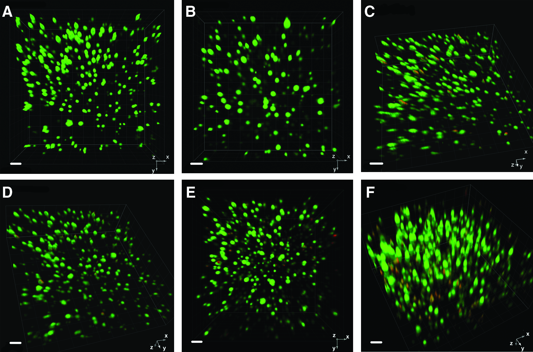
Three-dimensional projections of LIVE/DEAD images for agarose and IPN experimental groups. (
GAG content analysis
While normalized GAG content at 0 weeks was different between agarose and IPN gel groups (p < 0.05), the normalized GAG contents increased over time (p < 0.00005) and no significant differences were observed between the IPN and agarose groups at 3 weeks (Fig. 3). DNA content did not change significantly for agarose or for the IPN from 0 to 3 weeks.
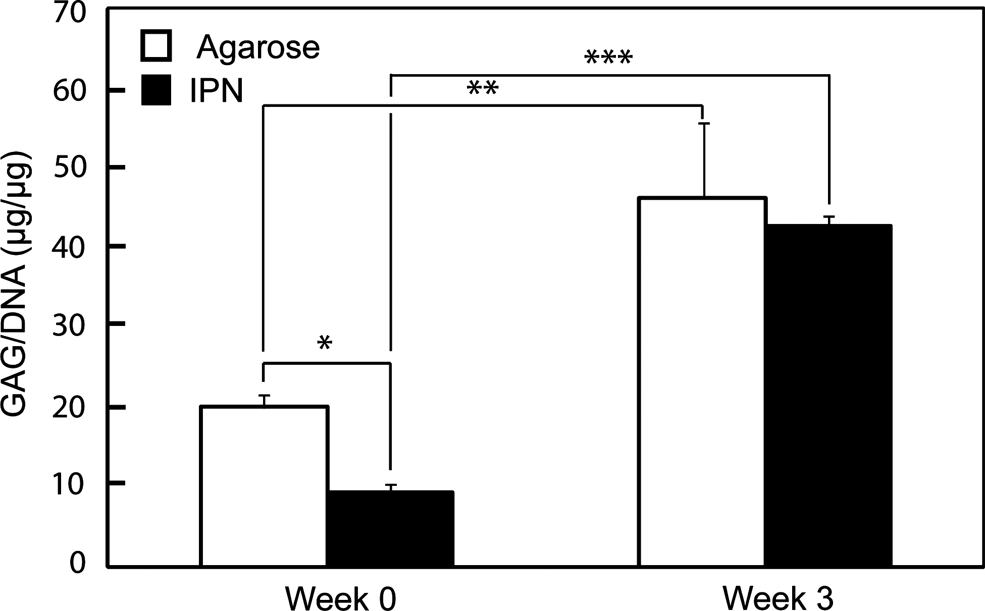
GAG content of agarose and IPN gels with encapsulated chondrocytes at 0 and 3 weeks, normalized to DNA content (mean ± standard deviation, n = 4). Note that normalized GAG content increased over time for the IPN gels, and that normalized GAG contents were not statistically different between the two groups after a 3-week period. Asterisks indicate statistically significant differences (*p < 0.05, **p < 5 × 10⁻⁵, and ***p < 5 × 10⁻⁶). GAG, glycosaminoglycan.
Mechanical analysis
Representative stress–strain curves for the various hydrogel groups are displayed in Figure 4. The initial slopes of these curves yield the Young's modulus, E. While mechanical failure of PEG and IPN gels occurred abruptly as they shattered during failure, agarose gels split gradually, and therefore compressive failure strain in agarose gels was difficult to determine precisely. Figure 5 displays the same trials as Figure 4 plotted according to the neo-Hookean model, and the slope of the curves in Figure 5 is equal to the gel shear stress, G. The curves were highly linear in neo-Hookean plots up to at least 50% strain (R2 > 0.99), and IPN gels displayed this linearity (demonstrating behavior as ideal elastomers) even in the presence of encapsulated cells. A full summary of mechanical property data is displayed in Table 2. Stress–strain curves increase exponentially after 50% strain as shown in Figure 4, and therefore the moderate standard deviation in failure strain causes much larger standard deviations in failure stress and gel toughness. Shear moduli for acellular IPN gels were 4.0 times higher than for pure PEG networks and 4.9 times higher than for pure agarose networks. These relationships are statistically significant and compared in Figure 6. As E ∼ 3G for incompressible materials such as hydrogels, similar increases in the Young's modulus, E, were observed (see Table 2). With regard to failure properties, IPNs were 2.3 times stronger and 3.3 times tougher than pure PEG gels, although differences were not statistically significant (Table 2). Commonly reported gel parameters calculated from measurements of G and Q such as mesh size and crosslink density are displayed in Table 3.12,49
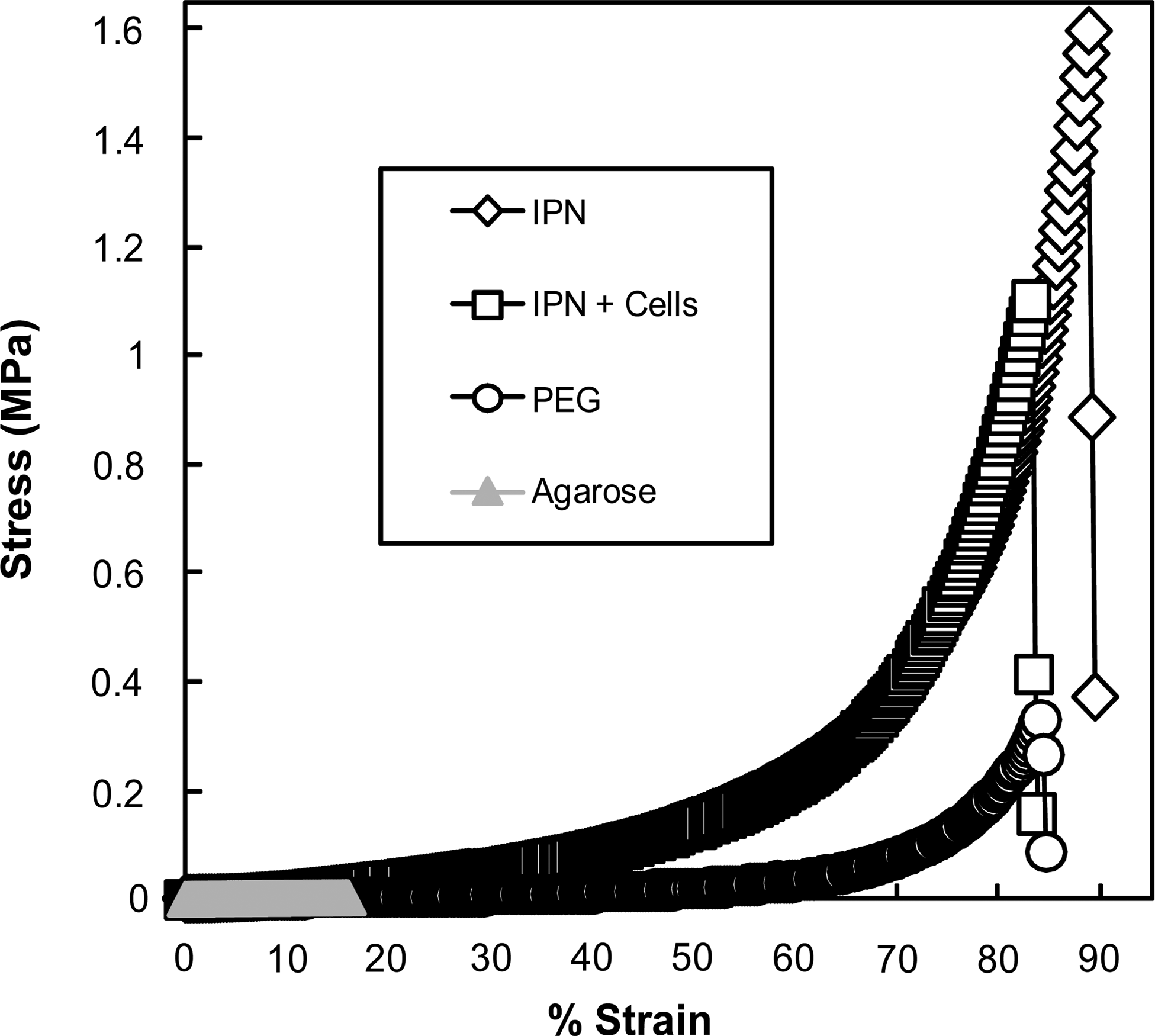
Representative stress versus strain curves. All groups have been truncated following failure. Note the extent of mechanical property enhancement in IPN hydrogels with and without encapsulated cells.

Representative neo-Hookean elasticity model plots of stress versus λ − 1/λ2 curves, where λ = L/L0, and the slope is equal to the shear modulus, G. Linearity up to moderate strains indicates ideal elastic behavior. These are the same samples shown in Figure 4. Left, 0%–57% strain; right, 0%–90% strain.
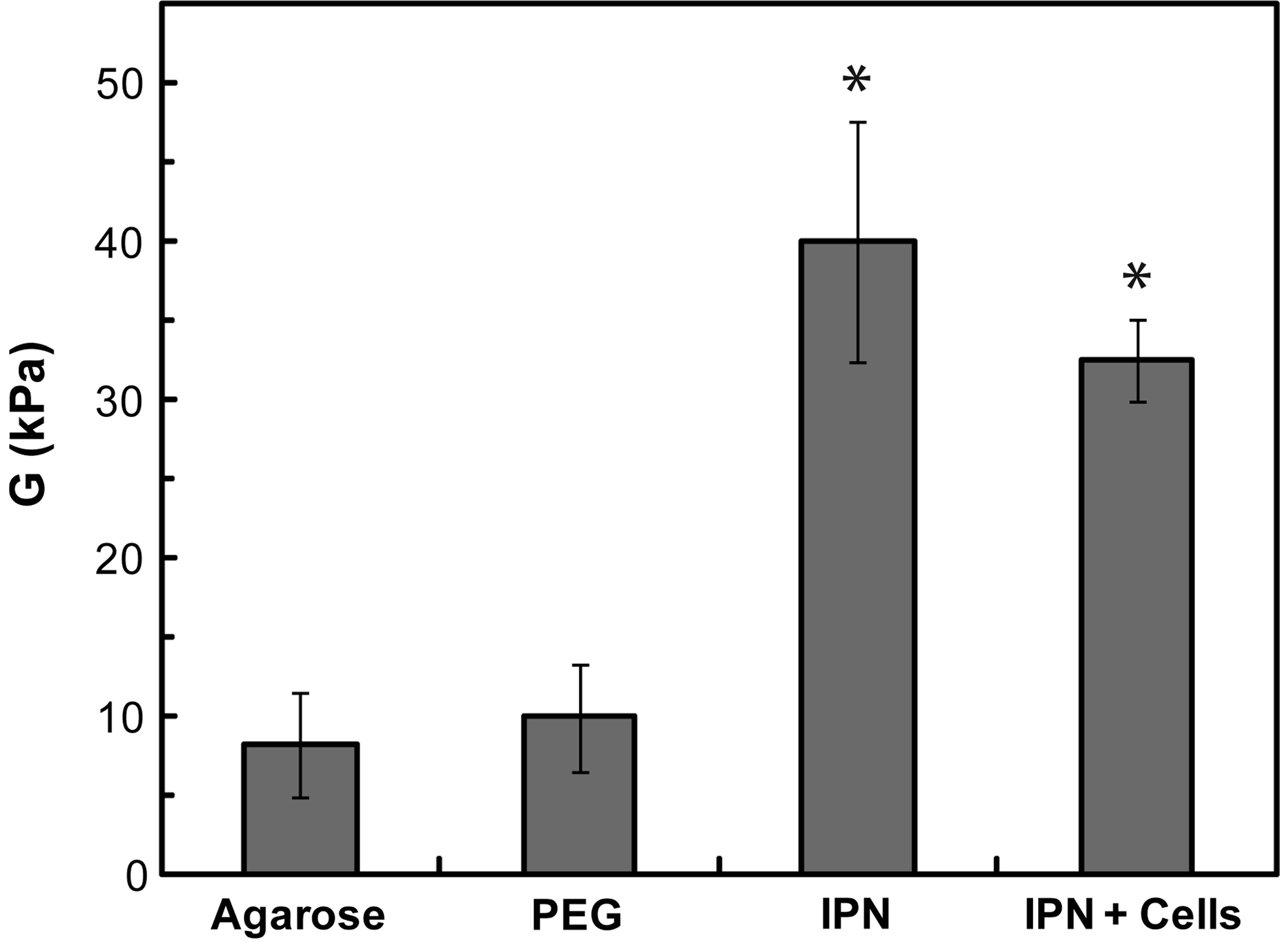
Comparison of the compressive shear moduli, G (mean ± standard deviation; n = 8 for PEG gels, n = 4 for all other groups). As G of IPN gels is so much greater than G of the pure components, a synergistic mechanism must exist between PEG and agarose networks. *Values statistically significant from agarose and PEG groups (p < 10−5). PEG, poly(ethylene glycol).
Agarose compressive failure was difficult to determine precisely, and approximate values have been presented for agarose failure.
IPN was statistically significant from agarose and PEG (p < 10−4).
IPN+cells was statistically significant from agarose and PEG (p < 10−4).
IPN was statistically significant from IPN+cells (p < 0.005).
No statistical significance among PEG, IPN, and IPN+cells groups.
E, Young's modulus; G, Shear modulus.
Crosslink density was calculated on a dry basis. Some calculations require Q and were not calculated for IPN+cells (no solids content data).
Mc, molecular weight between crosslinks; χ, polymer–solvent interaction parameter.
Discussion
By combining a brittle agarose network in low concentration with a more ductile PEG network at a higher concentration, an IPN hydrogel was formed with a fourfold improvement in shear modulus relative to the pure PEG gel, while maintaining the ability to encapsulate chondrocytes with a high degree of viability. As the IPN shear modulus is much greater than the additive sum of PEG and agarose shear moduli, it is clear that the two components act synergistically to enhance gel mechanical properties. This study demonstrated the potential of a rationally designed agarose-PEG IPN material as a high-strength cell carrier for tissue engineering applications.
Results of unconfined compressive mechanical testing corresponded well with theoretical correlations from the neo-Hookean elastic model, as linear fit R2 values up to 50% strain exceeded 0.998 for all PEG and IPN samples analyzed. Previous investigations with PEG and poly(ethylene oxide) have reported χ values of 0.47 under similar conditions,50,51 and E/G ratios from such compressive stress–strain tests have been previously reported to be ∼3.2 for hydrogels. 52 The good agreement of our results with polymer theory (linear neo-Hookean plots) and previous hydrogel studies (χ values and E/G ratios) suggested that gel properties were characterized accurately. The presence of encapsulated cells did not significantly compromise mechanical properties of IPN hydrogels. The small differences between acellular and cellular IPN constructs may have originated from a lower degree of PEG-DA incorporation caused by cells blocking light during the polymerization process and/or cells interfering with PEG-DA polymerization or the IPN microstructure through a yet-uncharacterized mechanism. Alternatively, at an encapsulated concentration of 10 million cells/mL, chondrocytes composed 1%–2% of hydrogel volume fraction and the mechanics of the cells themselves likely contributed to gel biomechanics. As single-cell relaxed moduli have been reported on the order of 1 kPa, 53 it is not surprising that the shear modulus was reduced with the inclusion of cells.
Agarose moduli are known to vary widely according to agarose type and thermal history, and agarose gels with elastic moduli from 25 to 40 kPa have been reported under similar experimental conditions. 54 PEG, IPN, and cellular IPN gels displayed distinct, often complete mechanical failure between 60% and 90% strain, while agarose gel failure occurred gradually and was indicated by a decrease in the slope of stress–strain curves concurrent with the appearance of macroscopic cracks between 10% and 20% strain. Mechanical failure is dependent upon both fundamental material properties and macro- and microscopic structural defects in particular samples. Previous IPN studies have reported similar deviations in failure properties within sample groups.16,55 Failure point determination in unconfined compressive testing can be difficult because a partially fractured gel still supports a compressive load, and therefore tensile mechanical data are usually preferred for analysis of failure properties. In spite of these drawbacks, compressive testing with characterization of failure strain is necessary for evaluation of potential materials for cartilage implantation. No radical differences in failure strain were observed between PEG and IPN groups, and we therefore conclude that the IPN represented a significant enhancement in total energy absorbed at failure by enhancing gel shear modulus (and therefore gel stiffness) while maintaining a comparable failure strain. The data collected in the current study support this conclusion, but the increases in average gel toughness reported were not statistically significant because small variations in failure strain cause much larger variations in gel toughness (energy to failure). Future studies by our group will specifically investigate failure properties of IPN gels compared to individual component networks, with an emphasis on fracture propagation and other failure tests such as notch or tear testing.
The exact mechanism of synergistic gel strengthening through IPN formation by the DN method remains unclear, yet the magnitude of the effect and its potential practical importance is such that this is under active investigation by several groups.20,22,56–62 It has been stated that the first network should be a polyelectrolyte for maximum property enhancement, though a significant effect has been seen even when both networks were nonionic, as in this study.19,21,22 Other theories maintained that increased toughness in DN-type IPN gels was the result of the ductile polymer's inhibition of crack propagation in incipient brittle failure of the high modulus network. 57 However, the exact mechanism of crack propagation inhibition in hydrogels is largely unknown. While the origin of the property enhancement in DN gels has yet to be fully elucidated, and only a subset of important properties have been measured, some empirical rules for selecting polymer combinations have been established and the impact upon mechanical properties is unmistakable.18,21 This study applied DN concepts outlined in previous investigations, including use of polymerizable monomers, in combination with the selection of synthetic bio- and cytocompatible polymers to rationally design a new high-strength hydrogel for tissue engineering applications, and data from the current study reinforced the empirical trends observed in earlier work.
The observed increases in GAG content per DNA of IPN gels demonstrated the capacity of IPN-encapsulated cells to synthesize extracellular matrix and exhibit productivity comparable to agarose-encapsulated cells. The limited cell death observed in the current study may have been caused by impurities in the PEG-DA reagent, which was used as received though not certified for cell culture applications. Elisseeff and coworkers have encapsulated chondrocytes in photopolymerized poly(ethylene dimethacrylate) semi-IPNs, and biochemical assays showed that proteoglycan and collagen contents increased over time, while mechanical testing revealed increases in equilibrium moduli and dynamic stiffness. 23 Numerous prior investigations have demonstrated the potential of encapsulated cells to enhance mechanical integrity in both agarose and PEG networks, and our results suggest that cells encapsulated in an agarose-PEG IPN may be able to exhibit similar productivity over time. Agarose has been shown to degrade hydrolytically in cell culture 63 and recent studies have outlined methods to create biodegradable PEG networks as well,64–67 so an agarose-PEG IPN implant can likely be optimized for in vivo degradation. Studies of various derivatives of PEG68–71 and agarose72,73 have demonstrated the biocompatibility of each, and we do not anticipate limiting complications with biocompatibility as we approach in vivo studies beyond this report. Finally, cell viability and chondrogenic differentiation have been enhanced through incorporation of specific adhesion peptides, and this approach may also be incorporated into a PEG-agarose IPN to yield a further improved cell scaffold.74,75
The current study outlined a technique for enhancing the mechanical properties of hydrogels for tissue engineering applications, and has yielded a gel with a notable combination of high mechanical strength, cytocompatibility, and good potential for biodegradability and biocompatibility. One limitation of this work is the use of 312 nm light, and future studies will move toward the use of 365 nm light. We also plan to investigate the mechanism of mechanical property improvement in agarose-PEG IPNs using combinations of tensile and dynamic mechanical testing and microscopic analysis. Additional studies that elucidate the nature of property enhancement will be of great benefit to tissue engineers and hydrogel scientists and contribute to this rapidly advancing field.
Conclusions
The relatively poor mechanical integrity of most synthetic hydrogels has limited their utility in cartilage tissue engineering applications with its mechanically demanding requirements. Therefore, enhancing hydrogel mechanical integrity is of the utmost clinical relevance. This study introduced a rationally designed agarose/PEG-DA IPN hydrogel that exhibited dramatically improved compressive moduli and shear moduli relative to its component networks. The gel synthesis methods were shown to be cytocompatible and cells encapsulated in the IPN exhibited GAG production comparable to that of agarose-encapsulated cells. Future studies will focus on methods to generate even higher viability and to increase biosynthesis. Mechanical property improvements were also observed in IPN gels with encapsulated chondrocytes. The end result was a hydrogel network with greatly enhanced mechanical integrity that was developed using DN concepts, yet designed specifically for cell encapsulation in tissue engineering applications. In addition to reporting the synthesis and characterization of a new hydrogel biomaterial, the method introduced in the current study may also provide a platform for the development of similar hierarchically designed hydrogel cell scaffolds for tissue engineering applications in which scaffold mechanical integrity is of great importance. DN concepts suggest that even greater improvement may be possible. We are hopeful that this technology in the long term will provide a more effective treatment for cartilage damage to enhance patient care.
Footnotes
Acknowledgments
The present work was supported by the National Institutes of Health (1 R21 EB008783-01, 5 P20 RR 16475-08) and the National Science Foundation (IOS 0726425 and DMR 0805264). We are also grateful to Dr. David Moore and Heather Shinogle of the University of Kansas Microscopy and Analytical Imaging Laboratory for their assistance with epi-fluorescence microscopic techniques.
Disclosure Statement
The authors do not have a conflict of interest with any commercial entities.
References
Supplementary Material
Please find the following supplemental material available below.
For Open Access articles published under a Creative Commons License, all supplemental material carries the same license as the article it is associated with.
For non-Open Access articles published, all supplemental material carries a non-exclusive license, and permission requests for re-use of supplemental material or any part of supplemental material shall be sent directly to the copyright owner as specified in the copyright notice associated with the article.