




Abstract
Bottom-up approach is an appealing strategy to build complex three-dimensional (3D) viable tissues in vitro starting from microtissue precursors (μTP). In this work we biofabricated a thick dermal-like tissue by sequentially combining two steps: a μTPs production and assembly followed by tissue maturation in a purpose-built bioreactor. The μTPs were produced by first seeding bovine primary fibroblasts on gelatine microparticles and then cultivating them in stirring conditions until a thick layer of ~80 μm of de novo synthesized extracellular matrix uniformly covered the microparticle surface. The μTPs were then loaded into a cylindrical chamber (2 mm in depth and 35 mm in diameter) and let to maturate and assemble into a 3D viable biohybrid tissue under specific fluid flow conditions. Several combinations of perfusion and/or tangential fluid flow were applied and their effect on the tissue formation and maturation was assessed. Results show that structural composition and mechanical features of the final 3D bioengineered tissue are strongly affected by the hydrodynamic environment and demonstrate that by optimizing culture conditions a 3D viable tissue with properties similar to that of native derma could be produced.
Introduction
Materials and Methods
Process description
General concept
The whole process consists of two stages: an initial phase of μTP production and their subsequent culture and assembly, leading to the production of a large 3D tissue equivalent (Fig. 1). The second stage took place in a maturation chamber (Fig. 2) designed to allow the introduction of μTPs, and their subsequent molding, assembly, and culture under different flow conditions (tangential and/or perfusion flow).
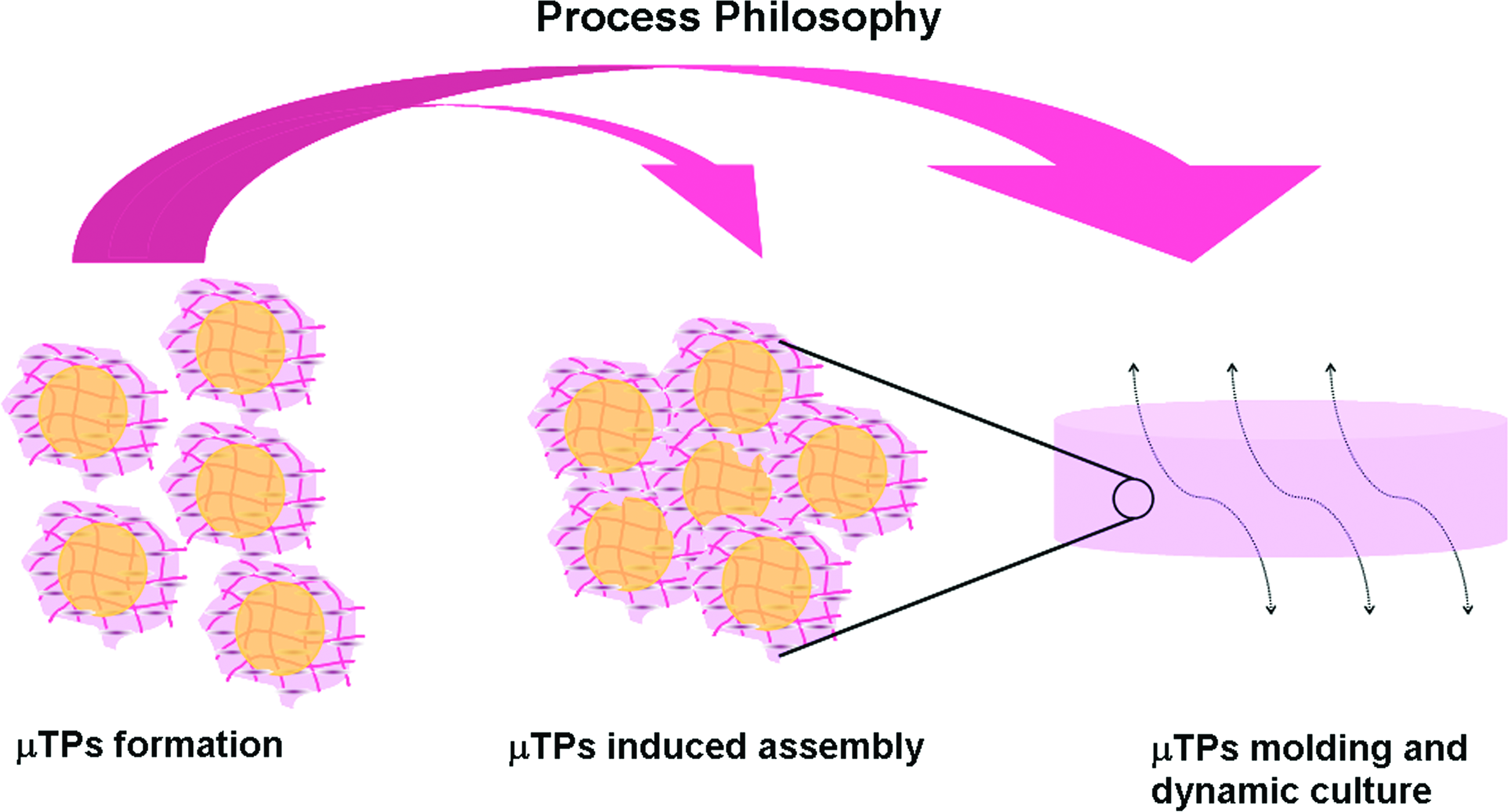
A schematic illustration of the whole process. Microtissue precursors (μTPs) were produced by means of dynamic cell seeding on macroporous gelatin microbeads. Then, their assembly is induced by confining them in proximity in a maturation chamber. The tissue maturation is then obtained under dynamic culture conditions, schematised by the arrows. Color images available online at

A picture of the bioreactor (center) and a diagram of bioreactor's cross sections (left and right) are shown. In particular, on the left are indicated the main components of the bioreactor: A, maturation space; B, meshes confining the maturation space that allow fluid and nutrient transport avoiding μTPs escape; C, spacer; D–E, endplates; F, screws; G, gaskets. Shown on the right is the bioreactor's cross-section; fluid gates and computational fluodynamic domain are highlighted: 1, perfusion flow inlet; 2–3, tangential flow inlets; 4, perfusion flow outlet; 5–7, tangential flow outlets; 6, gate to allow μTP entry in the maturation space; b, biohybrid domain; f, fluid domain. Color images available online at
μTP formation
Primary bovine dermal fibroblasts (BF-AG10385; Coriell) were propagated in monolayer culture in 150 mm Petri dishes at 37°C in 5% CO2 until a cell population density of 4.89 × 103 cm2 was reached. The culture medium (modified Eagle's medium high glucose concentration [4.5 g/L]) containing 20% heat-inactivated fetal bovine serum, 100 μg/mL L-glutamine, and 100 U/mL penicillin/streptomycin was changed every 2 days. Dry microcarriers (Percell Biolytica AB) were rehydrated in calcium-free and magnesium-free phosphate-buffered saline (PBS) for a minimum of an hour at room temperature. Without removing the PBS, the microcarriers were sterilized by autoclaving (15 min, 121°C) and stored at 4°C until used. Before use, PBS was removed and replaced with the culture medium. Cells obtained from the monolayer culture were added to the 1 g/L microcarriers suspension in the culture medium. The mixture was transferred into a 500 mL spinner flask (Integra Bioscience) and then incubated for 4 days at 37°C in 5% CO2. Microcarrier seeding was carried out at a cell concentration of 105 cell/mL in 250 mL of the culture medium using an intermittent stirring regime (30 min at 0 rpm followed by 5 min at 30 rpm) for 6 h. After seeding, the stirring speed was kept constant at 30 rpm and 50 μg/mL of ascorbic acid was added every day in the culture medium.
Internal geometry of the bioreactor vessel
As depicted in Figure 2 the bioreactor includes chamber A, created by placing two thin and rigid meshes B on the opposite sides of a spacer ring C. The void space of the spacer ring A is fashioned in desired shape of the final tissue (internal diameter [ID] = 35 mm, h = 2 mm). The space A, delimited by the surfaces of the meshes and by the inner surface of the spacer, forms the maturation space. The perforated meshes B used in this work have been chosen so that the opening size, 18 μm, is such that the microcarriers are retained within the chamber, while allowing the free circulation of the culture medium. The perforated meshes B are pressed against the spacer C by means of two opposite endplates D–E fastened to each other by means of screws F. Fluids are prevented to escape from the chamber and contaminants prevented from entering the culture environment, by means of gaskets located inside suitable seats G. The endplates D and E are recessed so as to provide a space for liquid flow above and below the chamber A. Moreover, they are provided with inlet 1 and outlet 4 ports (Fig. 2), which allow the introduction of a perfusing fluid into the space on one side of the chamber and its removal from the space on the opposite side. When port 1 is used as the medium inlet and port 4 as the medium outlet (Fig. 2), the perfusing fluid is forced through the mesh adjacent to port 1 (F, Fig. 2), then into the maturation chamber A, where it comes into contact with the tissue elements present within, and finally through the mesh delimiting the chamber on the opposite side, thus leaving the bioreactor through the outlet port 4 fitted to endplate D. This culture configuration is termed perfusion flow mode. The endplates (D–E, Fig. 2) are also fitted with inlet ports 2 and 3 and outlet ports 5 and 7 (Fig. 2), which allow the introduction of a culturing fluid into the respective cavities located within the thickness of the endplates, and the removal of the same fluid from the same cavity, without being forced through the perforated meshes and the maturation chamber (A, Fig. 2) delimited by them. This culture configuration is termed tangential-flow mode. An additional inlet port (6, Fig. 2) is fitted to the spacer ring (C, Fig. 2) and allows the introduction of μTPs into the maturation chamber A. The construction material of the meshes, the spacer, and the endplates is stainless steel AISI 316L (DIN 1.4404). All gaskets are made of pharmaceutical-grade silicone rubber.
Flow circuit, bioreactor loading, and operations
Figure 3 shows the bioreactor flow circuit, including the perfusion (P) and the tangential (T) loops as well as the μTPs loading line L. The μTPs produced during the seeding phase of the process, consisting of fibroblast-colonized gelatin microcarriers suspended in the growth medium at a concentration of 0.2 mg/mL, are introduced into the perfusion chamber through inlet port 6 (Fig. 2) by pressurizing the spinner flask S (Fig. 3), where the first stage had taken place. The spinner is pressurized at 0.2 barg with compressed air supplied through valve V (Fig. 3) and the μTPs suspension is transferred from the spinner flask to the maturation chamber (A, Fig. 2) via inlet port 6 (Fig. 2) through line L (Fig. 3). During the loading phase, fluid lines P1, P2, T1, and T2 (Fig. 3) of the flow system circuit are kept closed, while lines T3 and T4 (Fig. 3) are kept open to allow the escape of the suspending fluid through line R (Fig. 3), whereas the microcarriers are retained within the maturation chamber (A, Fig. 2). The maturation phase can be realized by operating the bioreactor under either perfusion flow or tangential flow conditions. Perfusion flow is performed by pumping a culture medium from a reservoir into port 1 (Fig. 2) while keeping all other ports closed, with the exception of port 4 (Fig. 2), through which the medium coming from the maturation chamber can leave the bioreactor. By switching from the perfusion flow mode to the tangential-flow mode using inlet lines T1 and T2 and outlet lines T3 and T4 (Fig. 3), the culture medium essentially moves along the meshes (B, Fig. 2) rather than across them. Circulation of the culture medium through the bioreactor during the perfusion flow as well as during the tangential-flow mode is obtained by means of a peristaltic pump, taking a fresh medium from reservoir. The medium emerging from the bioreactor then returns to the medium reservoir, thus closing the perfusion loop.
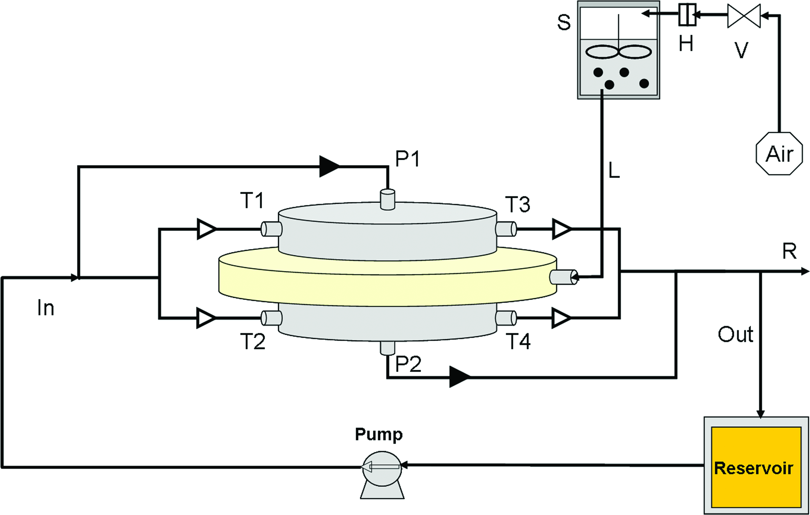
Drawing of the bioreactor and fluid flow loop. T1–T4, tangential loop; P1–P2, perfusion loop; L, μTP loading lines by means of μTP are transferred from spinner to maturation chamber; R, drain line; S, spinner flask in which the formation of μTP takes place; H, filter; V, valve to allow the pressurization of the spinner flask. Arrows indicate the main flow direction. Filled triangles indicate the direction of the perfusion flow. Empty triangles indicate the direction of the tangential flow. Color images available online at
3D dermal equivalent assembly, culture, and characterization
Culture conditions
At the end of the seeding phase, μTPs were transferred into the maturation chamber. To explore the effect of culture time and fluid dynamic conditions on construct maturation, two flow regimes (CF and MF) were tested over a period of 8 weeks. The volume of the medium reservoir was 200 mL. Both the bioreactor and the medium reservoir were placed in a cell culture incubator at 37°C, with an internal gas composition of 5% CO2 in air. The dissolved gases in the medium reservoir were equilibrated with the controlled atmosphere of the incubator by gas exchange through a 0.4 μm filter assisted by vigorous stirring to avoid gradient formation and settling of solids. Every 3 days the culture medium was entirely replaced and ascorbic acid added at a final concentration of 50 μg/mL. The medium flow rate (Q) was set at 0.5 mL/min for the perfusion flow mode and at 1 mL/min for the tangential flow mode. At the end of the incubation period the flow was stopped, the bioreactor dismantled, and the disk-shaped tissue samples (biohybrids) contained in the maturation chamber were carefully removed by means of a spatula, to be characterized further.
Biochemical analyses
Biochemical analyses were performed both on the perfusion medium and on the biohybrids directly. To evaluate the amount of glycosaminoglycan (GAG) and collagen released into the perfusion medium, aliquots were collected from the medium reservoir after 8 weeks of culture and under both CF and MF. For GAG analysis on the perfusion medium 1 mL aliquots were centrifuged, the supernatant was removed and the pellet was digested with 1 mL of papain digestion buffer (125 μg/mL of papain in 150 mM sodium chloride, 55 mM sodium citrate, 5 mM L-cysteine, and 5 mM ethylenediaminetetraacetic acid) for 16 h at 60°C. Total soluble collagen was quantified with the Sircol collagen assay kit (Biocolor Ltd.) according to the manufacturer's instructions. Accordingly, the culture medium was first reacted with Sircol dye reagent, containing Sirius red, which specifically reacts with the basic side-chain groups of collagens in a mechanical shaker for 30 min. After centrifugation for 10 min at 12,000 g, the unbound dye was removed, and the collagen-bound dye was recovered in the alkali reagent. Absorbance was measured at 540 nm using a microplate reader (Wallac Victor 3TM; Perkin Elmer). To evaluate the GAG deposition in the biohybrid, samples were weighed, lyophilized, and then digested in 1 mL/plug of papain digestion buffer for 16 h at 60°C. The content of sulfated GAG (either from the perfusion medium or from the biohybrid) was assessed by reaction of 40 μL digested portions with 250 μL of dimethyl methylene blue dye solution in 96-well microplates. Absorbance at 525 nm was read using the microplate reader; chondroitin sulfate (0–5 mg from shark cartilage; Sigma) was used as a standard. At each time point (1 and 8 weeks) samples from three biohybrids obtained from independent experiments were analyzed (n = 3).
Mechanical analyses (rheological study)
Three samples of 15 mm in diameters were punched from each of the biohybrids obtained. All samples were then tested in oscillatory shear in the 0.01–10 Hz frequency range at 37°C using a rotational rheometer (Gemini, Bohlin Instruments). Each sample underwent an amplitude sweep test before oscillation to check the linear range. An oscillation amplitude of 0.1% was selected since this value falls within the linear range for all the samples tested. The samples were clamped between two parallel plates with a compressive preload of ~10 g. As control the same test was performed on naked microbeads as well as on native bovine reticular dermis. In particular, a portion of the ventral section of bovine hide (24-month-old bovine) was collected from a local slaughterhouse. Samples where thoroughly rinsed in physiological solution and manually unhaired and defleshed before testing. The rheometer gauges were equipped with a chamber with humidity and temperature control to maintain the sample at 37°C and 95% relative humidity (RH). The mean value of the elastic modulus (G′) of the three samples was taken as the G′ value of the biohybrid which the samples originated. At each time point three biohybrids from independent experiments were tested (n = 3).
Histology
Samples of biohybrids were subjected to hematoxylin/eosin staining. First they were fixed in 10% formaldehyde for 24 h and then they were dehydrated by successive transfers into ethanol solutions of increasing strength (70%, 80%, 90%, and 100%), each step lasting for 10 min at room temperature, and finally they were embedded in paraffin. Five micrometer sections were cut with a microtome and stained with hematoxylin for 10 min, followed by eosin for 20 min, at room temperature. The coverslips were mounted and morphological features of constructs were observed with light microscope (Olympus CK 40).
Scanning electron microscopy
Samples of biohybrid were fixed with 2.5% (v/v) glutaraldehyde. Fixation was initiated at room temperature and was then followed by storage in the fixative solution for 3 days at 4°C. Samples were washed twice in 100 mM cacodylate buffer, pH 7.2, for 10 min at room temperature. A second fixation in 1% (w/v) osmium tetroxide, buffered in 100 mM cacodylate, pH 7.2, was done overnight at 4°C. Dehydration was carried out by gradually decreasing the water concentration and increasing the ethanol concentration (10%, 30%, 50%, 70%, 90%, 100%, and 100% again, each step 10 min at room temperature). Samples were then treated with liquid carbon dioxide using a Critical Point Dryer (Emitech K850). Dried samples were mounted onto metal stubs using double-sided adhesive tape and then gold-coated using a sputter coater at 15 mA for 20 min. Coated samples were then examined by scanning electron microscopy (SEM) (Leica S400).
Permeability measurements
The measurements of the hydraulic permeability (kb, [m2]) were performed by carrying out a permeation test on microbeads bed and by applying Darcy's law:
where Q [m3 s−1] is the volumetric flow rate, μ [Pa × s] is the dynamic viscosity of the mixture fluid–microbeads, A [m2] is the cross-sectional area, ΔP [Pa] is the pressure drop, H [m] is the thickness of the microbeads bed. A polycarbonate tube having 0.5 cm inner diameter was filled with microbeads reaching a final thickness (H) of 5 cm. By means of a peristaltic pump different cross flow rates ranging from 0.1 to 1 mL min−1 were then applied. At each flow rate the steady-state pressure drop between the inlet and the outlet of the polycarbonate tube was measured and the permeability kb of the system was obtained by linear regression of Equation 1.
CFD simulation within the bioreactor
Model equation
The fluid flow regime in the bioreactor was evaluated by means of CFD analysis. The system was divided into three different domains as shown in Figure 2. f is the domain where the fluid flows freely in the bioreactor chambers, and b is the domain where the fluid flows thorough the porous microbeads bed. The equation used to describe the fluid dynamics within domain f is the steady-state Navier-Stokes equation:
where μf is the dynamic viscosity, vf is the fluid velocity, and P is the hydrostatic pressure (the superscript f refers to the fluid). Within domains b the Brinkman equation has been used to describe the flow through the porous medium:
The superscript b refers to the fluid in the microbeads bed, kb is the hydraulic permeability, μb is the viscosity of the fluid within microbeads bed or the effective viscosity, and Pb the pressure.
Boundary conditions
The boundary conditions have been set to reflect the two flow configurations. Under perfusion conditions the fluid flows from the inlet port 4 to the outlet port 1 (Fig. 2) by keeping the other gate close. Fully developed Poiseuille velocity profile was specified at inlet, reference pressure has been considered at outlet 4, no slip condition was adopted at the fluid/bioreactor wall, and continuity of velocity and pressure was imposed at the Navier-Stokes/Brinkman interfaces ( f/b). Under tangential flow the boundary conditions were similar to the perfusion flow, but in this case the fluid flows from the inlet 2 and 3 to the outlets 5 and 7.
Perfusion flow
Tangential flow
Perfusion flow and tangential flow
where Q is the flow rate imposed by the peristaltic pump and A is the cross area at the inlets and outlets. The simulation was performed using commercially available software COMSOL MULTIPHYSICS, by means of Momentum Transport-Steady State/Navier-Stokes/Brinkman application mode.
Results
3D tissue equivalent assembly, growth, and characterization
Bioreactor loading
The μTPs (Fig. 4A) produced in spinner culture consisted of gelatine microbeads surrounded by cells and a thin layer of tissue about 80 μm thick. 6 After the μTPs' development phase the volume of the culture medium in the spinner flask was increased to 1000 mL to decrease the concentration of μTPs from 1 to 0.2 mg/mL. The spinner flask was then pressurized at 0.2 barg to transfer over a period of 30 min an aliquot of 800 mL of suspension into the maturation space, equivalent to 160 mg of μTPs. μTP concentrations >0.2 mg/mL resulted in the obstruction of the loading line, whereas injection pressures >0.2 barg induced a deformation of the microbeads inside the maturation chamber. By operating under the optimized loading conditions the fluid transfer spontaneously stopped when about 160 mg of μTPs had filled the maturation chamber. This procedure resulted in the production of a disc-shaped compact and homogeneous biohybrid cylindrical shaped (Fig. 4B, C).

μTP and biohybrid.
Mechanical properties
Rheological studies on the constructs were performed at each time point (after 1 and 8 weeks of maturation) and at different flow conditions. Reported in Figure 5 are the elastic moduli of the construct cultured for 1 and 8 weeks under the two flow conditions, CF and MF. As shown in Figure 6 the elastic modulus of the constructs slightly increases with the frequency. No significant variation with culture time was observed for the construct cultured under CF conditions as shown by the values of the elastic modulus at the frequency of 1 Hz of samples 1 and 8 weeks, that is, 2.8 × 10³ ± 617 and 3.2 × 10³ ± 594 Pa, respectively. These values are comparable with those of the construct cultured for 1 week under MF conditions, 2.9 × 10³ ± 532 Pa. In contrast, the elastic modulus of the construct cultured under MF conditions for 8 weeks was 4.6 × 10³ ± 630 Pa close to the typical elastic modulus of native bovine reticular dermis, that is, 5.6 × 10³ ± 1364 Pa. Mechanical tests performed on the naked microbeads did not detect any mechanical property.
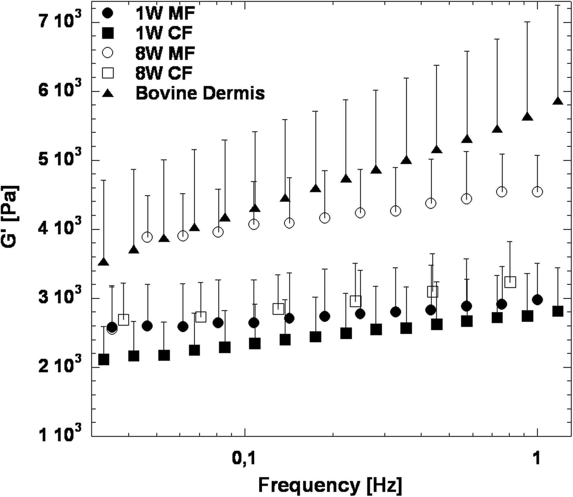
Mechanical properties. Biohybrid cultured under continuous perfusion flow (CF) 1 week (full square), CF 8 weeks (empty square), mixed flow (MF) 1 week (full circle), and MF 8 weeks (empty circle). Native bovine derma (triangle). Data were presented as average ± standard deviation (n = 3).
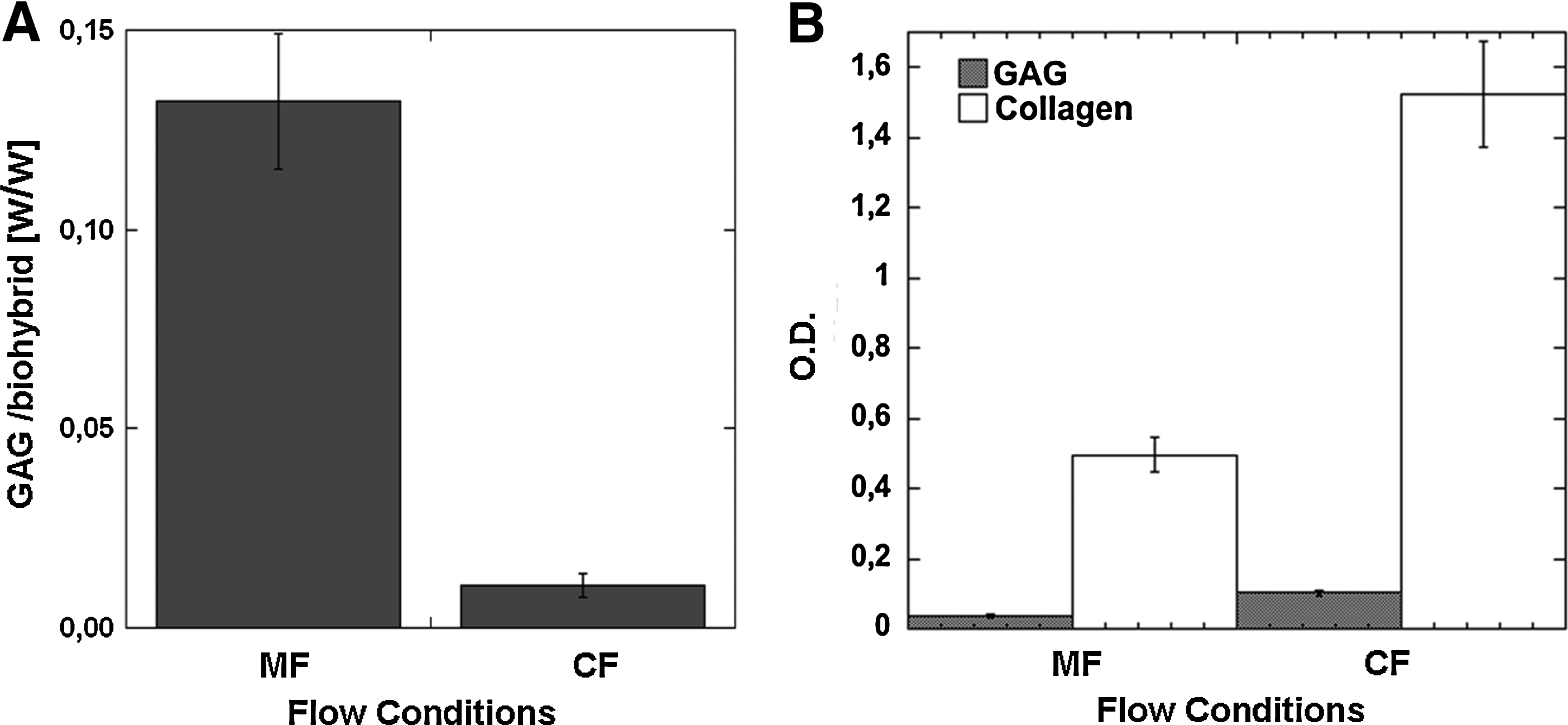
Biochemical analyses.
Biochemical analysis
Biochemical analyses have been performed both in the culture media and in biohybrids. In Figure 6A the GAG contents of the biohybrids at 1 and 8 weeks under the two flow conditions are reported. Under CF the GAG content (expressed as mg GAG/mg biohybrid) decreased from 0.025 ± 0.005 to 0.011 ± 0.003. The GAG content under MF increased from 0.027 ± 0.0015 to 0.132 ± 0.017. In Figure 6B data concerning the GAG and collagen released in the culture fluid at 8 weeks are shown. Clearly, the concentration of ECM components in the culture fluid under CF conditions is higher at both time points.
Tissue assembly
In Figure 7 SEM and histological images of a biohybrid cultured for 8 weeks under CF and MF conditions are shown. SEM images (Fig. 7A, D) show microbeads completely surrounded by cells and ECM forming large 3D tissue equivalents. In Figure 7E and F hematoxylin/eosin-stained histological sections derived from an 8-week construct kept under MF conditions are shown. ECM appears to be present both within and around the microbeads. In contrast, in a biohybrid cultured for 8 weeks under CF conditions ECM is present in much smaller amounts with respect to the MF condition.

Histological analysis.
CFD analysis
The values used for the simulations are listed in Table 1. The relationship between fluid viscosity μf and effective viscosity μb can be described as μb = μf/(1 − 2.5 × [1 − ɛ]), where ɛ is the porosity.21,22 Because the microspheres are highly porous the effective viscosity has been set equal to the medium viscosity. Figure 8 shows the results obtained by performing a numerical CFD simulation under tangential flow (Fig. 8A, C, E) and perfusion flow (Fig. 8B, D, F) conditions inside the bioreactor. Figure 8A and B shows the discretized domain of the maturation chamber as well as the boundary conditions used under tangential and perfusion flow, respectively. The flow rate was set at the value Qt = 0.5 mL/min (tangential flow) and Qp = 1 mL/min (perfusion flow), whereas the outlet pressure was Pref = Patm. Figure 8C and D contains the arrow plots of the fluid velocity field within the bioreactor operating under perfusion flow and tangential flow, respectively. Under tangential flow the velocity is mainly directed along the x-direction, whereas under perfusion flow the velocity is oriented along the z-direction (Fig. 8C, D). Figure 8E and F depicts the horizontal velocity (vx) along the biohybrid thickness (z) and the vertical velocity (vz) along the biohybrid diameter (x), respectively. Under both perfusion and tangential flow conditions a plug-flow regime is achieved. The maximum value of the velocity reached under perfusion flow mode is of the order of 10−4 ms−1 that decreases to 10−10 ms−1 under tangential flow mode.
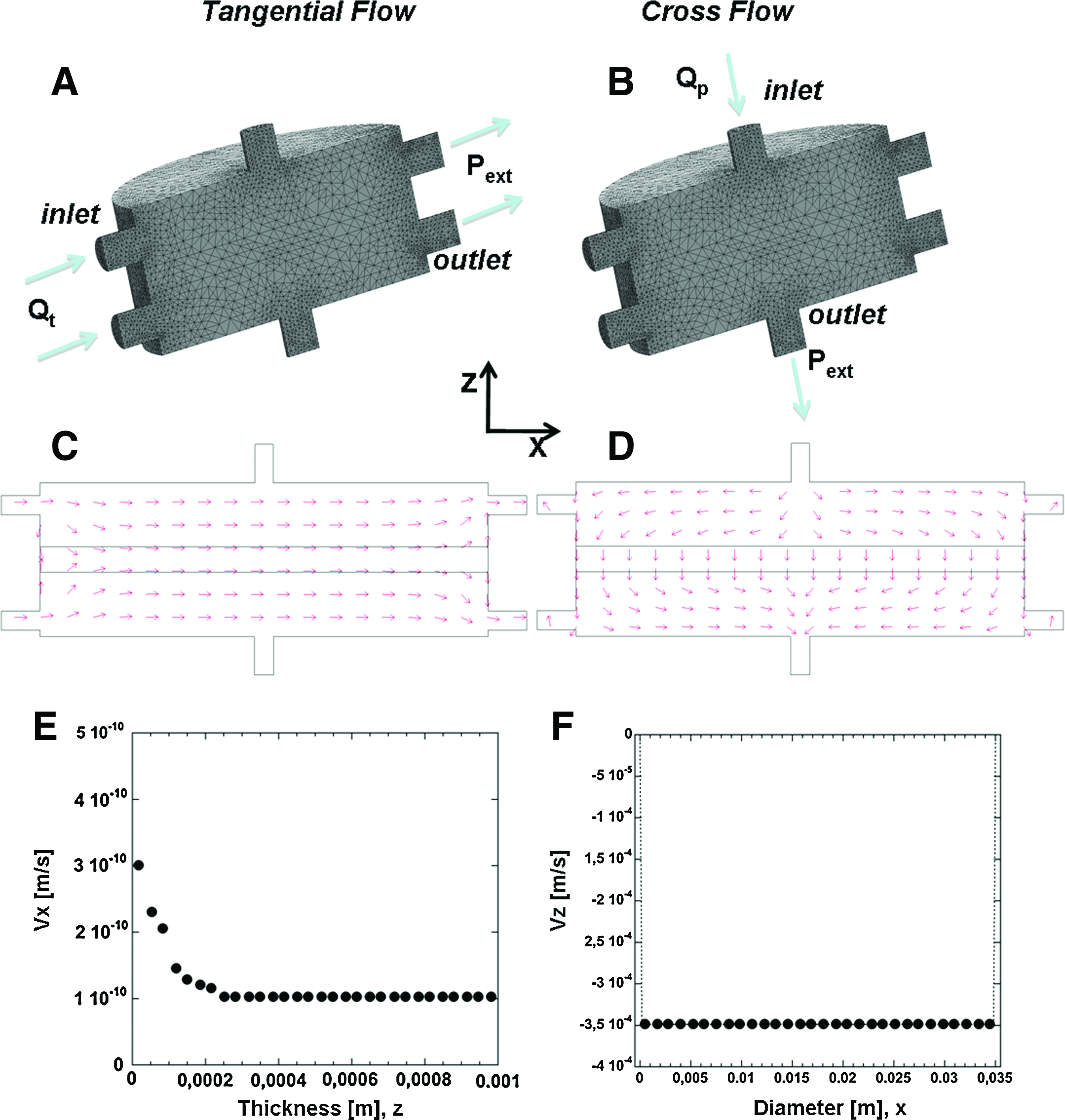
Computational fluodynamic simulations.
Discussion
Bottom-up tissue engineering based on modular tissue assembly is a biomimetic alternative to traditional scaffold-based strategies, which offers many advantages for engineering whole-organ and large tissue grafts and potentially transforms the conventional cell seeding/porous scaffold paradigm of tissue engineering. 18 Some of the major challenges of this strategy are to generate functional tissue modules as well as to find the optimal techniques to create larger 3D engineered tissues from modular microscale units. Another major challenge is the integration of bottom-up techniques with more traditional top-down approaches1,2 to create more complex tissue by optimizing the advantages of each technique. To our knowledge the use of a perfusion bioreactor to achieve the assembly of microtissue units and the maturation of a 3D tissue equivalent is reported for the first time. In the bioreactor realized in this study μTPs can be injected, assembled in a defined shape and size, and cultured under several culture conditions (Figs. 2 and 3). The μTPs (though on a much smaller scale) resemble packed beds, a common and well-understood component in chemical engineering, 23 and their injection in the maturation space has been optimized to avoid high μTPs packing that could lead to contact inhibition hindering tissue biosynthesis. 24 However, the development of the final 3D tissue equivalent requires a proper process design.1,2 The bioreactor described in this study allowed the exploration of different combinations of fluid flow conditions. The two regimes investigated herein demonstrated that culture conditions can affect and modulate the mechanical properties and tissue assembly of the final 3D macrotissue. By using CF conditions a biohybrid with inferior mechanical properties has been obtained irrespective of maturation time as compared to MF conditions. In fact, the elastic modulus of the biohybrid cultured for 8 weeks under MF conditions approached that typical of native derma. It is well known that the mechanical properties of engineered tissues are correlated with ECM deposition and organization.25,26 Our data confirmed the existence of a strong correlation between mechanical properties and ECM (GAG) neo-deposition within the construct. After 1 week of culture GAG accumulation within the biohybrid is similar for both flow conditions, whereas after 8 weeks the GAG content of the biohybrid cultured under MF conditions was almost 10-fold higher than that of the biohybrid cultured under CF conditions. The increased GAG content is an indication of major ECM deposition as confirmed by morphological analyses. Histological analysis shows the biohybrid structure to be composed of gelatine microbeads interconnected by abundant ECM. Further, a compact matrix is present in the biohybrid cultured for 8 weeks under MF conditions, which is absent from the biohybrid cultured under CF conditions, where matrix distribution is highly nonhomogeneous. The compact structure of the former biohybrid is confirmed by SEM observations, showing a surface completely coated by cells and ECM, whereas a fibrillar structure presumably made of collagen can be seen at higher magnifications. We can use these results to justify the superior mechanical properties of the biohybrid cultured under MF regime at 8 weeks. Moreover, according to previously reported surveys,27,28 we hypothesized that CF conditions could induce a “washing effect” of the neo-tissue, leading to an accumulation of ECM components in the culture medium. CFD analysis, utilized as tool to evaluate the influence of fluid-dynamic culture conditions on tissue assembly and deposition, seems to justify the hypothesis as under CF conditions the ratio between the thickness of the maturation space (2 mm) and the average fluid velocity (10−4 ms−1) gives an average residence time of fluid inside the biohybrid of about 20 s. This relatively low value could lead to a washing-out of the neo-synthesized ECM components. In fact, such residence time is shorter than the average tissue assembling time, which ranges from 103 to 105 s. 28 The CFD results obtained under tangential flow conditions show that the fluid moves parallel to the surfaces of the biohybrid avoiding the drag-down of the neo-synthesized ECM components. In addition, the lower value of the fluid velocity (10−10 ms−1) and the longer fluid path (maturation space diameter, 35 mm) give an average residence time of 3.5 × 10⁸ s. In the light of these observations it is possible to conclude that perfusion flow is necessary to assure a rapid nutrient supply as well as waste removal from the maturation chamber, but continuous perfusion does not allow a correct tissue assembly due to the high drag velocity acting on ECM-free components. On the other hand, under tangential flow the interstitial velocity is reduced, and therefore the ECM components can more easily assemble in the construct, but the lower velocities drastically reduce nutrient supply. By combining the two flow regimes, a satisfactory balance between ECM assembly and nutrient mass transfer can be achieved. In this study we have focused the attention on tissue remodeling under different process condition. In a similar fashion the role of the biomaterial on 3D tissue development should be investigated. The structural characterization of the biohybrid shows that gelatine microbeads persist after 8 weeks of culture. It is fair to say that the commercial gelatine microbeads used herein are characterized by a very low degradation rate. 29 To obtain a tissue construct completely made up of neo-synthesized tissue, a balance should be created between the degradation rate of the microbeads and the deposition of new ECM. 30 Moreover, we claim that the approach proposed herein could be used to produce human dermal equivalent tissue and in the light of this consideration the fabrication of μTPs by using human dermal fibroblast are currently under investigation.
Conclusion
In this work a combined strategy to produce large 3D dermal tissue equivalent is presented. The μTPs' biosintering property is exploited by inducing their biological fusion inside a maturation chamber, demonstrating that μTPs can be injected and molded to produce a tissue construct of desired shape and size. It has been found that the properties of the final tissue-equivalent are strongly affected by culture conditions. Future studies should explore different culture condition as well as the possibility of tuning the microcarriers' degradation rate. It is reasonable to expect that by using different cell types in combination with suitable engineered microcarriers having different morphology and shape, complex and heterotypic tissues with specific 3D architecture can be realized by means of the proposed approach.
Footnotes
Acknowledgments
The authors are grateful for financial support provided by the EU community, DERMAGENESIS U.E. COLL-CT2003-500224, and by Italian Public Instruction Ministry, Tissuenet n. RBPRO5RSM2. Moreover, the authors acknowledge Dr. Maurizio Ventre for his useful contribution for mechanical analysis and discussion of the results.
Disclosure Statement
No competing financial interests exist.