




Abstract
Human bone marrow mesenchymal stromal cells (MSCs) are considered a potential cell source for MSC-based bone regeneration, but improvements in the proliferation and differentiation capacity of MSCs are necessary for practical applications. Estrogen effectively improves MSC capabilities and has strong potential as a regulator of MSCs. The aim of this study was to develop a delivery system that provides intracellular release of estrogen and test its ability to improve osteogenic differentiation of MSCs. Biodegradable poly (lactic-co-glycolic acid) (PLGA) microparticles were developed that entrap 17-β estradiol (E2) and provide intracellular release of E2. The results show that we can prepare PLGA particles with efficient loading of E2 and maintain release of E2 up to 7 days. Surface modifying E2-loaded PLGA particles with cationic polyamidoamine dendrimers enabled increased uptake by human MSCs. Human MSC uptake of the E2-loaded PLGA particles significantly upregulates osteogenic differentiation markers of alkaline phosphatase and osteocalcin. In conclusion, cationic-modified PLGA particles can serve as a tool for intracellular delivery of estrogen to effectively execute estrogen regulation of MSCs. This approach has the potential to improve the osteogenic capabilities of MSCs and to develop appropriate environments of implantation for MSC-based bone tissue engineering.
Introduction
Estrogen, a multifunctional sex steroid substantially participates in the regulation of bone metabolism by inhibiting bone resorption and increasing bone formation. Its powerful capacity to regulate stem cells and bone marrow MSC proliferation and differentiation has been recently described.13–16 Estrogen effectively regulates the stemness characteristics of adult and embryonic stem cells.13,14 Supplements of estrogen increase human MSC proliferation and prevent MSCs senescence.15,16 Estrogen exerts an osteogenic function in bone formation via release or upregulation of a number of cytokines (interleukin-1 and 6), prostaglandin, and osteogenic growth factors in human MSCs (bone morphogenic proteins, transforming growth factor-beta1, and insulin-like growth factor).17–20 These cytokines, hormones, and growth factors further promote proliferation and differentiation of osteo-progenitor cells and MSCs through autocrine or paracrine mechanisms. Accordingly, estrogen may serve as an effective regulator to improve MSC capability of engineered grafts and recruit osteo-progenitor cells from implantation environments to accelerate bone regeneration. As estrogen executes the regulation on bone marrow MSCs primarily through nuclear receptors, 15 intracellular release of estrogen would be an appropriate approach to localize the estrogen effects and improve efficiency of estrogen regulation.
Biodegradable poly (lactic-co-glycolic acid) (PLGA) microparticles have been developed to deliver multiple growth factors and steroids, including estrogen, due to its safety and biodegradability.21,22 However, unmodified PLGA microparticles also display limited uptake in nonphagocytic cells. We have recently developed PLGA microparticles that are surface functionalized with cationic polyamidoamine (PAMAM) dendrimers. The modification of the PLGA particles results in a net surface positive charge that facilitates uptake in cells. 23 In this study, we develop a delivery system that provides intracellular release of 17-β estradiol (E2) for MSC regulations. The E2-loaded PLGA microparticles were surface modified with PAMAM dendrimers to facilitate uptake into the cells. The E2-loaded PLGA particles can provide sustained release of E2 for at least 1 week. After they are taken up by human MSCs, intracellular release of E2 from PLGA particles can effectively improve osteogenic differentiation of MSCs.
Materials and Methods
Poly (lactide-co-glycolide) (PLGA) with a carboxylic terminal (85:15, viscosity: 0.6 dL/g) was purchased commercially (Durect Corporation). E2, poly-vinyl alcohol (MW 30–70 kDa), and solvents were purchased from Sigma. All the solvents, including ethyl acetate and dichloromethane (DCM) and acetonitrile, were of high performance liquid chromatography (HPLC) analytical grade. Fresh human bone marrow was purchased from Allcells. All culture media and supplements were provided by Invitrogen.
Preparation of E2-loaded PLGA microparticles with a net positive surface charge
A modified emulsion-solvent evaporation method was used to prepare E2-loaded PLGA microparticles. In brief, 200 mg of PLGA and 10 mg of E2 was dissolved in a mixture of 2 mL ethyl acetate and 3 mL DCM. The mixture was then emulsified into 50 mL of 1% poly-vinyl alcohol solution and homogenized at 9500 rpm for 1 min. The mixture was continuously stirred at 40°C using a magnetic stirrer until complete removal of the organic solvents. The microparticles were collected by centrifugation, washed twice with deionized water, and lyophilized overnight. The size and zeta potential of particles were analyzed using dynamic laser light scattering (Zetasizer Nano ZS; Malvern Instruments). Briefly, the microparticles were suspended in deionized water at a concentration of 1 mg/mL. The size measurements were performed at 25°C at a 173° scattering angle. The mean hydrodynamic diameter was determined by cumulative analysis. The zetapotential determinations were based on electrophoretic mobility of the microparticles in the aqueous medium, which were performed using folded capillary cells in automatic mode. All the measurements were carried out in triplicate. To modify the surface of particles with cationic PAMAM dendrimers, we used 1-Ethyl-3-[3-dimethylaminopropyl] carbodiimide hydrochloride (EDC)- N-hydroxy-succinimide (NHS) chemistry to conjugate the cationic polymer on the surface as described previously. 23 Briefly, 20-fold molar excess of EDC and N-hydroxysulfo-succinimide (Sulfo-NHS) over PLGA was dissolved in 1 mL MES buffer each. They were slowly added to 3 mL MES buffer (pH 5.5) suspending 100 mg E2-loaded PLGA particles for 2 h. Un-reacted EDC-NHS was removed by washing twice with phosphate-buffered saline (PBS). The particles suspended in 3 mL PBS were added drop-wise into 2 mL PAMAM solution (generation 3) (Sigma-Aldrich) at a10-fold molar excess under constant stirring. The reaction was kept at room temperature for 3 h to render an overall positive charge to the E2-loaded PLGA particles. Zeta potential measurements were used to confirm PAMAM conjugation. The amount of un-entrapped E2 in the supernatant and washing buffers during microparticle fabrication were measured to analyze the entrapment efficiency of E2.
The surface morphology and shape of the cationic-modified PLGA microparticles was determined using scanning electron microscopy (SEM; Hitachi S-4000). Briefly, air-dried microparticles were placed on adhesive carbon tabs mounted on SEM specimen stubs. The specimen stubs were coated with ∼5 nm of gold by ion beam evaporation before examination in the SEM operated at 5 kV accelerating voltage.
Measurement of E2 using HPLC
E2 was quantified by a Waters 2690 separations module run by Empower Millennium Software. The UV spectrum of E2 in acetonitrile was recorded using a Spectramax spectrometer (Molecular Devices) and the λmax was found at 280 nm. The column for HPLC analysis was a Symmetry C18 column (5 μm, 150 × 3.9 mm I.D.). The mobile phase was a mixture of acetonitrile/water at 65:35 (v/v) at a flow rate of 1.0 mL/min. It was detected at 280 nm with a retention time of 4.2 min. To create a calibration curve, a stock solution of 500 μg/mL was prepared to generate the standard curve using HPLC. A standard solution of E2 at various concentrations was prepared by serial dilutions of the stock solution in the mobile phase as described above. The standard curve was generated from the linear relationship between peak area and the concentration of E2. The concentration of E2 was quantified by the peak area method from the calibration curve.
Influences of internal phase solvents on entrapment efficiency and in vitro release of E2
Table 1 summarizes the types and ratios of methanol or ethyl acetate used in combination with DCM to fabricate PLGA microparticles and to determine the effects of internal phase solvents on entrapment efficiency and in vitro release. After preparing E2-loaded PLGA microparticles with different solvents, we measured the entrapment efficiency of E2 and in vitro release of E2.
DCM, dichloromethane.
In vitro release of E2 from cationic PLGA particles
Five mg of PLGA particles loaded with ∼200 μg of E2 was suspended in 100 mL PBS and shaken at 100 rpm in a shaker bath at 37°C. At each time point, 1 mL of the sample was centrifuged to collect the supernatant. The concentration of E2 in the supernatant was analyzed using HPLC in triplicate.
The preparation of human bone marrow MSCs
Mononuclear cells of fresh human bone marrow were isolated using density gradient centrifugation (Histopaque-1.077; Sigma). The cells were cultured with Dulbecco's modified Eagle's medium (DMEM), 10% fetal bovine serum (Atlanta Biologicals), and 1% antibiotic-antimycotic (Sigma). On day 5, nonadherent cells were removed after medium exchange. The cells were collected when they reached 70%–80% confluence to test the uptake of E2-loaded PLGA particles and regulatory functions of intracellular release of E2.
Uptake of PLGA microparticles by human MSCs
Rhodamine-labeled PLGA microparticles were used to track the uptake of PLGA particles by human MSCs. To prepare rhodamine-labeled PLGA microparticles, 200 mg of PLGA and 200 μg of Rhodamine were dissolved in 2 mL of DCM, and Rhodamine-labeled cationic PLGA particles were prepared using the emulsion-solvent evaporation method as described above. To test uptake of particles in human MSCs, 104 MSCs were seeded on eight-well glass chamber slides overnight. About 75 nM of LysoTracker® Green DND-26 was used to track lysosomes according to the manufacturer's protocol (Invitrogen). After MSCs were treated with 200 μg of particles per well for 1 or 24 h, the cells were washed twice with PBS to remove suspended particles and fixed with 4% paraformaldehyde solution. PLGA particles that were ∼1.8 μm in size were selected for uptake studies on the basis of efficient uptake observed in alternative cell lines in previous studies. 23 After the cells were mounted with Vectashield containing 4′,6-diamidino-2-phenylindole (DAPI) to bind DNA and locate the nucleuses (Vector Laboratories Inc.), a confocal microscope (Zeiss-701) was used to assess the uptake of cationic-modified PLGA particles by human MSCs.
Osteogenic differentiation of human MSCs after uptake of E2-loaded PLGA microparticles
About 0.4 and 2 mg E2-loaded microparticles loaded with 17.4 and 87 μg of E2 respectively were suspended into DMEM and added to human MSCs cultured on a 100-mm Petri dish for 24 h. The MSCs were then washed twice with PBS to remove suspended microparticles. MSCs that had internalized particles were exposed to the osteogenic differentiation medium consisting of DMEM supplemented with 100 nM dexamethasone, 10 mM β-glycerophosphate, and 0.05 mM ascorbic acid-2-phosphate up to 3 weeks. The MSCs without treatment of particles served as controls. Osteogenic differentiation markers of MSCs, including alkaline phosphatase (ALP) and core binding factor alpha (Cbfa)-1 and estrogen receptors (ER), were quantitatively measured using real-time polymerase chain reaction with TaqMan Universal PCR Master Mix (Applied Biosystems). Primers and probes were designed using Primer Express software (Applied Biosystems). The forward and reverse primers and real-time probes for Cbfa-1 are 5′-CAACAAGACCCTGCCCGT-3′, 5′-TCCCATCTGGTACCTCTCCG-3′, and 5′-CTTCAAGGTGGTAGCCC-3′. The primers for ALP are 5′- GGGAACGAGGTCACCTCCAT-3′, 5′-TCGTGGTGGTCACAATGCC-3′, and 5′- TGGGCCAAGGACGCTGGGAAAT-3′. The primers of GAPDH and ERs are purchased from Applied Biosystems.
Statistical analysis
All quantitative data were expressed as means ± standard deviation. The osteogenic differentiation of MSCs with uptake of E2-loaded PLGA microparticles was analyzed by one-way analysis of variance with the use of commercially available statistical software (SPSS Inc.), and p-values <0.05 are considered significant.
Results
Characterization of E2-loaded PLGA microparticles and in vitro release
E2-loaded PLGA particles with an approximate diameter of 1.8 μm were prepared using ethyl acetate as a co-solvent and 1:1.5 ratios to DCM (Fig. 1A). Table 2 summarizes the characterization of E2-loaded PLGA particles with PAMAM dendrimer surface modification. With the conjugation of PAMAM to the surface of the PLGA particles, the zeta potentials of particles were changed from −37.1 ± 4.17 to +21 ± 3.42 (mV), whereas limited nonsignificant changes in the size of particles were observed. Particle sizes after PAMAM modification were 1.81 ± 0.81. The particles had a relatively broad size distribution. The polydispersity could be improved in future studies by improved control over formulation parameters such as homogenization rate and by particle size fractionation. The release profile of E2 exhibited an initial burst release phase followed by a slower lag phase. About 98% of the total amount of E2 was gradually released by 1 week (Fig. 1B). These release profiles were carried out under physiological conditions (pH 7.4, 37°C); however, we are aware that the presence of serum, cytoplasmic elements, and enzymes can potentially alter the release profile in vivo. Because the primary mechanism of degradation of PLGA is hydrolytic, we expect that the release profiles generated in vitro will broadly correlate with the release profiles observed in vivo and in the cytoplasm.
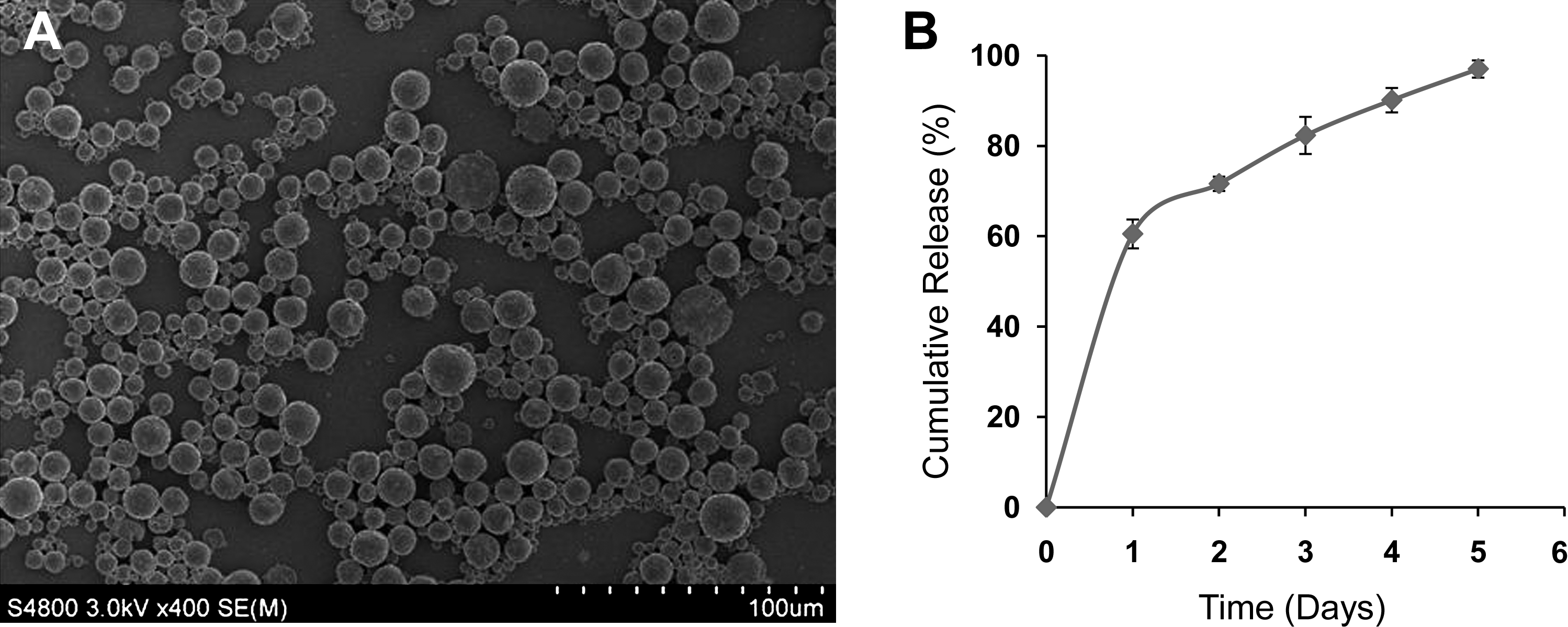
Microphotographs
E2, 17-β estradiol; PAMAM, polyamidoamine; SD, standard deviation.
Entrapment efficiency and release profile regulated by internal phase of solvents
Table 3 shows the entrapment efficiency of estradiol and their cumulative release within 24 h from PLGA microparticles prepared under conditions of varying types and ratios of co-solvents. Although the entrapment efficiency was high for all the groups ranging from 80% to 92%, the cumulative releases of E2 within 24 h are different among the groups using different types and ratios of co-solvents. Formulations using methanol resulted in complete release within a few hours. Formulation using ethyl acetate as co-solvent and 1:1 ratio to DCM exhibited about 70% release, whereas ethyl acetate in a ratio of 1:1.5 to DCM displayed the slowest release with only 55% estradiol release. These results highlight our capacity to manipulate the release profile of the E2 from cationic PLGA particles by careful control over the formulation parameters.
Uptake of E2-loaded PLGA microspheres by human MSCs
Figure 2 shows confocal microscopy images of MSCs after 1 and 24 h of incubation with rhodamine-loaded PLGA particles. Overlaying images of MSCs after 24 h incubation show a significant increase of uptake of particles compared to that after 1 h incubation, also highlighting that cellular uptake of the particles is time dependent. To confirm that rhodamine-labeled PLGA particles were inside the cell and not simply surface bound, we acquired confocal sections through the cell. The overlaying images of rhodamine and LysoTracker Green suggest that uptake of E2-loaded PLGA particles primarily occurs by endocytosis. We have also previously observed that preincubating cells with cytochalasin D (Sigma) at 100 μg/mL results in a 73% drop in cationic particle uptake by the cells. Cytochalasin D is a well-documented inhibitor of endocytosis that works by interfering with actin polymerization. 24 These observations are strongly consistent with the findings that rhodamine-labeled PLGA particles are co-localized with lysotracker-stained organelles. The remaining uptake of cationic PLGA particles observed when cells were treated with cytochalasin D may reflect internalization of particles at the smaller end of the size range through mechanisms that are alternative to endocytosis. This study, however, suggests that endocytosis is the dominant mechanism for cell uptake of cationic-modified PLGA particles. No significant toxicity was observed by incubating the cationic-modified PLGA particles with MSCs, and this is consistent with our previous studies evaluating the toxicity of PAMAM-modified PLGA microparticles for nonviral gene delivery applications in human embryonic kidney (HEK293) and Monkey African green kidney fibroblast-like cell lines (COS7). 25

Confocal microscopic images of human MSCs after 1 and 24 h incubation with rhodamine-loaded PLGA particles (formulation 4 from Table 3) observed by overlaying images obtained by Zeiss-701.
Intracellular release of E2 increases osteogenic differentiation of human MSCs
In confocal images evaluating particle uptake in MSCs, we observed diffusion of rhodamine from the cationic PLGA particles in sections through the cell that confirmed that the content of the PLGA particles was being released intracellularly. Uptake of E2-loaded PLGA particles increases the osteogenic differentiation capacity of MSCs (Fig. 3). After exposure to osteogenic medium, osteogenic differentiation markers ALP, Cbfa-1, of MSCs with intracellular release of E2 are upregulated earlier and significantly higher than that of controls (Fig. 3A, B). Expression of ER-α was upregulated in MSCs after taking up E2 particles (Fig. 3C), whereas limited expression of ER-β was observed (data not shown).
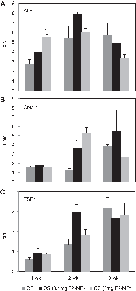
Osteogenic differentiation markers and estrogen receptor of MSCs improved by uptake of E2-loaded PLGA particles (formulation 4 from Table 3). Fold changes of ALP
Discussions
Estrogen improves proliferation and osteogenic differentiation of human bone marrow MSCs and upregulates expression of osteogenic growth factors.17,26,27 This suggests that estrogen can serve as a regulator for MSC-based bone tissue engineering. In the present study, we successfully developed biodegradable cationic PLGA microparticles that entrap estrogen with high loading efficiencies. Entrapped estrogen is released over several days. With the conjugation of cationic PAMAM dendrimers to the surface of the particles, E2-loaded PLGA particles are efficiently taken up by MSCs and improve osteogenic differentiation of MSCs in vitro. These data demonstrate that PLGA particles can serve as an effective tool for intracellular delivery of estrogen. Intracellular release of E2 using the PLGA particles can effectively execute the estrogen regulation of MSCs and potentially improve osteogenic capability of MSC differentiation and environments of implantation for MSC-based bone tissue engineering.
An ideal delivery system capable of providing intracellular release of estrogen should have relatively high entrapment efficiencies, be easily taken up by human MSCs, and be able to provide a controllable release prolife. Among the many factors that affect the release profile,28,29 two important parameters are organic solvent and stabilizer solution, which have a very strong influence on particle formation and release profiles. To optimize entrapment efficiency of estrogen into PLGA particles, we investigated different types and ratios of co-solvents, including methanol and ethyl acetate on entrapment and release of estrogen. While methanol and ethyl acetate in combination with DCM exhibit high entrapment efficiency of estrogen, they have different effects on the release prolife. The difference in release rates can be attributed to the co-solvent used in the internal phase. Methanol has high aqueous solubility and is completely miscible with water. During solvent evaporation, it rapidly diffuses to the external phase and volatilizes at the DCM/water interface. This results in extraction of estradiol from the emulsion droplet core and accumulation of the entrapped drug closer to the surface of the microparticle. Therefore, the drug releases immediately within a few hours of incubation owing to the extremely short diffusion path length. Conversely, ethyl acetate possesses lower aqueous solubility in comparison to methanol (13.0 g/100 mL). However, ethyl acetate exhibits higher water solubility over DCM (8.3 g/100 mL and 1.3 g/100 mL, respectively). The higher solubility of ethyl acetate results in a greater extent of diffusion of estradiol to the interface during particle fabrication, resulting in most of the estradiol being present closer to the surface than being uniformly dispersed within the microparticles. This surface-associated estradiol releases within a day of incubation in PBS (pH 7.4) and the remaining E2 is released quickly within the next 3–4 days. This problem can be overcome by the use of a smaller ratio of ethyl acetate to DCM (10:90) or (5:95) that can prolong the release of estradiol and result in a slower release. Alternatively, estradiol can be dispersed in the organic phase during the microparticle fabrication process, eliminating the complete use of ethyl acetate in the process. The release profile can be further modified by the use of PLGA (85:15) or PLGA (75:25) with a higher inherent viscosity (higher MW grade) that results in the formation of a more viscous internal phase during the emulsification step of the microparticle fabrication. The high viscosity internal phase can be used to provide an increased resistance barrier to the diffusion of estradiol to the interface. The ability to modulate the release profile of E2 by varying the formulation parameters is expected to generate a valuable tool that can be utilized to characterize the relationship between the release rate of E2 and the degree of osteogenic differentiation.
The next step was to covalently attach cationic PAMAM dendrimers to the surface of the PLGA microparticles using EDC/NHS chemistry. We selected G3 PAMAM dendrimers based on a previous optimization study showing that this group generated a sufficient net positive charge that significantly increased cell uptake without adding toxicity when compared to unmodified PLGA microparticles. 23 Zetapotential measurements of −37.1 ± 4.17 before surface modification transitioned to +21 ± 3.42 confirming that PAMAM conjugation had occurred (Table 2). The strategy of conjugating PAMAM dendrimers to the surface of E2-loaded PLGA particles is also useful because the cationic PLGA particles display some buffering capacity that can be used to promote release of the PLGA particles from the endosome into the cytoplasm through the proton sponge mechanism. 30 In addition, the net positive surface charge of the E2-loaded PLGA particles could be used to electrostatically bind DNA and siRNA molecules that work synergistically with the E2. Further, binding of such agents still allows for the PLGA particle to retain a net positive charge although reduced.
The in vitro release of E2 from PLGA particles in this study is about 7 days. Intracellular release of E2 from PLGA particles results in improvement of osteogenic differentiation of MSCs. It significantly increases ALP and Cbfa-1 expressions after 1 and 2 weeks. While the osteogenic differentiation markers cannot directly represent the capability of bone formation, ALP activity of MSCs in vitro has been reported to correlate to bone regeneration capability. 31 Cbfa-1 is also a key transcription factor associated with osteoblast differentiation and bone formation. MSCs with intracellular release of E2 expressed an earlier and significant upregulation of Cbfa-1 compared to the control. These results demonstrate the effect of intracellular release of E2 on improvement of MSC osteogenic differentiation. Future studies will focus on in vivo studies on bone formation from MSCs treated with E2-loaded particles and creating a tool-box of PLGA particles with varying release periods and doses of estrogen to match the varying regulation of osteogenic capacity of MSCs.
Footnotes
Acknowledgments
We thank anonymous reviewers for their insightful comments. We gratefully acknowledge support from Start-up funding from the Dows Institute for Dental Research, College of Dentistry, University of Iowa, the American Cancer Society (RSG-09-015-01-CDD), the National Cancer Institute at the National Institutes of Health (1R21CA13345-01/1R21CA128414-01A2/UI Mayo Clinic Lymphoma SPORE), and the Pharmaceutical Research and Manufacturers of America (PhRMA) Foundation. Y. Krishnamachari acknowledges support from a Guillory Fellowship.
Disclosure Statement
No competing financial interests exist.