
Abstract
Within the scope of developing an in vitro culture model for pharmacological research on human liver functions, a three-dimensional multicompartment hollow fiber bioreactor proven to function as a clinical extracorporeal liver support system was scaled down in two steps from 800 mL to 8 mL and 2 mL bioreactors. Primary human liver cells cultured over 14 days in 800, 8, or 2 mL bioreactors exhibited comparable time-course profiles for most of the metabolic parameters in the different bioreactor size variants. Major drug-metabolizing cytochrome P450 activities analyzed in the 2 mL bioreactor were preserved over up to 23 days. Immunohistochemical studies revealed tissue-like structures of parenchymal and nonparenchymal cells in the miniaturized bioreactor, indicating physiological reorganization of the cells. Moreover, the canalicular transporters multidrug-resistance-associated protein 2, multidrug-resistance protein 1 (P-glycoprotein), and breast cancer resistance protein showed a similar distribution pattern to that found in human liver tissue. In conclusion, the down-scaled multicompartment hollow fiber technology allows stable maintenance of primary human liver cells and provides an innovative tool for pharmacological and kinetic studies of hepatic functions with small cell numbers.
Introduction
Although different types of clinical-scale liver cell bioreactors showed improved liver cell function,8,9 scale down of these systems to a size useful for application in preclinical drug testing was not performed. We previously described a clinically used multicompartment hollow fiber membrane bioreactor technology for 3D perfusion high-density culture of human liver cells within a capillary network based on three independent interwoven capillary systems for arterio-venous medium perfusion, oxygen supply, and carbon dioxide removal. 10 Previous studies have shown that parenchymal and nonparenchymal liver cells cocultured in the bioreactor cell compartment reassemble and form tissue-like structures, including neo-biliary channels and neo-sinusoidal endothelialized structures.11,12 Since the clinical 800 mL bioreactor system using primary human liver cells showed a stable metabolic activity over several weeks and was successfully used in a pilot study to support patients with liver failure,13,14 this technique would be of interest for kinetic drug metabolism and toxicity studies. The interweaving of the capillaries allows the creation of small repetitive capillary units independent of the scale of the device. Thus, we hypothesize that scaling down of the technology for pharmacological research is possible with comparable cell support and preserved tissue functions.
We investigated the performance of primary human liver cells in two smaller bioreactors (2 and 8 mL), based on the same technology as the clinical-scale bioreactor (Figs. 1 and 2). The down-scaled technology was contrasted with the large-scale system with respect to the metabolic activity of human hepatocytes over 2 weeks and showed comparable results, indicating its usefulness for applications in pharmacological research. To confirm the suitability of the small-scale 2 mL bioreactor for drug metabolism and toxicity studies, the activities of several human-relevant cytochrome P450 (CYP) activities (CYP1A, CYP2C9, and CYP3A) were analyzed over prolonged culture intervals up to 23 days. In addition, the tissue organization in the 2 mL bioreactor and the expression of three major canalicular transporters, namely, multidrug-resistance-associated protein 2 (MRP2), multidrug-resistance protein 1 (MDR1, P-glycoprotein), and breast cancer resistance protein (BCRP), were analyzed by immunohistochemistry.
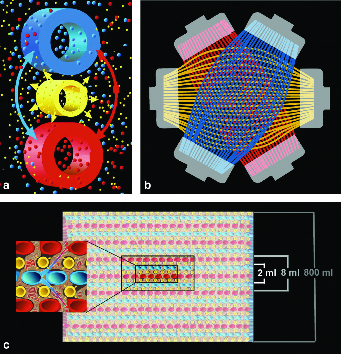
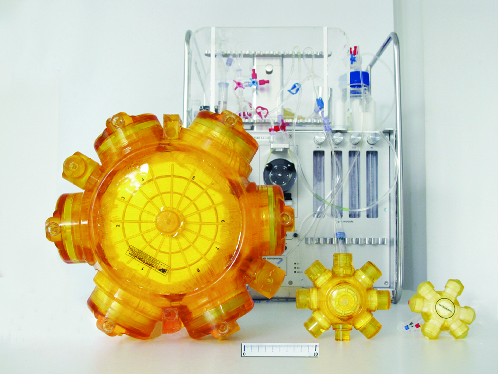
The photograph shows the clinical-scale 800 mL bioreactor (left) and two laboratory-scale variants with a cell compartment volume of 8 mL (middle) or 2 mL (right). The scale bar corresponds to 10 cm. In the background, the perfusion device for bioreactor operation can be seen. The device disposes of pump heads for medium recirculation and medium substitution with automated pressure control, an electronically regulated heating unit, rotameters for regulation of the flow rates for air, oxygen, CO2, and for the total gas mixture, and a display for digital monitoring and regulation of system parameters.
Materials and Methods
Bioreactor technology
The multicompartment bioreactor consists of three independent but interwoven hollow fiber capillary systems that serve for counter-current medium perfusion (two medium compartments) and decentralized oxygenation (gas compartment) of the cells located in the extracapillary space (cell compartment), as shown in Figure 1a and b. A detailed description of the technology can be found elsewhere. 12 The clinical-scale bioreactor with a cell compartment volume of 800 mL was scaled down to smaller variants with a cell compartment volume of 8 or 2 mL for in vitro research on liver cells (Figs. 1c and 2). Bioreactors are integrated into a perfusion device that is designed to include all equipment required for long-term cell maintenance (Fig. 2). Bioreactors and perfusion devices were manufactured by Stem Cell Systems, Berlin, Germany.
Cell preparation
Primary human liver cells were isolated from donor organs excluded from transplantation due to organ injury or from liver tissue remained after partial resection as described previously.15,16 Cells were isolated from whole organs or tissue pieces in accordance with European and national regulations and with the approval by the local ethics committee. The viability of cells obtained from discarded donor organs was between 55% and 65%, and that of cells derived from resected material was 75% to 85%, as determined by trypan blue staining.
Cultivation of primary human liver cells in bioreactors
To compare the metabolic activity of primary human liver cells in differently sized bioreactors, cells were cultivated in bioreactors with a cell compartment of 800 mL (n = 14), 8 mL (n = 6) or 2 mL (n = 6) over a period of 14 days. Some of the 2 mL bioreactors were maintained over prolonged periods, up to 23 days, for analysis of CYP activities. The number of inoculated cells was 1.42 ± 0.12 × 10¹⁰ for the 800 mL bioreactor, 2.6 ± 1.5 × 10⁸ for the 8 mL bioreactor, and 1.2 ± 0.5 × 10⁸ for the 2 mL bioreactor. A modification of Williams' Medium E medium (Heparmed, Biochrom, Berlin, Germany) specifically developed for high-density culture of primary liver cells was used, which was further supplemented before use with 20 IU/L insulin, 3 μg/L glucagon, 0.8 mg/L transferrin (ITG), and/or 2.5% fetal calf serum, all provided by Biochrom. Bioreactors were maintained at 37°C, and at a pH value of 7.30–7.45. Medium recirculation and feed rates, and also gas (air/O2/CO2) perfusion rates were adapted to the different bioreactor sizes.
Concentrations of urea, ammonia, glucose, lactate, lactate dehydrogenase, aspartate aminotransferase, and albumin in samples from the culture perfusate and/or the medium outflow vessel were analyzed as described previously. 13 Human alpha-fetoprotein was measured with a chemiluminescent microparticle immunoassay (Abbott Diagnostics Division, Sligo, Ireland).
Analysis of CYP activities
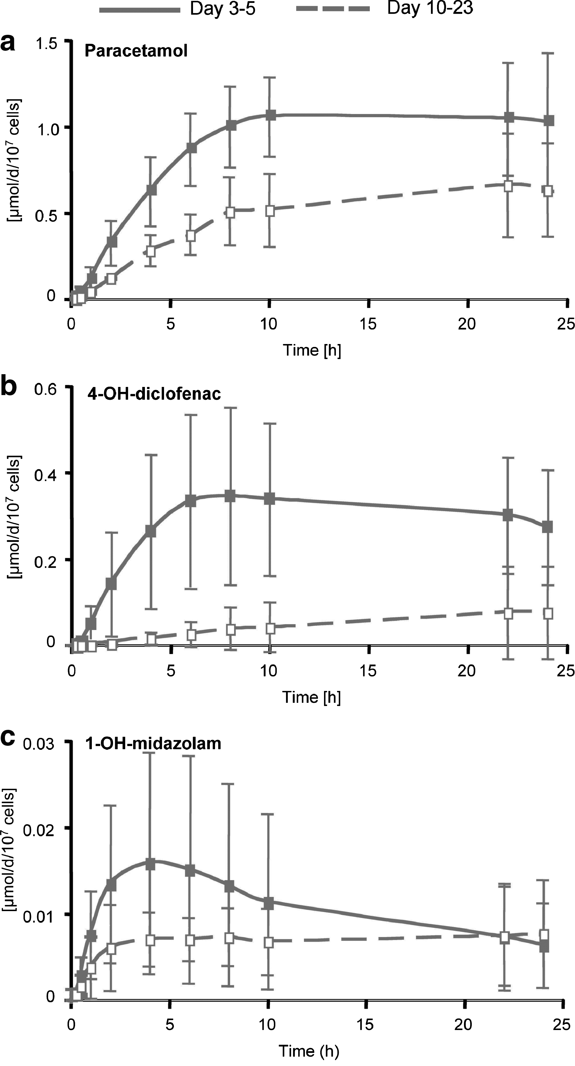
CYP activities were determined in 2 mL bioreactors run with primary liver cells by injecting a mixture of the probe substrates phenacetin (CYP1A), diclofenac (CYP2C9), and midazolam (CYP3A). Phenacetin and diclofenac (both from Sigma-Aldrich, Taufkirchen, Germany) were applied at final concentrations of 26 and 9 μM, respectively, whereas midazolam (Dormicum®) was applied at a final concentration of 3 μM. Concentrations of the corresponding metabolites, paracetamol, 1-OH-midazolam, and 4-OH-diclofenac, in samples taken at 0.25, 0.5, 1, 2, 4, 6, 8, 10, 22, and 24 h from the perfusion circuit were measured by liquid chromatography/mass spectrometry, as described previously. 17
Areas under curve (AUC), maximal concentrations (Cmax), and initial formation rates were calculated for paracetamol, 1-OH-midazolam, and 4-OH-diclofenac using the noncompartment model 220 in WinNonlin 5.2.1 software (Pharsight, Mountain View, CA). The AUC were calculated between 0 and 24 h, and initial formation rates were calculated from the linear phase of the slope in the concentration–time curve.
Immunohistochemistry
Samples from cell culture material from different locations within the capillary network were taken from 2 mL bioreactors upon culture termination. After fixation in 5% paraformaldehyde, the material was dehydrated and embedded in paraffin and cut into 5 μm sections. Immunohistochemical staining was performed on the staining module Discovery XT (Ventana® Medical Systems Inc., Tucson, AZ), using antibodies against CK19 (Santa Cruz Biotechnology, Santa Cruz, SA), von Willebrand factor (DAKO A/S, Glostrup, Denmark), CD68 (DAKO A/S), MRP2 (Abcam, Cambridge, United Kingdom), MDR1 (P-glycoprotein; Sigma-Aldrich, St. Louis, MO), and BCRP (Abcam). The immunohistochemical staining was observed with diaminobenzidine chromogen, and the counterstaining of nuclei was performed with hematoxylin.
Statistics
Values of biochemical parameters are given as means ± standard deviation in the text and in figures. The level of significance of the correlation between different bioreactor groups with respect to the time-course of biochemical parameters was analyzed with PASW v18 statistic software (SPSS Inc., Chicago, IL) using the Spearman's rank correlation coefficient (Spearman's rho) followed by two-tailed significance calculation.
Results
Metabolic activity of primary human liver cells cultivated in 800, 8, and 2 mL bioreactors
The metabolic activities of primary human liver cells cultured in differently sized bioreactors (800, 8, or 2 mL) over 14 days were contrasted. The results showed comparable time-course profiles of most biochemical parameters analyzed in bioreactor perfusates (Fig. 3). Differences between different size variants were observed mainly for urea and albumin production (Fig. 3a, b). However, there was still a significant correlation in the time course of urea production between individual bioreactor types. Albumin synthesis rates were stable over the culture period in both 800 and 8 mL bioreactors, whereas a distinct increase during the first week was observed in 2 mL bioreactors (Fig. 3b). Levels of lactate dehydrogenase and aspartate aminotransferase activities showed almost equal time-course profiles in the three different bioreactor types (Fig. 3c, d). Typically, an initial increase in enzyme release during the first days of culture, presumably due to the process of cell isolation, was followed by stabilization of values to a basal level. Glucose metabolism showed a parallel time-course profile in all size variants, characterized by an initial peak in glucose release that decreased during further cultivation (Fig. 3e). A significant correlation was only detected between 800 and 2 mL bioreactors. The time profile of lactate secretion was similar in 800 and 2 mL bioreactors with constant production rates during the whole culture period (Fig. 3f). In contrast, 8 mL bioreactors showed no significant production during the first week of culture, whereas increasing lactate production was observed during the second week of culture.

Time-courses of urea production
Analysis of CYP enzyme activities in the miniaturized 2 mL bioreactors
The stability of CYP activities in the small-scale 2 mL bioreactor inoculated with primary human hepatocytes was evaluated for up to 23 days. CYP-dependent metabolism of phenacetin (CYP 1A), diclofenac (CYP2C9), and midazolam (CYP3A) was determined on days 3–5 (early culture phase) and on days 10–23 (late culture phase). The time profiles of metabolite formation over 24 h in bioreactors are shown in Figure 4. All enzyme activities were detected in both the early and the late culture phase, with a high variability between cultures from different donors. In the late culture phase, the AUC for paracetamol (Fig. 4a), 4-OH-diclofenac (Fig. 4b), and midazolam (Fig. 4c) reached an average of 53%, 16%, and 60% of the values from early culture days, respectively. Initial formation rates of paracetamol, 4-OH-diclofenac, and midazolam were maintained at 43%, 8%, and 35%, and the Cmax was 60%, 35%, and 69%, respectively, in the late culture phase as compared with values from the early culture phase. The values for the AUC, formation rates, and Cmax can be found in Table 1.
Time profile of metabolite formation from
AUC, areas under curve; Cmax, maximal concentrations.
Immunohistochemical characterization of primary human liver cell structures in the 2 mL bioreactor
Immunohistochemical staining of markers for different liver cell types was performed in the 2 mL bioreactors to evaluate the cell arrangement and composition in 3D liver cell cocultures (Fig. 5). Hepatocytes and nonparenchymal cells formed tissue-like aggregates between the capillary fibers. Biliary cells characterized by cytokeratin (CK) 19 immunoreactivity regularly formed channel-like structures that were similar in size to those found in intact liver tissue (Fig. 5, upper row). Endothelial cells detected by von Willebrand factor expression were arranged in strains between hepatocytes (Fig. 5, middle row). Further, the presence of Kupffer cells was revealed by immunostaining of CD68 antigen that showed a similar distribution pattern as compared with that found in intact liver tissue (Fig. 5, lower row).
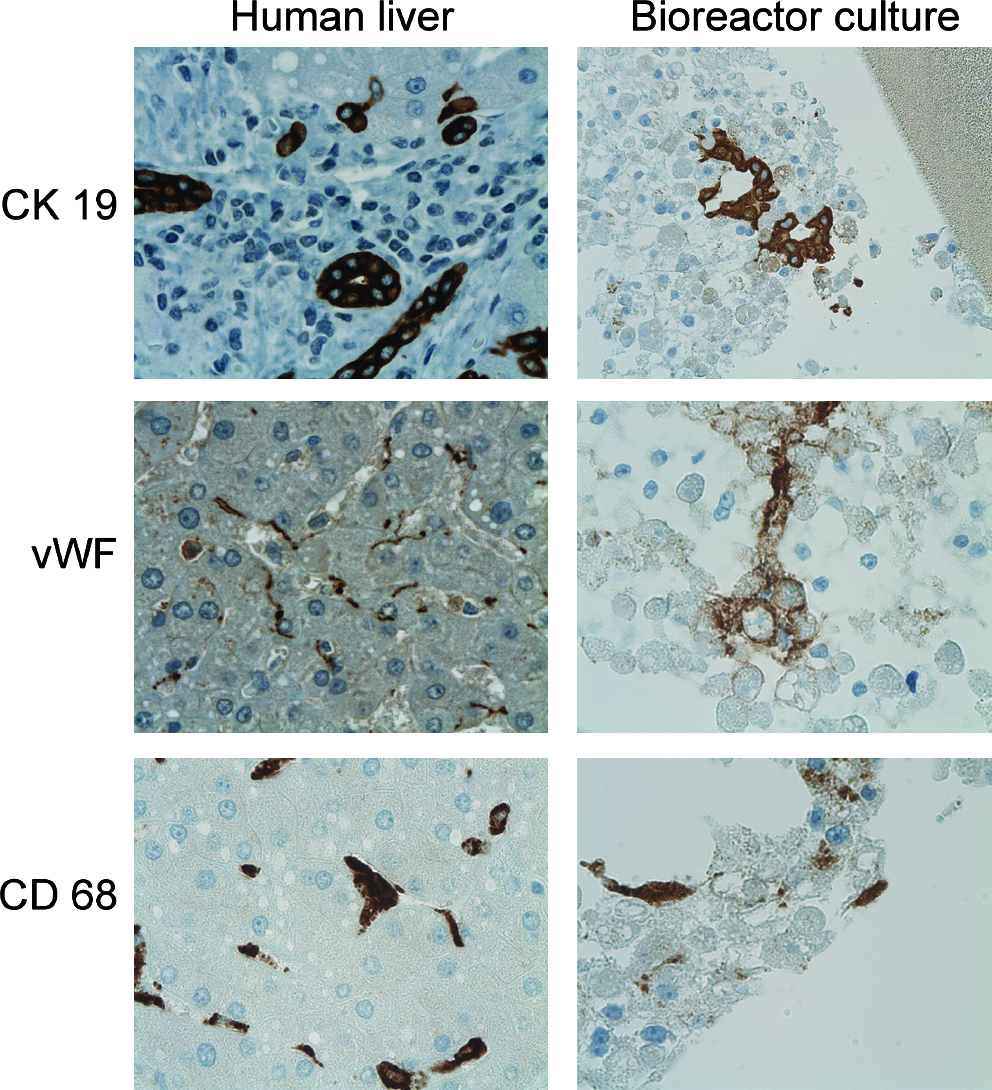
Immunohistochemical characterization of primary human liver cells cultivated in the miniaturized 2 mL bioreactor over 2 weeks (right lane). Staining of intact human liver is shown for control (left lane). Hepatocyte aggregates in the bioreactor cell compartment contain several biliary cells (CK19 positive) arranged in partly open channel-like structures (upper row). In addition, endothelial cells characterized by von Willebrand factor (vWF) pervade the hepatocyte aggregates (middle row). CK68 staining characteristic for Kupffer cells is detected in single cells, which typically line hepatocyte aggregates, similar to the in vivo situation (lower row). Magnification: 200-fold (bioreactor culture, CK19) or 400-fold.
Immunohistochemical staining of the canalicular transporter proteins MRP2, MDR1, and BCRP revealed the expression of all three transporters in 3D bioreactor cultures (Fig. 6). The localization and distribution of transporter proteins in the bioreactor was similar to that found in human liver tissue used as a control. Both MRP2 (Fig. 6, upper row) and MDR1 (Fig. 6, middle row) were confined to the apical (canalicular) membranes of hepatocytes. BCRP was detected both in the apical and the basolateral membranes in human liver tissue and the staining showed a similar distribution in the bioreactor cultures (Fig. 6, lower row).
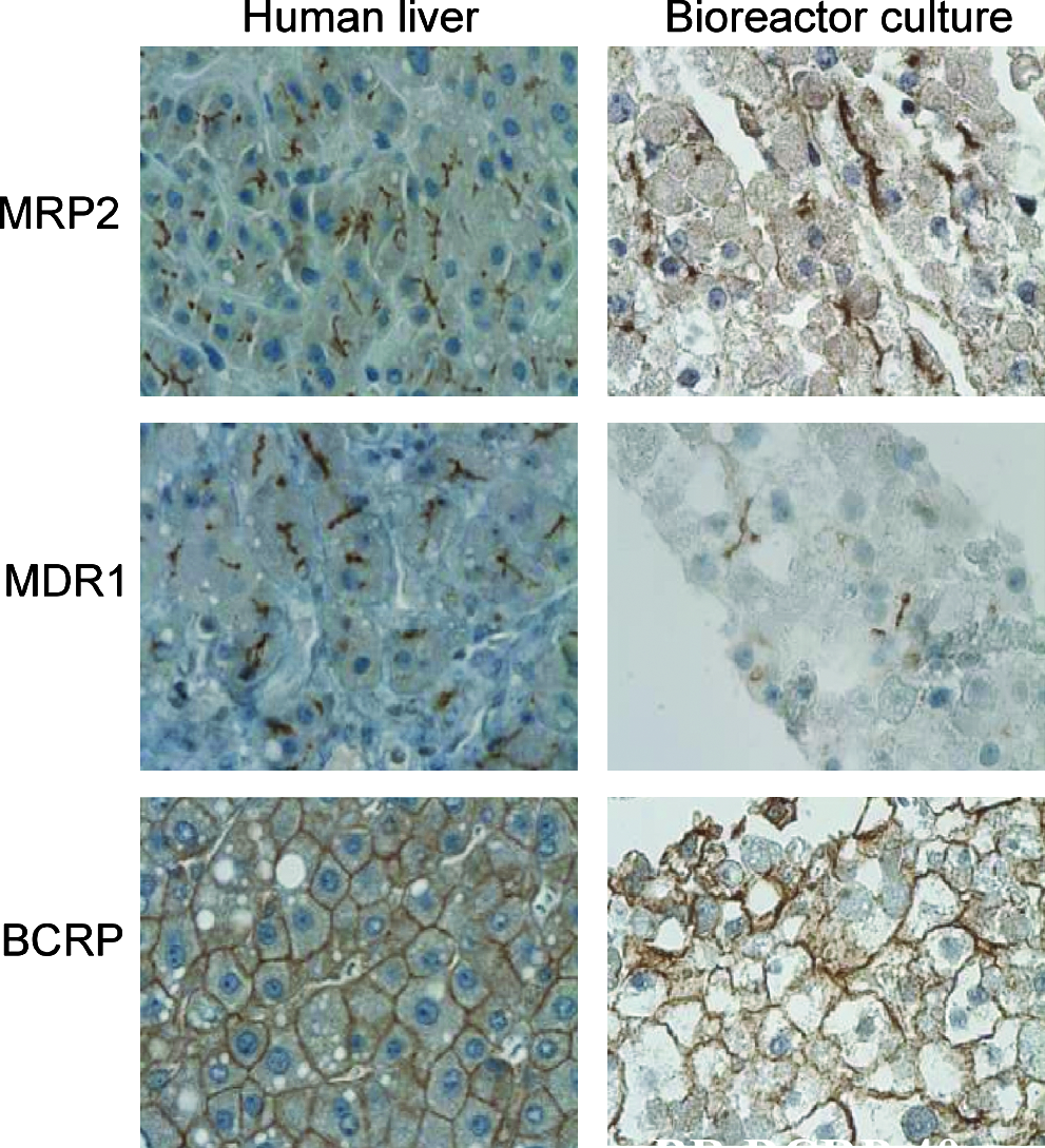
Immunohistochemical detection of the hepatic transporters multidrug-resistance-associated protein 2 (MRP2), multidrug-resistance protein 1 (MDR1), and breast cancer resistance protein (BCRP) in primary human liver cells cultivated in the miniaturized 2 mL bioreactor over 2 weeks (right lane). The staining pattern of these transporters in the bioreactor shows a similar distribution than that observed in human liver tissue used as a control (left lane). Both MRP2 (upper row) and MDR1 (middle row) are localized in the apical (canalicular) membranes of hepatocytes, whereas BRCP immunoreactivity is detected throughout the whole hepatocytic plasma membrane (lower row). Magnification: 400-fold.
Discussion
A major challenge in preclinical drug development and discovery research is the development of human in vitro liver models that (1) reflect the complex functions of the intact liver, (2) allow stable long-term maintenance of hepatocytes, and (3) can be adapted to a screening mode useful in drug discovery settings.
In this study we show the feasibility of scaling down a previously introduced clinical 3D perfusion multicompartment bioreactor technology from a cell compartment volume of 800 mL to small-scale laboratory sizes with cell compartment volumes of 8 or 2 mL for in vitro pharmacological studies.
The results from bioreactor studies with primary human liver cells, which are considered as the gold standard for in vitro drug metabolism studies,18,19 show that all different size variants of the bioreactor technology tested support the hepatic functions of the cells in vitro. While most parameters showed a similar performance in the 800, 8, and 2 mL bioreactor, some differences in time profiles were observed between the different bioreactor types. These variations can probably mainly be attributed to inter-donor variability, which is well known from both in vivo and in vitro studies. Interestingly, we observed a much higher albumin production in the 2 mL prototype than in the other bioreactor types, which cannot be explained by inter-donor differences alone. It remains to be investigated whether this is due to differences in protein biosynthesis rates or to noncellular factors, for example, adsorption depending on scale/material surface of the different bioreactor circuits.
The activity of major human-relevant CYP enzymes (CYP1A, CYP2C9, and CYP3A) in primary human hepatocytes cultured in the 2 mL bioreactor showed that these model substances were metabolized into their primary products. Metabolism rates varied between cell preparations from different donors, which probably reflects human variation in drug metabolism due to human pheno- and/or genotypes.20,21
Maintenance of CYP activities over culture periods of up to 23 days was shown in the 2 mL bioreactor, indicating a stable time period of at least 3 weeks, which may vary due to variations in the donor tissue quality. These results suggest that the system could be used for studies requiring long-term performance of cultures, for example, to study kinetic profiles, slowly metabolized drugs, drug–drug interactions, induction of drug metabolism, and toxicity. Further studies are needed to explore the maximum period suitable for pharmacologial studies in the bioreactor. In addition, a more detailed investigation of factors influencing the metabolic performance of the cells in the bioreactor would be useful to further improve the stability and reliability of the system.
The immunohistochemical studies confirmed the tissue-like arrangement of parenchymal and nonparenchymal liver cell populations in the small-scale 2 mL device, similar to the observations reported earlier for the large-scale clinical bioreactor.11,12 Hepatocyte aggregates contained biliary cells forming channel-like structures, endothelial cell linings, and Kupffer cells as detected by the staining of specific antigens. Structures observed in the bioreactor resembled those found in human liver tissue, which indicates that a physiological cell reorganization process occurs in the 3D bioreactor perfusion system. The importance of heterotypic cell interactions for the preservation of hepatocyte functionality has been shown in several studies. For example, an increased sensitivity for drug-induced toxicity was observed in a coculture model of liver cells and monocytes. 22 In addition, models using perfused flow and heterotypic cell–cell interactions have been shown to provide improved conditions for drug discovery and safety assessment studies. 23
The results from immunohistochemical analysis of the canalicular transporters MRP2, MDR1 (P-glycoprotein), and BCRP, which are involved in the active transport of xenobiotics and/or their metabolites across the canalicular membrane of the hepatocyte into the bile canaliculus, 24 further confirmed physiological tissue restructuring in the 2 mL bioreactor. The distribution pattern of all three transporters was similar to that observed in human liver tissue. Both MRP2 and MDR1 were localized to the apical membranes of hepatocytes, confirming liver-typical polarization of the cells in the bioreactor. The localization of BCRP to both basolateral and apical membranes observed in human liver tissue is in accordance with findings on human liver biopsies that showed partly basolateral expression of BCRP, especially in chronic biliary diseases. 25 A similar distribution of BCRP over the hepatocyte plasma membrane was observed in the bioreactor. Since the expression of drug transporters is a major determinant for drug metabolism and toxicity, the physiological distribution of these proteins observed in this study supports the suitability of the bioreactor for pharmacologial studies.
The results from metabolic and histological studies suggest that the small 2 mL bioreactor provides similar high-performance culture conditions as the larger devices, while enabling a significant reduction of required cell amounts. The option of long-term maintenance of the bioreactor system also opens up the possibility for its use in subchronic to chronic toxicity testing. In addition, the structure of the perfusion circuit allows applying different drug regimens, including repeated dosing and continuous infusion. Further, the model could enable studies on hepatic functions that involve different liver cell populations, which is particularly interesting for studies of human liver drug toxicity. Due to technological features, further size variants would be possible for specific applications.
In conclusion, our results demonstrate the feasibility of scaling down the multicompartment hollow fiber technology from a large clinical device to small laboratory models. The performance of the devices was retained in the small models as shown for primary human hepatocytes. Specifically, the small-scale 2 mL bioreactor introduced in this study opens up a range of applications in preclinical drug testing, since it allows for reduction of reagents and cell amounts needed while supporting cell metabolism similar to the larger devices. The availability of innovative in vitro models that can be used for prolonged studies applying relevant drug concentrations would therefore increase the quality of the assessment of drug candidates selected for clinical testing.
Footnotes
Acknowledgments
The study was performed with support from the European Commission (EU, STREP-CT-2005-018940) and from the German Federal Ministry for Education and Research (BMBF, 01GG0731/-732). We thank Wolfgang Mudra for illustrations and Dr. Eva Wönne for the photograph.
Disclosure Statement
J.C.G. licensed technology to StemCell Systems, Berlin, Germany.