
Abstract
All human organs consist of multiple types of cells organized in a complex pattern to meet specific functional needs. One possible approach for reconstructing human organs in vitro is to generate cell sheets of a specific pattern and later stack them systematically by layer into a three-dimensional organoid. However, many commonly used cell patterning techniques suffer drawbacks such as dependence on sophisticated instruments and manipulation of cells under suboptimal growth conditions. Here, we describe a simple cell patterning method that may overcome these problems. This method is based on magnetic force and photoresponsive poly (ethylene glycol) diacrylate (PEG-DA) hydrogels. The PEG-DA hydrogel was magnetized by mixing with iron ferrous microparticles and then fabricated into blocks with a specific pattern by photolithography. The resolution of the hydrogel empty space pattern was approximately 150 μm and the generated hydrogel blocks can be remotely manipulated with a magnet. The magnetic PEG-DA blocks were used as a stencil to define the area for cell adhesion in the cell culture dish, and the second types of cells could be seeded after the magnetic block was removed to create heterotypic cell patterns. Cell viability assay has demonstrated that magnetic PEG-DA and the patterning process produced negligible effects on cell growth. Together, our results indicate that this magnetic hydrogel-based cell patterning method is simple to perform and is a useful tool for tissue surrogate assembly for disease mechanism study and drug screening.
Introduction
Many complex human organs display an orderly arrangement of small repeating units containing multiple types of cells. Such an organization provides accurate cell-to-cell and cell-to-extracellular matrix microenvironments to ensure proper functions of the organ.3,4 Unfortunately, current scaffold-based tissue engineering technologies lack the capability to deposit cells at desired positions on the scaffold, and are incapable of reconstructing such a complex organ pattern. Therefore, development of a technology that can arrange cells in the right place and provide proper microenvironments is one of the major trends in current tissue engineering research.5–7
One possible approach to overcome the limitation of scaffold-based methods is to generate cell sheets of a specific pattern and later stack them systematically by layer into a three-dimensional (3D) organoid.8–10 Generation of cell patterns mimicking repeating units in an organ usually requires the ability to manipulate cells in microscale, as well as a method for immobilizing and maintaining the pattern. A variety of cell manipulation technologies, such as optoelectronic tweezers, 11 laser-guided direct writing, 12 electrokinetic, 13 and their combination with a microfluidic system,14,15 have been employed for generating cell patterns in an active manner. Alternatively, passive cell patterning methods such as those operating by defining the area for cell adhesion using photolithography 16 or microcontact printing 17 have also been tested successfully. Recently, magnetic force-based cell manipulation has been applied in cell patterning.18–22 These methods utilize magnetic force to manipulate cells to form a desired pattern and reconstruct 3D multilayered tissues from cell sheets, demonstrating the feasibility of using magnetic force in tissue engineering. However, some of these techniques require sophisticated instruments to perform. Labeling cells with magnetic materials may influence cell viability and the presence of magnetic materials in the tissue surrogate may be undesirable for certain subsequent analyses. Using microcontact printing to pattern cells is easy to conduct, but fabricating the printing stamp relies on highly specialized facility. Thus, a simple-to-perform cell patterning technique that does not depend on cell labeling and expensive equipments will be very useful in tissue engineering.
We describe a magnetic hydrogel-based cell patterning (MHCP) method possessing several favorable properties such as ease of fabrication and operation. The magnetic microparticle-containing hydrogel blocks with defined patterns were first generated by using photoresponsive poly (ethylene glycol) diacrylate (PEG-DA) hydrogel combined with photolithography techniques. 23 These magnetic hydrogel blocks can be easily manipulated using a magnet and applied as stencils, occupying specific areas and leaving the nonhydrogel covered areas for cell adhesions. Repeating the process using different types of cells can also achieve heterotypic cell patterning. 24
Materials and Methods
Materials
All tissue culture media, antibiotics, sera, and reagents were purchased from Invitrogen. Other chemicals were purchased from Sigma-Aldrich unless indicated otherwise. Cell lines were purchased from American Type Culture Collection. The magnetic particles (Mn-Zn WDP magnetic powders; average core particle size, 9.5 μm; bulk density, 2 g/mL) were purchased from HIMAG Magnetic Corporation. Permanent neodymium iron boron (NdFeB) magnets were obtained either from Super Electronics or local bookstores. The magnetic field intensity of magnets was measured by a Gauss meter (F.W. Bell 5180).
Cell culture, fluorescence dye staining, and image acquisition
HepG2 cells and Balb/3T3 cells were maintained in Dulbecco's modified Eagle's medium supplemented with 10% fetal bovine serum (Gibco) and 1% penicillin/streptomycin in a 5% CO2 humidified incubator at 37°C. Fluorescent marker dyes used in this study were DiO (green), DiI (red), 5-chloromethylfluoroscein diacetate (CMFDA), and 5-(and-6)-(((4-chloromethyl)benzoyl)amino)-tetramethylrhodamine (CMTMR). The working concentrations of DiO, DiI, CMFDA, or CMTMR were 5–25 μM. All cell manipulation images in this study were captured using an epi-fluorescence upright microscope (BX-51; Olympus) equipped with a digital camera and analyzed using the SPOT Advanced Plus Imaging software (Version 4.6).
Generation of magnetic hydrogel blocks
The 50% PEG-DA precursor solution was prepared by dissolving PEG-DA (Mw=575) in phosphate-buffered saline (PBS) supplemented with 1% ultraviolet (UV) photoinitiator (2, 2-dimethoxy-2-phenylacetophenone) and 10 mg/mL magnetic particles. The PEG-DA solution was loaded into a thin chamber (height=256 μm) and then exposed to UV light through a photomask to form magnetic PEG-DA blocks. This study employed a mercury lamp on a fluorescent microscope (BX-51; Olympus) as the light source and the exposure time was between 4–6 s. All of the photomasks were designed using L-Edit v10 software (Tanner Research) and printed on emulsion film with a resolution of 20,000 dpi (Taiwan Kong-King). After photolymerization, blocks were immersed in PBS for 1 day to remove excessive photoinitiator and unpolymerized PEG-DA.
Cell viability assay and proliferation measurement
The effects of MHCP on cell viability were assessed by the LIVE/DEAD Cell Viability kit (Invitrogen), and the Alamar Blue reagent (Biosource). 25 Briefly, cells were patterned and cultured using MHCP and stained with LIVE/DEAD dye after 24, 36, and 48 h. Live cells were stained with calcein AM in the kit showing green fluorescence (excitation/emission: 495 nm/515 nm); and dead cells were stained with ethidium homodimer-1, emitting a red fluorescence (excitation/emission: 495 nm/635 nm). Images of the cell viability assay were obtained using a fluorescence microscope (BX-51; Olympus) equipped with a digital camera. Alternatively, the metabolic activity of cells grown in the presence or absence of a hydrogel block with or without a magnet for 1–4 days was determined using the Alamar Blue at an excitation wavelength of 530 nm and emission wavelength of 590 nm with a Wallac 1420 multilabel counter (Wallac).
Process of MHCP
The flowchart for MHCP is shown in Figure 1. A cylindrical NdFeB magnet (diameter, 11 mm; height, 3 mm; 2000 G) was fixed under a well of the 24-well culture plate. The magnetic PEG-DA blocks were loaded into the 24-well plate and attracted to the bottom of the culture plate by an NdFeB magnet. Cells were seeded and allowed to attach to the areas not occupied by the hydrogel blocks. After cell attachment, the magnetic hydrogel blocks were removed using a magnet. To achieve heterotypic cell patterning, the second type of cell was seeded into the same cell culture plate after the hydrogel block was removed. The cells would adhere preferentially to areas not previously occupied by the first type of cell.
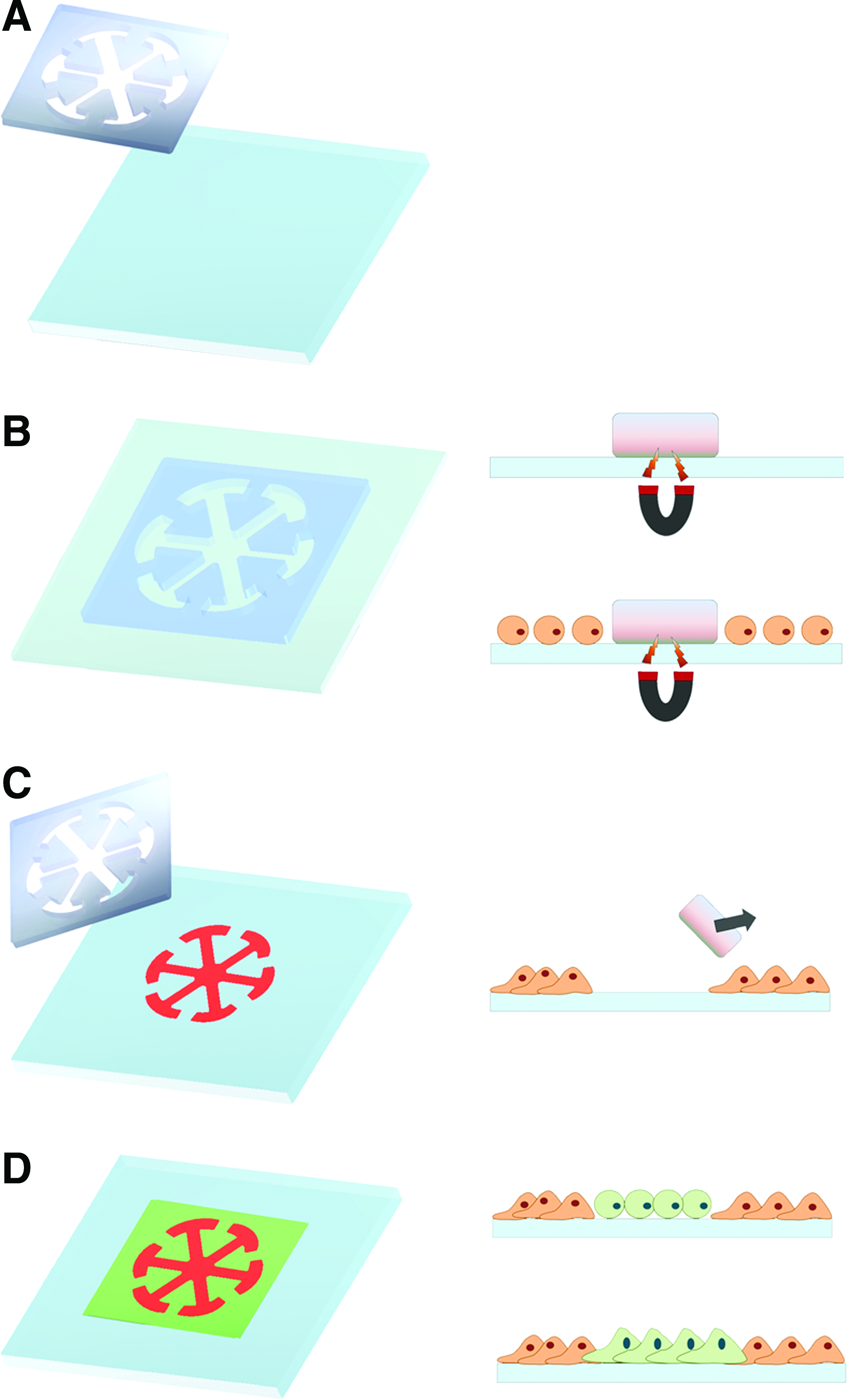
Schematic illustration of the process for MHCP.
Statistical analysis
Data for Alamar Blue assays presented as bar graphs are the means±standard deviation of three independent experiments (n=3). Statistical analysis was performed using Student's t-test (p<0.01 is considered significant).
Results
Resolution of the pattern formed on hydrogel blocks
The PEG-DA precursor solution (Mw=575 Da) containing 1% of UV photoinitiator and 10 mg/mL magnetic particles formed hydrogel blocks within several seconds under a standard microscope mercury lamp exposure. A higher PEG-DA precursor or photoinitiator concentration could reduce the exposure time, although the precise hydrogel patterns are more difficult to control precisely and the elasticity of PEG-DA blocks is also reduced. Figure 2A shows a photomask with a line width ranging from 50 to 400 μm that was used to test the resolution of the pattern formed on the hydrogel blocks. Generation of empty lines with a width above 150 μm is relatively straightforward using this method. Further reduction of line width depends on precise control of the light exposure duration and rapid removal of non-reactive PEG-DA solution. As demonstrated in Figure 2A, the limitation using this technique to generate line shape space is approximately 100 μm. A photomask with line widths above 200 μm could easily generate PEG hydrogel blocks with comparable line width. The actual line width on hydrogel blocks may be 150 μm smaller than the corresponding line width on the photomask if the line width is below 200 μm.
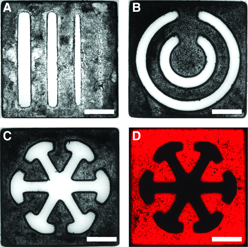
Fabrication of magnetic PEG-DA blocks and determination of the resolution.
Fabrication of magnetic PEG-DA blocks
This study further attempted to fabricate magnetic hydrogel blocks with various geometric shapes and patterns, particularly those mimicking the repeating units in real organs. As shown in Figure 2B–D, hydrogel blocks with two concentric circles and a snowflake-like shape, designed to imitate blood vessels and liver-lobules, could be produced successfully.
Effects of MHCP on cell proliferation
A basic requirement for successful application of MHCP in cell patterning is that the magnetic PEG-DA hydrogel blocks must be nontoxic to cells. To assess whether the magnetic blocks or patterning process can cause cell damages, we employed LIVE/DEAD cell staining assay to investigate cell viability after MHCP and cultured for 24, 36, and 48 h. As shown in Figure 3, almost all the cells exhibited green fluorescence after MHCP, indicating that the process resulted in negligible cell damages. In the Almar Blue reduction assay, both HepG2 and Balb/3T3 cells were cultured individually with a magnetic PEG-DA block in the presence of a permanent magnet for 4 days. As shown in Figure 4, the metabolic activity of both HepG2 and Balb/3T3 cells co-incubated with PEG-hydrogel blocks, either in the presence or in the absence of magnetic force, showed no inhibition on cell growth comparing to those without any treatment. The result is consistent with that of the LIVE/DEAD staining assay, indicating that the procedure is not harmful to cells.

Effect of MHCP on cell viability. The viability of Balb/3T3
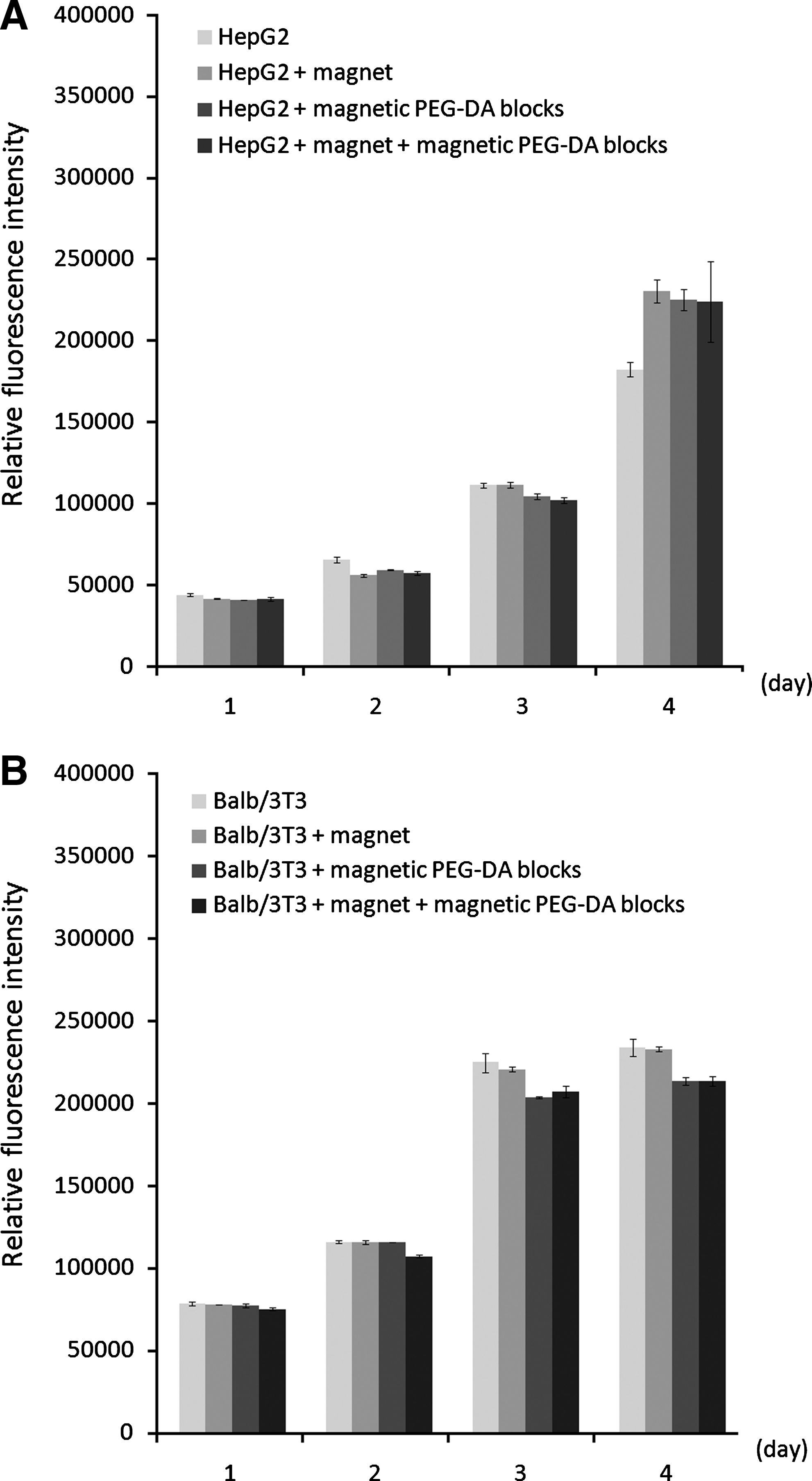
Effect of MHCP on cellular metabolic activity. HepG2
Homotypic and heterotypic cell patterning by MHCP
The homotypic and heterotypic cell patterning were performed following the MHCP procedures illustrated in Figure 1. To demonstrate the complex heterotypic cell patterning by MHCP, we first generated the magnetic PEG-DA blocks with a snowflake pattern (Fig. 2) to fabricate the liver lobule-like tissue. The overall size of the pattern is approximately 2 mm, and the width of the empty line space is approximately 150 μm. After immobilizing the PEG hydrogel block tightly to the culture plate by magnetic attraction, the red fluorescence dye-stained HepG2 cells were seeded into the plate and allowed to attach to the substratum by culturing in a 5% CO2 humidified incubator at 37°C. After 3–8 h of incubation, the hydrogel block was removed using a magnet and non-adhered cells were washed away gently with PBS to reveal the cell patterns. As demonstrated in Figure 5B, a red fluorescent cell-formed snowflake pattern corresponding to the pattern on the hydrogel block can be clearly observed. The cell pattern can be made heterotypic by seeding approximately 2×10⁵ Balb/3T3 fibroblast cells, stained previously with a green fluorescence dye, into the culture plate. Figure 5 shows the pattern formed by HepG2 cells and Balb/3T3 fibroblasts, separately in green and red fluorescence, using the MHCP technique.
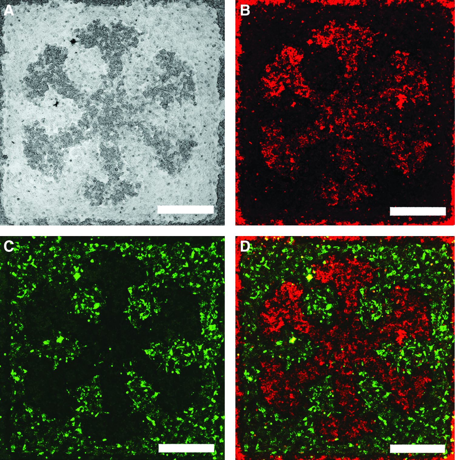
Heterotypic cell patterning using the MHCP technique.
Discussion
Cell patterning is a key step before generation of cell sheets for fabrication of complex organ through the layer-by-layer assembly strategy. Numerous methods are being developed to improve the quality and speed of cell patterning. Magnetic manipulation of cells has gained much attention recently due to its remote controlling capability and simplicity in operation. The magnetic force-based tissue engineering was demonstrated first by Ito and colleagues in 2004, 18 who utilized magnetite cationic liposomes to label and enable cells to be manipulated by magnetic force. Subsequently Akiyama et al. developed a cell patterning method using magnetically patterned PEG-modified magnetite particles to block cell attachment selectively. 22 These studies have demonstrated the feasibility of using magnetic particles and magnetic force in cell patterning. In contrast to these methods that rely on complex magnetic field gradients, MHCP adopts a passive cell manipulation strategy with hydrogel blocks and a simple magnet to produce complex cell patterns. In addition, the most specialized equipment for MHCP is a fluorescence microscope, which can be found in many cell biology laboratories, and thus the procedure is considered inexpensive to perform.
An advantage in fabricating cell patterning templates using photoresponsive hydrogels is the simplicity. PEG-DA hydrogel exhibits more favorable biocompatibility and high mass transfer than other commonly used polymers such as polydimethylsiloxane. Moreover, PEG-DA blocks are sufficiently strong when they are manipulated in the solution using magnetic force. It also has potential to generate a cell sheet with a large-area pattern mimicking repeating units in organs. One problem in using PEG-DA hydrogel is its relatively poor resolution that prohibits some highly complex cell patterns to be fabricated by MHCP. Several strategies may be adopted to improve the pattern resolution with MHCP. One possible approach is to use a higher light intensity to reduce the exposure time and the diffusion of the polymerization-initiating free radicals. Another possibility is to integrate microfabrication techniques, such as maskless photolithography,26–29 into this method. Finally, magnetic polydimethylsiloxane may also be used to replace PEG hydrogel blocks to achieve high resolution cell patterning, although the production procedure is much more sophisticated than generation of PEG-DA hydrogel blocks.
Microfluidic systems, capable of providing continuous dynamic perfusion and complex drug treatment schemes on cells by automatic fluidic control, have become important tools in many biomedical studies. Many microfluidic cell culture systems involve co-culture of multiple cell types and are generally critical to have a proper control of cell number of each cell type and their relative position. However, generation of cell patterns in microfluidic chip is difficult and sometimes can cause cell damages. For example, microcontact printing is hard to perform when the microfluidic devices are completely fabricated. Dielectrophoresis typically need to be performed under a condition not optimal for cell growth. 30 Furthermore, certain cell labeling procedures have been reported to be cytotoxic and can reduce cell viability. 31 In contrast, the MHCP technology described in this article is label-free, cause negligible cell damages, and therefore is suitable for cell patterning in microfluidic systems.
The MHCP technique can be applied in many biomedical studies. First, because of its capability in generating well-defined empty spaces, MHCP can be used to study cell migration as in the conventional wound-healing assay. 32 Second, MHCP can produce cell culture arrays with desired cell numbers and cell–cell interactions to mimic the in vivo conditions and therefore is useful for drug screening. Third, MHCP can be used to generate cell sheets with predesigned patterns. Subsequent systematically layered tissue reconstruction technology can be applied to assemble the cell sheets into 3D tissues.
In conclusion, we have developed a method termed MHCP using magnetic PEG-DA blocks to generate cell patterns. The technique offers advantages including simple to operate and low cell damage, and therefore is a good alternative to current cell-patterning technologies. As magnetic tissue assembly technology matures and more data are accumulated, we believe that the technology will find many applications in biomedical research in the near future.
Footnotes
Acknowledgments
The authors thank Dr. Ruei-Zeng Lin for critical reading of this article. This work was supported by the National Science Council of Taiwan, R.O.C.
Disclosure Statement
No competing financial interests exist.