
Abstract
The application of cell-biomaterial systems in tissue engineering and regenerative medicine is an important challenge in biomedicine, which preserves not only cells, but also tissue-engineered constructs. In this study, the constructs and cryoprotectant parameters were optimized, and it was evaluated whether the characteristics of dental pulp stem cells (DPSCs), which have high proliferation ability as stem cells, were maintained during encapsulation and cryopreservation. The optimal cell-biomaterial gel constructs with the gelation rate of 2% alginate: 100 mM CaCO3: 200 mM glucono-δ-lactone (GDL)=4:1:1 and suitable cryoprotectants (CPAs) used for cryopreservation were Dulbecco's modified Eagle's medium (DMEM) supplemented with 10% ethylene glycol (EG), 1.0 M sucrose and 0.00075 M polyvinylpyrrolidone (PVP). Optimality was confirmed by cell viability (trypan blue, live/dead analysis), the proliferation of DPSCs, and the microstructure using scanning electron microscopy (SEM) in the constructs, and surface epitope by flow cytometric analysis before and after cryopreservation. There were no visible differences in the structure. In conclusion, this study indicates that the optimal cell-biomaterial gel constructs and the cryoprotectant are promising biomaterials. The defined encapsulation/thawing system offers an excellent option for cell-banking therapy to be developed with ready-to-use viable biomaterials and patient-specific products as drug delivery systems.
Introduction
The biomaterials used as scaffolds are natural or synthetic polymers such as polysaccharides, hydrogels, or thermoplastic elastomers. In particular, hydrogel provides a three-dimensional environment similar to that in vivo and, therefore, allows cells to maintain their characteristics and function well in the body. The hydrogels used most frequently in TERM are agarose, alginate, collagen, fibrin, gelatin, and hyaluronic acid. Among these, the sources of collagen, fibrin, and gelatin are animal/human proteins; and the source of hyaluronic acid is animal/human polysaccharide, but the source of agarose and alginate is seaweed. The alginate gelation method uses ionotrophic cross linking and is degradable by ion exchange, whereas the agarose gelation method uses thermal change and is nondegradable. 9 In this study, alginate, known for its safe, low-cost, biodegradable, and easy-to-manipulate properties, was examined as a scaffold.
The role of cryoprotectants (CPAs) is to replace water in cells/tissue and to form an amorphous state at low temperature. Ideal CPAs can be achieved using penetrating cryoprotectants with the additional usage of nonpenetrating cryoprotectants. 10 The problem with conventional CPAs is that they contain dimethyl sulfoxide (DMSO) and animal substances. DMSO has long been used as a cryoprotectant for its high penetrating property; however, due to its cytotoxicity, an alternative agent is required. Serum or protein additives have also been used in CPAs to protect cells, but the possibility of contamination remained.11–14 CPAs that do not contain DMSO or protein additives need to be developed but are highly expensive.
Next, as a cell source of important TERM elements, we focused on dental pulp stem cells (DPSCs), which have high proliferation and differentiation ability and have been considered appropriate candidates not only for dental tissue regeneration but also for the treatment of general diseases.15–18 We encapsulated the DPSCs in alginate gel and optimized the gelation rate containing CaCO3 and glucono-δ-lactone (GDL). 19 After fabricating the cell-biomaterial constructs, we cryopreserved them in CPAs. For medical approval, we examined the optimized condition of ethylene glycol (EG) as a penetrating cryoprotectant and sucrose and polyvinylpyrrolidone (PVP) as nonpenetrating cryoprotectants without DMSO and animal substances. EG, sucrose, and PVP have the merit of being cost-effective CPAs.
Taken together, the approach in this study contributes to the development of effective cryopreservation methods and novel cell banking therapy with ready-to-use viable biomaterials, which reduce the time to fabricate cell-biomaterial constructs and are easy packaging treatments in tissue engineering and medicine technology.
Materials and Methods
Culture of DPSCs and formation of the cell-biomaterial gel encapsulation
Isolation and culture of DPSCs
Human dental pulp tissues were obtained from clinically healthy extracted deciduous teeth. The experimental protocols were approved by the ethics committee of Nagoya University. DPSCs were isolated and cultured as previously described.15,17 Briefly, the pulp was gently removed and digested in a solution of 3 mg/mL collagenase type I and 4 mg/mL dispase for 1 h at 37°C. After filtration using 70-mm cell strainers (Falcon; BD Labware, Franklin Lakes, NJ), cells were cultured in Dulbecco's modified Eagle's medium (DMEM) (GIBCO, Rockville, MD) containing 20% mesenchymal cell growth supplement (Lonza, Inc., Walkersville, MD) and antibiotics (100 U/mL penicillin, 100 μg/mL streptomycin, and 0.25 μg/mL amphotericin B; GIBCO) at 37°C under 5% CO2. After primary culture, cells were subcultured at about 1×10⁴ cells/cm2. The cells were used in the experiment from one to five passages.
Optimization of cell encapsulation with alginate gel (cell-biomaterial gel constructs)
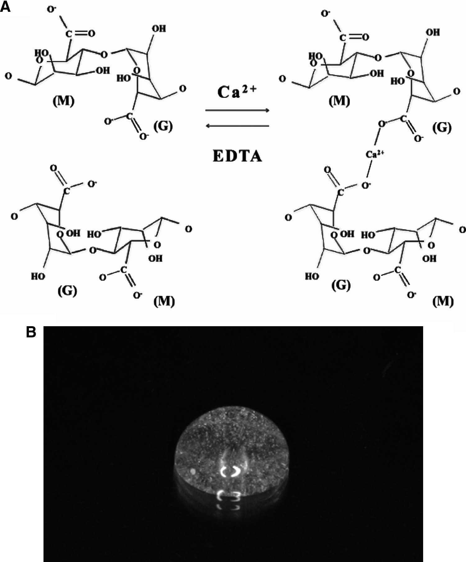
DPSCs were encapsulated in alginate gel, the gelation rate of which was controlled using CaCO3 (Sigma-Aldrich, Tokyo, Japan) and GDL (Sigma-Aldrich, St Louis, MO). The CaCO3 to GDL molar ratio of 0.5 was maintained to achieve a neutral pH. The DPSCs were mixed with the 5 w/v sodium alginate solution (Kaigen, Hokkaido, Japan) to obtain a final density of 5.0×10⁶ cells/mL before the gelation process. Cell-biomaterial gel constructs were fabricated by pipetting CaCO3 solution and GDL solution into the cell-alginate solution (Fig. 1). The gelation rate of experimental groups was as follows: 2% alginate: 100 mM CaCO3: 200 mM GDL: (1) 1:1:1, (2) 2:1:1, (3) 3:1:1, (4) 4:1:1, (5) 5:1:1, (6) 6:1:1, respectively, and the formation and viability of the encapsulated DPSCs were investigated. The average gel diameter was 5 mm. After fabrication, cell-biomaterial gel constructs were cultured for up to 7 days at 37°C under 5% CO2.
Cryopreservation
Optimization of CPAs
The optimal CPAs were determined by investigating CAPs of various mixture rates (Fig. 2). The agents tested were (1) DMEM+10% EG (Wako, Osaka, Japan), (2) DMEM+10% EG+1.0 M sucrose (Wako), (3) DMEM+10% EG+1.0 M sucrose+0.00025 M PVP (Sigma-Aldrich, St. Louis, MO), (4) DMEM+10% EG+1.0 M sucrose+0.0005 M PVP, (5) DMEM+10% EG+1.0 M sucrose+0.00075 M PVP, (6) DMEM+40% EG+0.6 M sucrose, (7) DMEM+10% fetal bovine serum (FBS)+12% DMSO (Wako), and (8) Banbanker (Genetics, Tokyo, Japan). Among these agents, the most optimal CPAs were determined by cell viability. All materials used in this study were analytical grade.

Schematic illustration of the preparation of cell-biomaterial gel constructs and experimental protocol.
Cryopreservation procedures and subsequent culture of cell-biomaterial gel constructs
Cell-biomaterial gel constructs were divided into two groups: (1) control: cell-biomaterial gel constructs without undergoing cryopreservation-thawing process; (2) cryopreservation: cell-biomaterial gel constructs treated with cryopreservation-thawing process. The cell-biomaterial gel constructs were cultured for 24 h at 37°C under 5% CO2, and then 1 gel was added to each 1.8 mL cryotube (Nunc, Rochester, NY). For the cryopreservation process, cell-biomaterial gel constructs were left at 4°C for 5 min, −30°C for 30 min, and finally stored at −80°C for 1, 7, 14, 21, and 28 days. For the warming process, cell-biomaterial gel constructs were thawed directly in a water bath at 37°C. The cell-biomaterial gel constructs were lysed using 0.5 M ethylene diamine tetraacetic acid (EDTA) (Gibco, Auckland, NZ), and the resulting cell suspension was cultured at 37°C under 5% CO2 or was analyzed without it. The cells were cultured for 7 days after thawing and before analyzing.
Analysis of cell viability with trypan blue and live/dead assay
Cells in the gel suspension were stained with 0.4% trypan blue (Invitrogen, Tokyo, Japan) to investigate the dead cells. The number of surviving and dead cells was counted, and the survival rate was calculated using Vi-CELL XR (Beckman Coulter, Tokyo, Japan). Noncryopreserved cell-biomaterial gel suspension was used as a control. The cell viability of cell-biomaterial gel constructs in both control and cryopreserved groups was also assayed by confocal laser microscopy using the LIVE/DEAD Viability/Cytotoxicity Kit (Invitrogen, Eugene, OR). Images were obtained with a Nikon confocal razor microscope A1Rsi (Nikon, Tokyo, Japan) with excitation wavelengths of 488 nm and 543 nm for calcein-AM and ethidium homodimer, respectively. Z stacks of images composed of 17–24 optical slices with z axis steps of 5 μm were obtained for each cell-biomaterial gel construct, and maximum projection images were made with the aid of NIS-Elements AR 3.0 software.
Analysis of cell proliferation
The proliferation rates of cultured DPSCs in cell-biomaterial gel construct suspensions of both control and cryopreserved groups were assessed by bromodeoxyuridine (BrdU) incorporation for 24 h using a BrdU staining kit according to the manufacturer's instructions (Invitrogen, Carlsbad, CA).
Assessment of microstructure using scanning electron microscopy
Cell-biomaterial gel constructs from both control and cryopreserved groups were rinsed three times with phosphate-buffered saline (PBS) and fixed in 10% formalin solution (Wako) for 30 min. The constructs were dehydrated with an increasing gradient of ethanol solutions (70%, 80%, 90%, 95%, and 100%), treated with t-butyl alcohol, and freeze dried with liquid nitrogen. Each sample was mounted, sputter-coated with osmium, and examined using scanning electron microscopy (SEM; JEOL JSM-7600; JEOL Ltd, Tokyo, Japan) with 2 kV accelerating voltage.
Analysis of surface epitope by flow cytometric analysis
Cultured DPSCs from both control and cryopreserved groups were analyzed by flow cytometric analysis according to a previous method. 20 Fluorescein isothiocyanate (FITC)-conjugated mouse antibodies against human CD13, CD14, CD29, CD31, CD34, biotin-conjugated mouse antibody against human CD44, CD45, CD73, CD146 (BD Biosciences, San Jose, CA), and CD105 (Ancell Corporation, Bayport, MN) were used for analysis of specific surface antigens. PerCP-conjugated streptavidin (BD Biosciences) was used as the secondary antibody to detect biotin-conjugated mouse antibody against human CD44.
Statistical analysis
Statistical differences were evaluated using the Tukey-Kramer test after one-way analysis of variance. p<0.05 was considered significant.
Results
Optimization of cell-biomaterial gel constructs
The optimal gel constructs were determined by the homogeneity of the gel and the viability of the encapsulated cells. The gelation rate of the alginate gels that would be easy to manipulate was investigated (Fig. 1). Homogenous alginate gels were investigated by controlling the gelation rate to 2% alginate: 100 mM CaCO3: 200 mM GDL=1–6: 1:1. At a rate higher than 5:1:1, full gelation did not occur, and the gel obtained was not homogeneous with the rate lower than 1:1:1. Next, the optimal gelation rate was investigated by analyzing the survival rate of DPSCs cultured in the gels. The optimal cell-biomaterial gel constructs were determined with a gelation rate of 2% alginate: 100 mM CaCO3: 200 mM GDL=4:1:1 (Fig. 3). The percentage of surviving cells at a gelation rate of 4:1:1 was 92.3±5.4, 90.9±6.2, 85.3±5.5, and 82.5±5.4 at 24, 48, 72, and 168 h, respectively (Fig. 3). Cell survival at this gelation rate showed statistically significant differences between 4:1:1 and 1:1:1 or 2:1:1 at 24, 48, 72, and 168 h; between 5:1:1 and 1, 2, 3:1:1, or between 6:1:1 and 2:1:1 or between 3:1:1 and 2:1:1 at 48 h; between 6:1:1 and 1, 2, 3:1:1 at 72 h; or 5, 6:1:1 and 2:1:1 at 168 h. However, there were no significant differences between 4, 5, 6:1:1 and the positive control (only the cells without gelation) at all times (Fig. 3).

Cell viability at different gelation rates of cell-biomaterial gel constructs using 2% alginate solution, CaCO3 solution, and GDL solution after culture (h). Bar: standard deviation (n=3). GDL, glucono-δ-lactone.
Optimization of CPAs
The optimal CPAs were determined by the survival rate of cells (trypan blue staining, live/dead analysis) after the cryopreservation-thawing process. CPAs containing EG, sucrose, and PVP were revealed to have better cell viability than the existing commercial product (Banbanker). Further analysis was performed by controlling the mixture rate of EG, sucrose, and PVP. The percentage of surviving cells (cell viability) using CPAs containing DMEM+10% EG+1.0 M sucrose+0.00075 M PVP group was 92.1±3.4, 91.8±1.4, 90.3±0.9, 89.4±1.3, and 77.8±2.5 at 1, 7, 14, 21, and 28 days, respectively. The cell viability of CPAs showed statistically significant differences between DMEM+10% EG+1.0 M sucrose+0.00075 M PVP group and DMEM+10% EG group, DMEM+10% EG+1.0 M sucrose group, DMEM+10% EG+1.0 M sucrose+0.00025 M PVP group, DMEM+40% EG+0.6 M sucrose group, DMEM+10% FBS+12% DMSO group, and Banbanker group at 1, 7, and 14 days, and all groups at 21 and 28 days; therefore, DMEM supplemented with 10% EG, 1.0 M sucrose, and 0.00075 M PVP was the most optimal CPA (Fig. 4). There were no significant differences among these days, and they corresponded with the data from trypan blue staining.

Characterization of DPSCs before and after cryopreservation-thawing process
Microstructure of the cell-biomaterial gel constructs after thawing process by SEM images
In the thawing process, the microstructure of cell-biomaterial gel constructs with and without cells (DPSCs) was examined. Low-magnification SEM images before and after cryopreservation showed no visible differences in the structure (Fig. 5A, B). Even at low magnification, DPSCs were found in cell-biomaterial gel constructs before cryopreservation and after 1, 7, 14, 21, and 28 days (Fig. 5C, E, G, I, K, M). The pore structures of the constructs were retained. At higher magnification, the cell morphology was intact in the constructs with no differences (Fig. 5D, F, H, J, L, N). Representative images revealed that the integrity of constructs was maintained during the process (cooling and warming), and the cells survived.
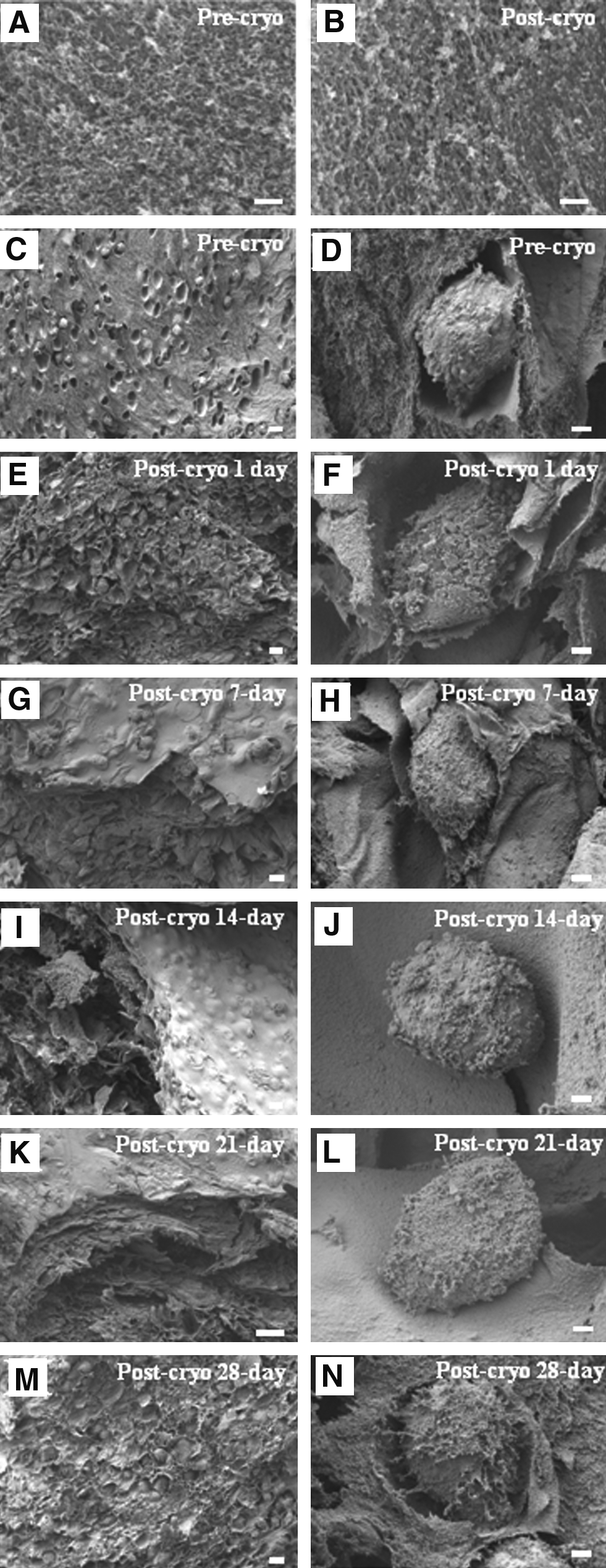
Scanning electron microscopy images of alginate and cell-biomaterial gel constructs showing the impact of cryopreservation. Sections of alginate gels without cells: precryopreserved constructs
Effects of DPSCs proliferation ability and cell-surface antigen in the constructs on cryopreservation time
The proliferation rate of DPSCs cultured in the optimized cell-biomaterial gel constructs in both control and cryopreserved groups was assessed using BrdU staining. The percentage of BrdU-positive cells in control and cryopreserved groups at 1, 7, 14, 21, and 28 days was 82.0±5.0, 80.7±3.9, 78.2±6.3, 77.8±2.1, 76.2±4.0, and 76.0±6.3, respectively (Fig. 6A). DPSCs proliferation showed no significant difference between control and cryopreservation groups. Consequently, there was no cryopreservation impact on cell proliferation.
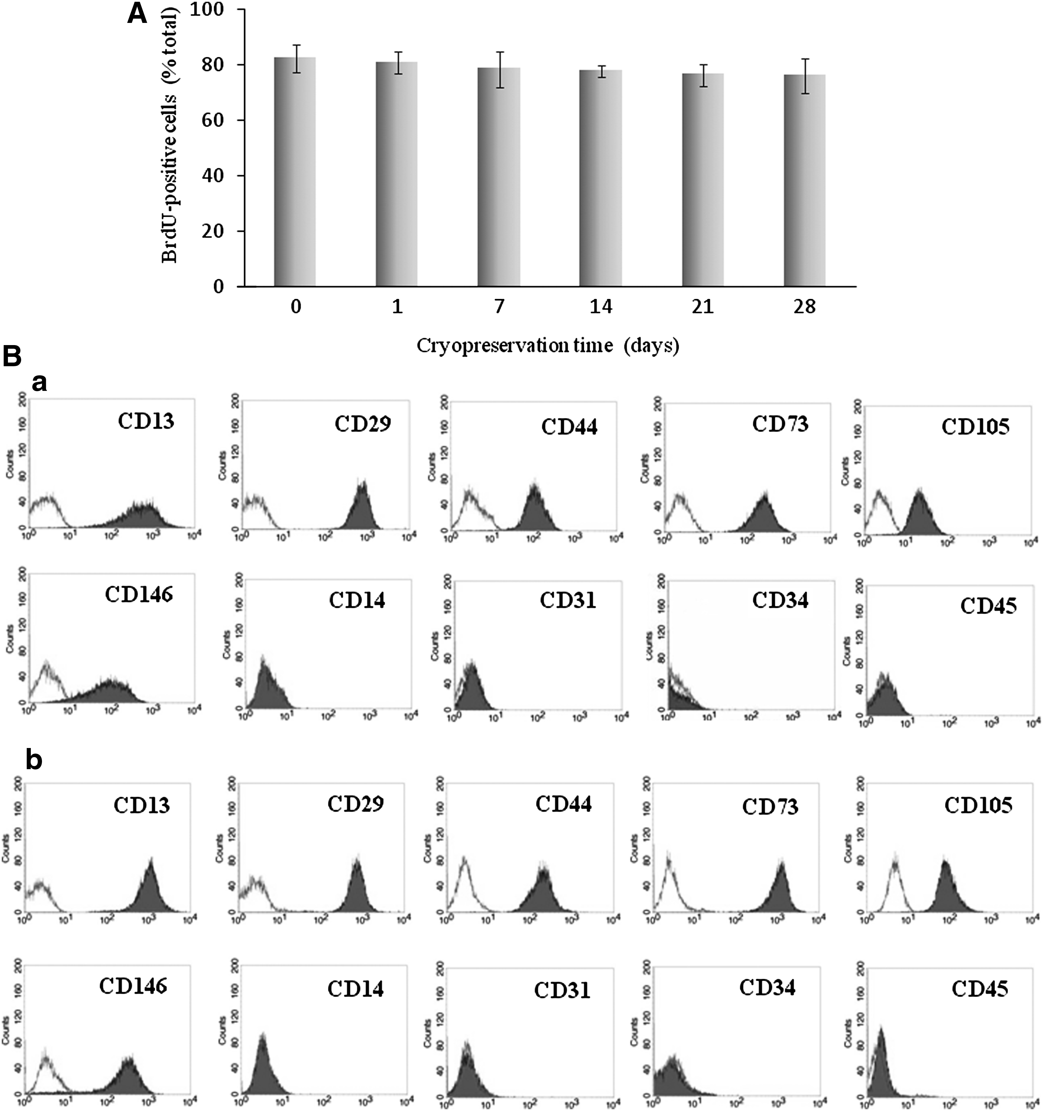
We have previously confirmed that DPSCs exhibit the characteristics of mesenchymal stem cells (MSCs).18,20 In the present study, the expression pattern of DPSCs was investigated and was found to be comparable to that of MSCs. The expression pattern of DPSCs encapsulated in cell-biomaterial gel constructs and cryopreserved in CPAs was also investigated. It was found that encapsulated and cryopreserved DPSCs also preserved the expression pattern of markers even after cryopreservation (Fig. 6B).
Discussion
Alginate gels are easy to manipulate, and the gels used in this study, which are used clinically to protect cells and tissues, are safe. They were solvated using EDTA as a chelating agent and GDL as a pH reducing agent and formed gels in the presence of aqueous divalent cations, such as Ca2+. 19 The alginate gel with a gelation rate of 2% alginate: 100 mM CaCO3: 200 mM GDL=4:1:1 was found to be optimal by investigating the survival rate of cells cultured in the cell-biomaterial gel constructs (Fig. 3). The viability of the encapsulated cells might have been maintained because of the mild internal environment of the gel.
An ideal CPA is a solution that is nontoxic to cells/tissues even after prolonged exposure. Strategies to avoid toxic effects of the solutions require component selection and adjustment of the solute concentration. This minimizes damage caused by ice formation and encourages the formation of an amorphous state in cells/tissues. 21 Currently, effective CPAs commonly consist of a minimum of two (penetrating cryoprotectant+sugar/polymer) or three (penetrating cryoprotectant+sugar+polymer) components. 16 The most traditional penetrating cryoprotectants, including DMSO, glycerol, and EG, have been tested alone and in combination. 22 EG is known for its detrimental effects on developmental potential, membrane integrity and cytoskeletal structure, which has made it the principal penetrating cryoprotectant. 22 As a nonpenetrating cryoprotectant, sugar is used as an important component of osmotic buffers. In this study, we used sucrose, a monosaccharide that can be dissolved more efficiently in solutions of penetrating CPAs, than either the disaccharides or polysaccharides. In terms of polymers, we used PVP for its ability to cryopreserve delicate tissues. 23 This is used in a wide variety of applications in medicine, pharmacy, cosmetics, and industrial production and is also known to be a more effective CPA than other polymers, such as dextran and Ficoll. 24 In this study, the cell viability of DPSCs was investigated in various CPAs. The viability of CPAs in DMEM supplemented with 12% DMSO and 10% FBS, commercial CPAs (Banbanker), DMEM supplemented with 10% EG, and 1.0 M sucrose and 0.00075 M PVP, which were the optimized CPAs, was 49.8%±4.5%, 77.5%±6.5%, and 96.1%±0.4% on day 1, respectively (Fig. 4A). No visual difference between the cell viability of control and cryopreserved gel was found when observed through a confocal microscope with trypan blue staining after 1, 7, 14, 21, and 28 days of cryopreservation (Fig. 4B). Moreover, the maintenance of structure integrity is a prerequisite in the cryopreservation of cell-biomaterial constructs, and the cryopreserved constructs were undamaged, such as the cell surface of SEM images showed no significant difference in microstructure between the control and cryopreserved groups (Fig. 5). The cryopreserved structure of the constructs appeared to have more pores and wrinkles on the surface (Fig. 5). This optimized CPA is DMSO, serum, and animal substance free; and the cryopreserved-thawed DPSCs had normal morphology (data not shown), maintained the properties of multipotent cells with high proliferation ability, and expressed the corresponding marker of MSCs (Fig. 6). Therefore, the matricellular environment of the DPSCs in our cell-biomaterial gel and CPAs constructs also remained unaltered. These results imply that the constructs were well preserved during the cooling-warming cycle and the DPSCs would be well protected.
On the other hand, culturing and expanding cells ex vivo is lengthy and costly work, and obtaining a sufficient cell number for tissue engineering applications would take some months 25 ; therefore, if well-preserved viable cell-biomaterial constructs could be prepared, it would eliminate the lengthy waiting period and bring down the medical fees. An attractive aspect of applying cell-biomaterial gel constructs and their successful long-term storage lies in the potential of providing immediate solutions to patients with acute diseases. Moreover, cell-biomaterial constructs and the application of CPAs are a step toward the application of stem cells such as DPSCs in TERM and provide novel tissue-engineering products, such as ready-to-use and patient-specific products, for cell-based services or drug delivery systems.
Conclusion
In this study, we optimized cell-biomaterial gel constructs and cryoprotectant parameters, and it was found that the characteristics of DPSCs were maintained during encapsulation and cryopreservation. In conclusion, this study indicates that the optimal cell-biomaterial gel constructs and the cryoprotectant could be promising biomaterials. The defined encapsulation/thawing system offers an excellent simple option for cell-banking therapy.
Footnotes
Acknowledgments
The authors are grateful to the members of the Department of Oral and Maxillofacial Surgery, Nagoya University Graduate School of Medicine, for discussions. This work was partly supported by Grants-in-Aid for Scientific Research (Nos. 20659297, 21390507, 21791848) from the Japan Society for the Promotion of Science.
Disclosure Statement
No competing financial interests exist.