




Abstract
The biological function of adherent cell populations strongly depends on the physical and biochemical properties of extracellular matrix molecules. Therefore, numerous biocompatible cell carriers have been developed to specifically influence cell attachment, proliferation, cellular differentiation, and tissue formation for diverse cell culture applications and cell-based therapies. In the present study, we evaluated the mechanical and the cell biological properties of a novel, thin, and planar collagen scaffold. The cell carrier is based on fibrillar bovine collagen type I and exhibits a low material thickness coupled with a high mechanical stability as measured by tensile tests. The influence of this new biomaterial on cell viability, proliferation, and cell differentiation was analyzed using 5-bromo-2-deoxyuridine (BrdU) proliferation assay, immunocytochemistry, water-soluble tetrazolium salt-1 assay (WST-1), live cell imaging, and electron microscopy. Cell culture experiments with the human osteosarcoma cell line Saos-2, human mesenchymal stem cells, and rodent cardiomyocytes demonstrated the in vitro biocompatibility of this chemically noncrosslinked scaffold. Both the mechanical characteristics and the in vitro biocompatibility of this collagen type I carrier facilitate the engineering of thin transferable tissue constructs and offer new possibilities in the fields of cell culture techniques, tissue engineering, and regenerative medicine.
Introduction
Despite of synthetic polymeric scaffolds such as polyglycolic or polylactic acid, 4 natural biomaterials based on extracellular matrix molecules like hyaluronan, fibrin, or collagen play a prominent role as substrates for cells in culture. 2
Collagen type I is the major component of the extracellular matrix in mammals, particularly strongly expressed in several tissues with specific mechanical and structural properties like tendons, ligaments, dermis, bone, dentin, or blood vessels.5–7 Additionally, it is a highly conserved protein that is ubiquitously expressed among mammalian species. 8 For these reasons, purified porcine or bovine collagen I also represents appropriate biocompatible sources for degradable scaffolds in the human system. Mostly used collagen type I compounds were derived from animal tissues, for example, from rat tail, 9 bovine skin, 10 or porcine skin. 11 However, collagen proteins were also isolated from human tissue 12 or produced by recombinant technologies. 13
In basic culture applications as well as in the fields of bioreactor technology and tissue engineering collagen I-based materials were extensively used as cell carrier for various primary cells such as hepatocytes, 14 mesenchymal stem cells, 15 chondrocytes, 16 keratinocytes, 17 smooth muscle cells, 18 cardiomyocytes, 19 or neural cells. 20 Many of the applied solid collagen scaffolds are generated in form of tubes, 21 sponges, 22 fibers, 23 or films and membranes9,24 with different physical and biochemical characteristics. Nevertheless, the availability of standardized chemically noncrosslinked collagen scaffolds that combine a low material thickness with a high mechanically stability is limited.
In the present study, we evaluated a novel, thin, and mechanically stable collagen scaffold for cell culture applications. This cell carrier is based on fibrillar bovine collagen I and was manufactured in form of thin planar sheets. The mechanical properties of the new material were measured by tensile tests. The in vitro biocompatibility of this scaffold was analyzed with different cell populations using 5-bromo-2-deoxyuridine (BrdU)-proliferation assay, immunocytochemistry, water-soluble tetrazolium salt-1 (WST-1) assay, live cell imaging, and electron microscopy.
Materials and Methods
Manufacturing of collagen cell carrier
The collagen I-based cell carrier (CCC) was purchased from Viscofan BioEngineering. Bovine hide was procured under conditions that meet the requirements of ISO 22442–2:2007 (Medical Devices Using Animal Tissues and Their Derivatives—Part 2: Controls on Sourcing, Collection and Handling) in terms of tissue safety and traceability. The hide was dehairred and split into three layers: grain split (located on the former hair side), central split, and flesh split. The central split was selected for further processing. It was submitted to a purification process based on an alkaline treatment (Ca(OH)2/NaOH, pH 13 for 120 h at 20°C) and a subsequent acidic treatment with HCl to yield purified swollen hide splits (pH 2.9), which were mechanically comminuted under strict temperature control (<23°C) into a fibrous collagen gel. The gel was diluted to a collagen concentration of 2.0% (wt/wt), the pH was adjusted with HCl to 2.8 and deaerated. Through a slit nozzle the final gel was extruded onto a conveyor belt that passed through a tunnel drier, which resulted in a thin collagen film. The membranes were adjusted to a pH of 7.3 by a Sørensen buffer/glycerol mixture, cut into rectangular sheets (21×14.8 cm), and finally sterilized by gamma irradiation (25 kGray).
CCC pretreatment for cell culture experiments
For cell culture experiments, CCC sheets were equilibrated overnight in distilled water (200 mL per sheet) at 37°C. Afterward, the wet biomaterial was stamped out into circular discs with a diameter of 21 mm. Discs were transferred into 12-well plates (Corning) preloaded with distilled water. Care was taken that discs were positioned free from creases to the center of the bottom of the well. After removal of residual water, CCC containing culture plates were dried overnight at room temperature under sterile conditions in a laminar air flow. Before cell seeding, dried CCCs were equilibrated with culture medium for 10 min at 37°C. Due to the drying process, collagen discs firmly attached to the plastic well bottom without gaps even after subsequent wetting with culture medium. Thus, seeded cells could only adhere to the upper surface of CCC. Nevertheless, after culturing, the attached CCC could be mechanically detached residual-free from the well bottom with forceps. To culture cells on both sides of the scaffold, attached CCC was removed from the bottom of the well after the preincubation step with culture medium.
Tensile test
To investigate the mechanical performance of the CCC, the load-bearing behavior of dry and wet collagen films was examined using a tensile test apparatus (UTS Testsysteme). Thus, test specimens with a defined shape of 114 mm in length and 14 mm in width in the middle of the specimen were stamped out from CCC sheets. The test samples were reinforced with stainless clamps at the chuck area to connect the specimens to the test apparatus.
After mechanical fixation, the specimens were preloaded up to a load of 0.1 N with a loading speed of 10 mm/min. The measured length between the clamps after preloading was 60.7±0.04 mm (mean±standard derivation [SD], n=5) for the dry specimens and 62.4±1.6 mm (mean±SD, n=5) for the wet specimens. The testing speed was set to 100 mm/min. Under load conditions all tested collagen films failed in the middle of the specimens, which excludes an influence of the clamps. The tests were carried out at a temperature of 22°C±1°C and a relative humidity of 65%±2%. The wet specimens were stored submerged in distilled water for at least 3 h before analysis.
Cultivation of Saos-2 cells
Human sarcoma cell line Saos-2 was cultured in HEPES buffered Dulbecco's modified Eagle's medium (DMEM, PAA Laboratories, Inc.) supplemented with 10% (v/v) fetal calf serum (FCS; Biochrom), L-glutamine (2 mM, PAA), penicillin (100 U/mL, PAA), and streptomycin (100 μg/mL, PAA). Cells were seeded onto CCCs and on plastic surfaces of conventional cell culture plates (Corning) at a density of 15,000 and 25,000 cells per cm2 and cultured for up to 14 days in a humidified incubator at 37°C and 5% CO2. The culture medium was renewed every 3–4 days.
Isolation and cultivation of human mesenchymal stem cells
Human mesenchymal adult stem cells (hMSC) were kindly provided by Wilhelm Aicher (Tuebingen Medical Center, University of Tuebingen). Cells were prepared from human adult bone marrow after approval of the local ethical committee at the University of Tuebingen and with the consent of patients. After density gradient centrifugation, hMSC were isolated by plastic adherence, grown in DMEM containing human serum and platelet extract, and characterized by flow cytometry and differentiation assays as previously described. 25 After further expansion in HEPES buffered DMEM containing 10% (v/v) FCS, L-glutamine, penicillin, and streptomycin, cells of passages 3–5 were seeded onto CCC (50,000 cells/cm2) and cultured for up to 40 days in a humidified incubator at 37°C and 5% CO2. The culture medium was renewed every 3–4 days.
Osteogenic differentiation was performed in basic culture medium supplemented with glycerol-2-phosphate disodium salt hydrate (10 mM, Sigma), ascorbate-2-phosphate (50 μM, Sigma), and dexamethasone (100 nM, Sigma).
Isolation and cultivation of rodent cardiomyocytes
Cardiomyocytes were prepared from fetal mice (C57BL/6) at embryonic day 18 and from neonatal rats (Wistar). Animals were handled in accordance with local guidelines for the ethical use of animals in research at the University of Tuebingen. Isolation of cardiomyocytes was carried out as previously described. 26 Ventricles were dissected and incubated with papain 0.2% (w/v; Sigma) digestion solution containing 0.05% (w/v) DNAse I (Sigma) for up to 60 min at 37°C. During incubation tissues were carefully triturated every 15 min with a fire-polished blue tip. After completion of the incubation period, FCS was added to the enzyme solution to reach a final concentration of 10% (v/v). Tissue was then triturated with a fire-polished yellow tip to obtain a single cell suspension. After centrifugation at 220 g for 5 min, cell pellet was resuspended in culture medium (DMEM/F12 [PAA], 10% [v/v] FCS, penicillin, streptomycin, L-glutamine, insulin/transferrin/selenite mix [1:100, Invitrogen, Darmstadt, Germany], Albumax [1 mg/mL, Invitrogen], hydrocortisone [1 μM, Sigma], glucagon [14.3 nM, Sigma], 3,3′,5′-triiodo-L-thyronine [1 nM, Sigma], ascorbate-2-phosphate [200 μM, Sigma], linoleic acid [20 μM, Sigma], and estradiol [10 nM, Sigma]). Cells were seeded onto CCC at a density of 100,000 cells per cm2 and cultured for up to 4 weeks in a humidified incubator at 37°C and 5% CO2. Culture medium was renewed every 3 days.
Fluorescent labeling and time-lapse monitoring of vital Saos-2 cells
Saos-2 cells were labeled with the lipophilic fluorescence cell tracker FM DiI (Cell Tracker FM-DiI, Invitrogen) for time-lapse monitoring of dividing cells on CCC. Cells were incubated in DiI solution (4 μg/mL in Hank's buffered salt solution, PAA) for 20 min at 37°C. Thereafter, Saos-2 cells were washed three times with prewarmed L-15 Leibovitz medium (PAA). Analysis of living cells was carried out in L-15-medium supplemented with 10% (v/v) FCS, penicillin, streptomycin, and L-glutamine using a confocal laser scan microscope (LSM Exciter Zeiss). Laser scans were recorded every 2 min for up to 12 h.
Fluorescent labeling and real-time monitoring of beating cardiomyocytes
Beating cardiomyocytes were labeled with the nucleus fluorescence tracker Hoechst 33342 (Invitrogen). After incubation in Hoechst solution (5 μg/mL in cell culture medium) for 45 min at 37°C, cells were washed two times with prewarmed cell culture medium. Real-time fluorescence monitoring of Hoechst 33342–labeled cells was performed using a microscope video system (Olympus IX 50).
WST-1 assay
WST-1 cell viability assay was performed with the WST-1 Roche kit (Roche) according to the manufacturer's recommendations. Cells were washed twice with prewarmed cell culture medium to remove putative nonattached cells. Thereafter, cell cultures were incubated with WST-1 solution (dilution 1:11 in culture medium) for 30 and 60 min. After careful stirring, 100 μL of medium was transferred into a 96-well plate. Absorbance was measured at 450 nm against 630 nm as reference with an ELISA reader (Dynatec, MR 5000; Dynatech Laboratories).
Immunocytochemistry
Immunocytochemistry was performed on cell cultures as described previously. 27 Cells were fixed with 4% phosphate buffered paraformaldehyde for 20 min. After washing three times with Tris buffer (TBS, 25 mM Trizma base, 140 mM NaCl, pH 7.4; Sigma) for 5 min, cultures were blocked with TBS containing 0.1% bovine serum albumin, 10% (v/v) swine serum (Biochrome) and 0.3% (v/v) Triton® X-100 (Roth) for 30 min. Primary antibodies vimentin (monoclonal, mouse, 1:100; Dako), vimentin, (polyclonal, rabbit, 1:1000; Abcam), alpha-actinin (monoclonal, mouse, 1:1000; Sigma) were applied overnight at 4°C. After washing three times with TBS for 10 min, cells were incubated with fluorochrome-linked secondary antibodies (goat anti-rabbit, Alexa 488, 1:500, Invitrogen; goat anti-mouse IgG, Cy3, 1:200; Jackson ImmunoResearch) for 30 min at room temperature.
Cell nuclei were stained with DAPI (4′-6-diamidino-2-phenylindole, 200 ng/mL; Roth) or Sytox Green (100 nM; Invitrogen) solution. Cultures were washed three more times for 10 min with TBS, briefly rinsed in distilled water, dried, and covered with Kaiser's gelatine solution (Merck).
BrdU labeling
The rate of proliferation of Saos-2 cells was determined by BrdU-proliferation assay. BrdU (10 μM) was added to the culture medium for 1 h. After fixation with ethanol (70% [v/v], 50 mM glycine) at −20°C, BrdU-positive cells were observed using immunofluorescence with the BrdU Detection Kit I (Roche) as previously described. 28
Scanning electron microscopy
Cell cultures were fixed with a solution containing 2% glutaraldehyde and 3% formaldehyde in cacodylate buffer (0.1 M cacodylate, 0.09 M sucrose, 0.01 M MgCl2, and 0.01 M CaCl2, pH 6.9) for 1 h on ice and washed with cacodylate buffer. After washing five times in TE buffer (0.02 M TRIS and 1 mM ethylenediaminetetraacetic acid, pH 6.9), cells were dehydrated on ice with a series of ascending acetone solution (10%, 30%, 50%, 70%, 90%, and 100% [v/v], 15 min each), followed by critical-point drying with liquid CO2. Samples were sputter coated with a gold film of approximately 10 nm thickness before examination with a field emission scanning electron microscope (DSM982 Gemini, Zeiss) at an acceleration voltage of 5 kV using the Everhart Thornley SE detector and the inlens-SE detector in a 50:50 ratio. Extracellular matrix compounds were measured by energy-dispersive X-ray (EDX) analysis (ISIS 300 EDX-system; Oxford Instruments).
Transmission electron microscopy
Cell cultures were fixed with 2% glutaraldehyde (Sigma) and 5% paraformaldehyde (Sigma) in HEPES buffer (100 mM HEPES, 90 mM saccharose, 10 mM MgCl2, and 10 mM CaCl2, pH 7.0) at 4°C for 2 h. Samples were postfixed with 1% osmium tetroxide in 100 mM phosphate buffer pH 7.2 for 1 h on ice, washed with H2O, treated with 1% aqueous uranyl acetate for 1 h at 4°C, dehydrated in a graded series of ethanol, infiltrated with ethanol/resin mixtures, and finally embedded in Epon (using glycid ether 100, Roth). Ultrathin sections were stained with uranyl acetate and lead citrate and viewed under a Philips CM10 electron microscope at 60 kV using a 30 μm objective aperture.
Quantification of cell numbers and statistics
To quantify the number of DAPI and BrdU-positive cells, each CCC or control well was microphotographed with a 10×objective at four defined areas using a fluorescence microscope (Axiovert 200, Zeiss). Each area was located at the half radius in the x- and y-axis relating to the center of the well. The nuclei number of analyzed areas per well (2384 mm2) was calculated to 1 mm2.
Statistical significance was determined by Student's t-test or by nonparametric Mann–Whitney U test. A p-value <0.05 was considered to be statistically significant.
Results
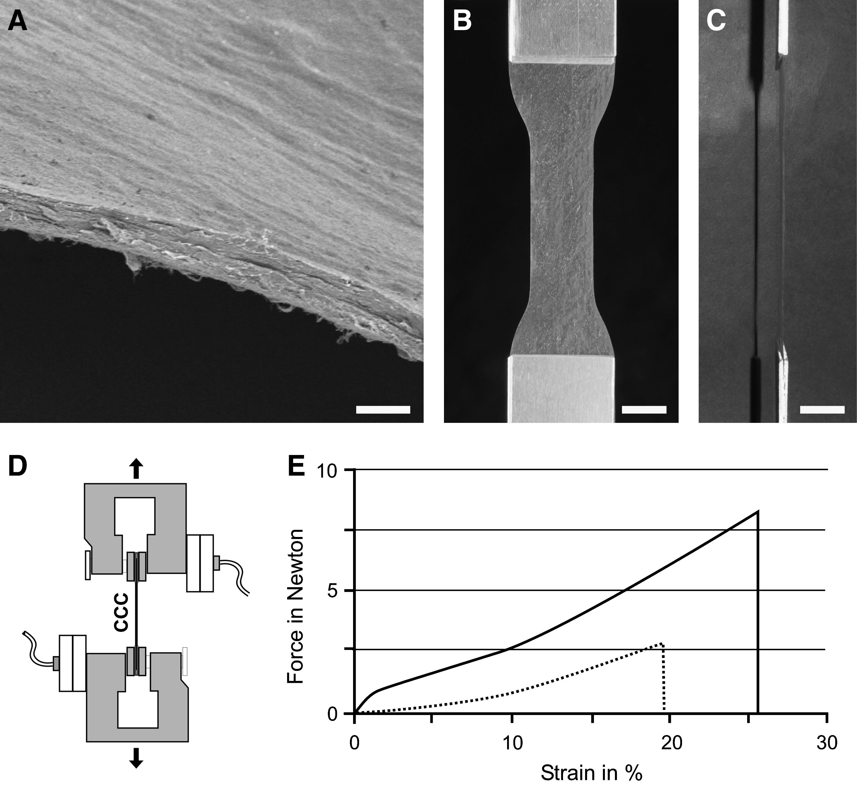
In this study, we analyzed the mechanical properties and the in vitro biocompatibility of a novel CCC. The scaffold is based on chemically noncrosslinked fibrillar bovine collagen type I and was delivered in form of thin planar and translucent sheets. For mechanical testing and cell culture experiments, sheets were pretreated and stamped out in different defined forms as described in the materials and methods part. The thickness of CCCs was measured using electron and laser confocal microscopy (Figs. 1A, 6, 7). The dry CCC sheet exhibited a thickness between 15 and 25 μm dependent on the relative humidity of the environment. The thickness of wet CCC was determined between 35 and 45 μm.
Mechanical properties of CCC
To investigate the mechanical characteristics of dry and wet CCCs the load-bearing behavior of defined collagen I specimens was examined using a tensile test apparatus (Fig. 1B–D). The maximum load at fracture for the applied specimens was measured 8.1 N±1.8 N (mean±SD, n=5) for the dry and 2.7 N±0.3 N (mean±SD, n=5) for the wet specimens (Fig. 1E). To compare the mechanical strength of CCC with data from the literature the strength at break for the specific specimens was related to the assumed mean area cross section of dry (0.28 mm2) and wet (0.56 mm2) CCC. The strength at break can be calculated to 38.6±8.6 MPa (mean±SD, n=5) under dry condition and 7.7±0.9 MPa (mean±SD, n=5) under wet condition. The mean value of the measured strength at failure was determined 25.7%±3.1% (mean±SD, n=5) for the dry and 19.2%±1.8% (mean±SD, n=5) for the wet CCC. Although tensile fracture strength showed higher values under dry conditions, the ductility of the wet collagen specimens at a specific load was increased. At a load of 2.5 N wet specimens were elongated by 19%, while dry CCC elongated by only 9%.
Cell biological evaluation of CCC with Saos-2 cells
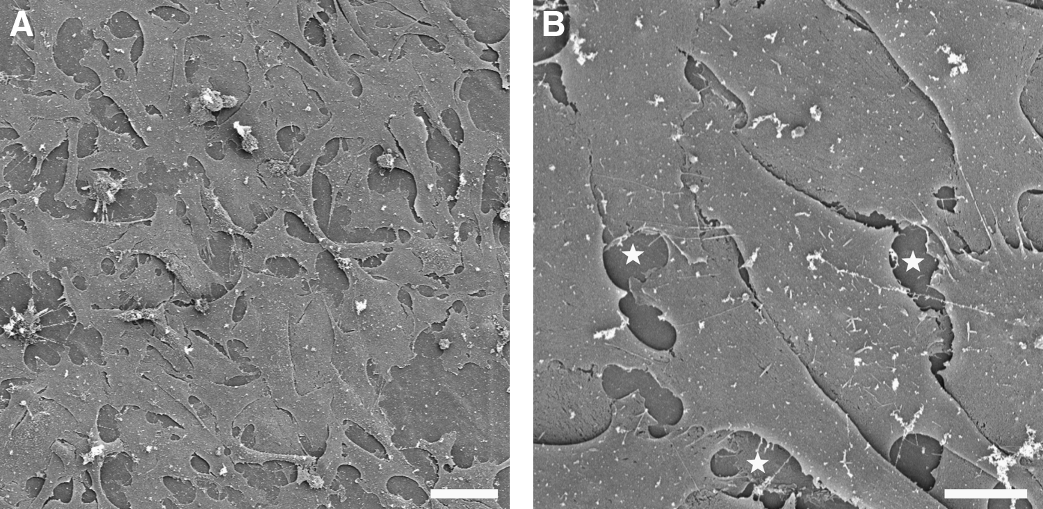
The human osteosarcoma cell line Saos-2 was cultured on CCC to evaluate the cell biological characteristics of collagen scaffold. Viability, proliferation, and adherence of Saos-2 cells cultured on CCC were demonstrated by vimentin immunocytochemistry, BrdU-proliferation assay, and scanning electron microscopy (Figs. 2–4). Further, time-lapse fluorescence imaging of DiI-labeled Saos-2 cells circumstantiated their proliferation on the CCC (Supplementary Movie S1; Supplementary Data are available online at

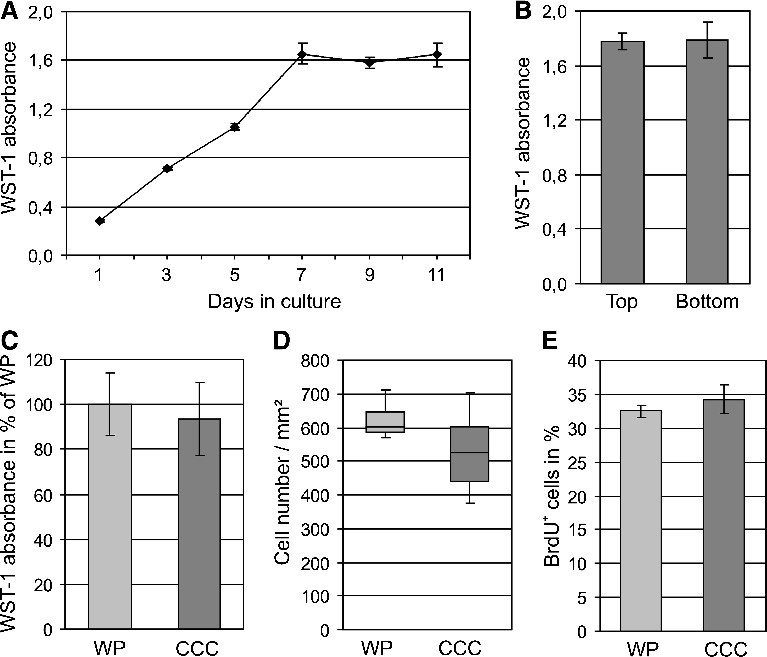
In addition to the WST-1 analysis, cell proliferation was determined with a BrdU-proliferation assay. Quantification of total cell numbers revealed a median cell density of 526 cells/mm2 (range 439–603, n=6) in the CCC group and 601 cells/mm2 (range 586–644, n=6) in the culture plate group (p=0.18, Mann–Whitney U test; Fig. 4D). The percentage of BrdU-positive cells was 34.2%±2.1% (mean±SD, n=6) on collagen scaffolds and 32.5%±0.9% (mean±SD, n=6) on culture plates (p=0.08, Student's t-test; Fig. 4E).
Differentiation of human mesenchymal stem cells into osteogenic direction
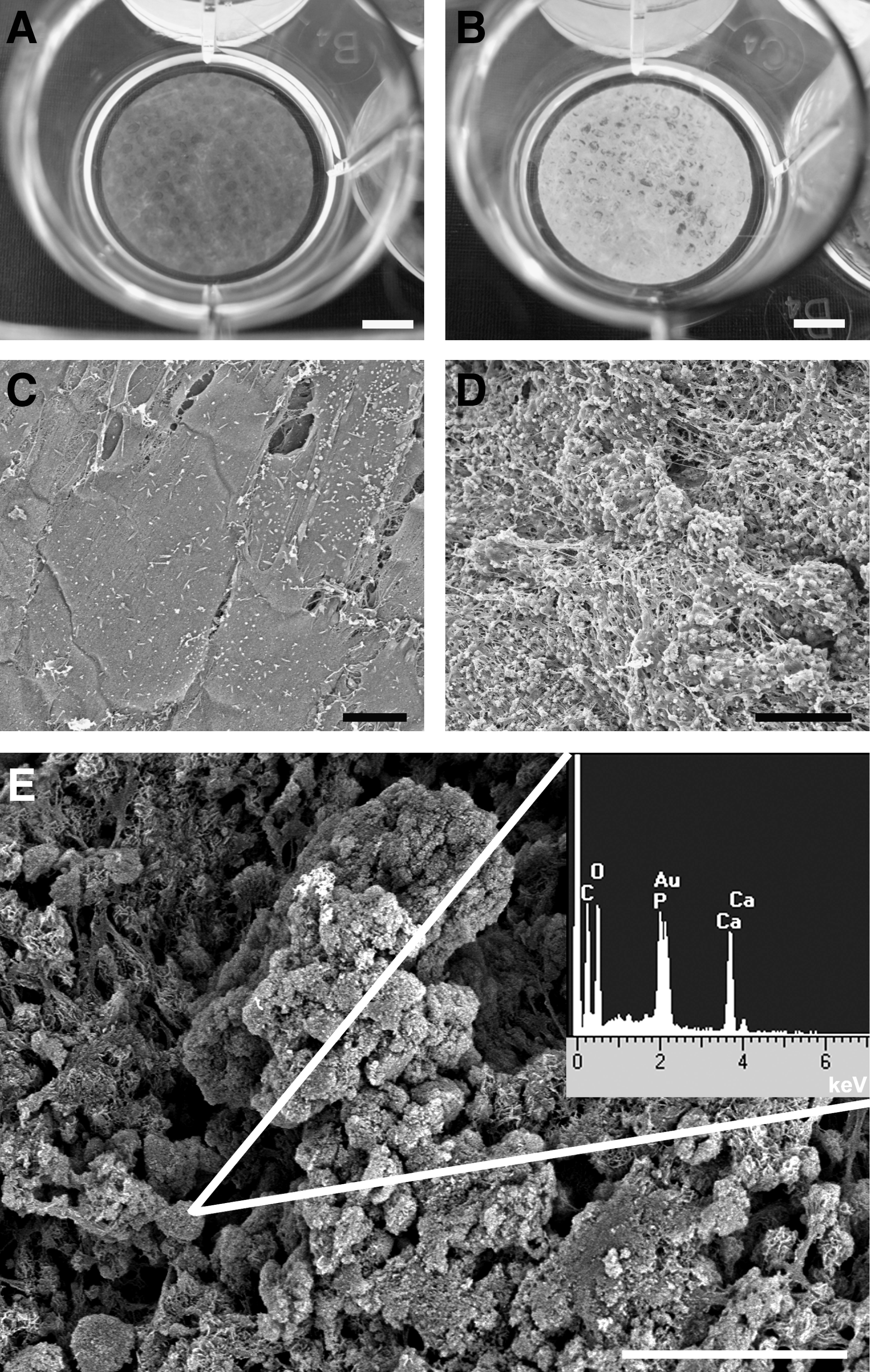
Human mesenchymal stem cells were taken into culture to test their osteogenic differentiation capacity on CCC. Thus, we seeded mesenchymal stem cells derived from human bone marrow on collagen scaffolds and cultured them using normal or osteo-inductive cell culture medium for up to 40 days. Under osteo-inductive culture conditions mesenchymal stem cells differentiated into the osteogenic lineage (Fig. 5B, D). Scanning electron microscopy with EDX analysis confirmed the calcium phosphate mineralization (Fig. 5E). Calcification was not detected, neither in stem cell cultures missing osteo-inductive factors (Fig. 5A, C), nor in cell-free scaffold controls incubated with osteo-inductive cell culture medium (data not shown).
Cultivation of rodent cardiomyocytes on CCC
Primary cardiomyocytes prepared from fetal murine and neonatal rat hearts were cultured for 14 days on CCC. In the first 24 h of culturing, heart cells adhered to the scaffold surface and started their spontaneous contractile activity. The cultures maintained their autonomous beating over the entire 16-day observation period (Supplementary Movie S2). Immunocytochemical staining for the striated muscle-specific protein alpha-actinin and scanning electron microscopic views also exemplified differentiation and cell adherence of cardiomyocytes on CCC (Figs. 6A, B; 7A, B). Striated muscle-specific contractile elements and cellular contact zones could be clearly identified by transmission electron microscopy (Fig. 6C–E).


In further experiments, CCC was seeded onto both sides with cardiomyocytes, and subsequently cultured for 14 days as free-floating cell culture constructs. Three-dimensional reconstruction of the immunolabeled cardiomyocytes using laser confocal microscopy emphasized the adherence of myocytes to both surfaces of collagen scaffold (Fig. 7C–F, Supplementary Movie S3). Under these cell culture conditions, adherent myocytes did not only show their autonomous beating but were also able to rhythmically contract the whole CCC-cell construct. Interestingly, the force of contractions was sufficient that the construct could move through the entire cell culture plate (Supplementary Movie 4).
Discussion
In cell culture systems, the physical and the biochemical characteristics of scaffolds can strongly influence the attachment, proliferation, differentiation, and physiological function of cultured cells. Collagen is the major component of the extracellular matrix of most soft and hard connective tissues in mammals. The natural cell biological and mechanical properties of native collagen are therefore one of the major reasons that collagen-based biomaterials are widely used in cell culture and tissue engineering approaches.2,6,7 The mechanical behavior, degradation, shrinkage, and water uptake of collagen based scaffolds depend on the hierarchical structure, collagen network, and length of collagen fibrils, fibers, and/or fiber bundles that are influenced by the purification and the manufacturing process.6,10,24,29 To increase the mechanical strength of the collagen material is often modified by physical crosslinking techniques such as UV-irradiation, 30 dehydrothermal treatment, 31 or by chemical crosslinking methods.10,24 These modifications do not only change the biochemical and physical characteristics of collagen molecules but might also influence the cellular response and cell–matrix interactions.
In the present study, we evaluated a novel chemically noncrosslinked scaffold based on fibrillar bovine collagen I. This solid and planar cell carrier exhibits a low material thickness coupled with a high mechanical stability. We analyzed the mechanical properties of dry and wet CCCs using a tensile test apparatus. In our study, the strength at break of CCC specimens was calculated to 38.6 MPa under dry and 7.7 MPa under wet condition. Gentleman et al. extruded and crosslinked bovine achilles tendon collagen fibers with diameters of 125 μm. 29 The strength at break of fiber containing scaffolds ranged from 2.9 to 36.0 MPa. In our study, the CCC showed similar material properties, but without chemical crosslinking treatment. Angele et al. produced collagen crosslinked and noncrosslinked scaffolds derived from bovine and equine collagen. 32 The strength at break of noncrosslinked bovine scaffolds was determined 0.12 MPa under dry conditions and 0.033 MPa under wet conditions. However, it has to be taken into consideration that the different testing speed of 127 mm/min by Gentleman et al., and only 1 mm/min by Angele et al. can strongly influence the test results for visco-elastic materials like collagen scaffolds.
The mechanical characteristics of CCC make this material suitable for mechanical manipulation without destroying the three-dimensional structure of the biomaterial. For this reason, the CCC-cell constructs could be easily transferred after cell culturing for histological analysis or for cell-based applications in vivo. In the field of tissue engineering, the mechanical properties of standardized CCC sheets could be also interesting for mechanical stimulation of biomaterial–cell constructs and for analysis of the mechanobiological response of cultured cells.
In the present study, the biocompatibility of CCC in vitro was demonstrated with the human osteosarcoma cell line Saos-2, human mesenchymal stem cells, and rodent cardiomyocytes. Analysis of cell morphology by fluorescence microscopy and electron microscopy emphasized the high affinity of flattened cells to the scaffold surface and the homogenous cell distribution on the CCC. The growth rate of Saos-2 cells on CCC was comparable to conventional culture plates. Quantification of total cell number of Saos-2 cells cultured on CCC revealed a relative median cell density of 87.5% compared to conventional culture plates. These data were strongly different from those analyzed on other solid collagen scaffolds. 33 In that study, four different commercial available collagen membranes were evaluated with Saos-2 cells. The percentage of cell density on these scaffolds compared to conventional culture plates only ranged between 0% and 20.8%. However, it is noteworthy that our results are not exactly comparable with those data due to the different cell seeding density, cell culture period, and cell culture medium.
To further evaluate the cell biological properties, human mesenchymal stem cells were cultured on CCC. These stem cells differentiate into osteogenic direction when osteoinductive cell culture medium is supplemented. 34 Collagen type I is the main organic extracellular matrix compound in bone and is therefore regarded as a suitable biomaterial for bone tissue engineering.1,35 In our study, we could confirm this cell biological nature of collagen. Under osteo-inductive culture conditions human mesenchymal stem cells cultured on CCC strongly calcified in a time-dependent manner.
Primary rodent cardiomyocytes were the third type of cells we used to analyze the in vitro biocompatibility of CCC. Functional cardiomyocyte tissue constructs using various biomaterials offer interesting perspectives for pharmacological test systems and for basic research to investigate cellular mechanisms that play major roles in cardiac cell differentiation, apoptosis, or electrophysiological behavior.19,21,36 In our study, we cultured fetal and neonatal rodent cardiomyocytes either on one or on both CCC sides. Within 24 h after cell seeding, cardiomyocytes adhered to the CCC surface, and started their spontaneous contractile activity. The cardiomyocytes exhibited cardiac-specific markers and showed in all applied methods a pronounced adherence to surfaces of collagen scaffolds. Further, the adherent and autonomously beating cardiomyocytes were able to rhythmically contract the whole CCC-cell construct due to the low thickness, the elasticity, and mechanical stability of the CCC.
The in vitro experiments showed that cells did not penetrate the matrix even after 6 weeks in culture. Thus, it is of primary interest in future studies to analyze the time-dependent scaffold degradation in animal grafting experiments. With respect to surgical approaches and cell-based therapies the standardized and planar collagen sheets might be particularly appropriate for reconstruction of hard and soft connective tissue. In addition, the CCC will be useful to place cells like stem cells and progenitors precisely at the required location of the donor tissue. Due to its low thickness of about 40 μm the amount of material necessary is almost negligible. In this context, it is noteworthy that purified collagen is generally considered as a biodegradable and nontoxic biomaterial in tissue engineering applications.2,6,7
Besides the biocompatibility of CCC in vitro, the cell culture experiments demonstrated an important analytical advantage of this scaffold. In contrast to many thicker scaffolds, the low thickness of CCC in combination with its only minimal autofluorescence makes this biomaterial suitable for fluorescence microscopy. Thus, fluorescence-labeled cells could be microscopically live-monitored during the cultivation process. Additionally, whole CCC-cell construct seeded on both sides could be analyzed throughout the matrix without sectioning using laser scan microscopy.
In conclusion, both, the in vitro biocompatibility and the mechanical characteristics of CCC open new possibilities for basic cell culture, complex bioreactor systems, and pharmacological in vitro models. It will be of interest to modify this CCC, for example, by the integration of pores or by combinations with tissue-specific extracellular matrix molecules and growth factors. In a next step, based on our initial cell culture experiments, the in vivo biocompatibility and scaffold degradation has to be evaluated in detail in animal models to reveal the potential of CCC in the fields of implantology, tissue engineering, and regenerative medicine.
Footnotes
Acknowledgments
This research was supported by University of Tuebingen Fortuene Grant 1557–0-0 and by Viscofan BioEngineering. We would like to thank Andrea Wizenmann for her helpful comments on the article, Wilhelm Aicher for kindly providing the human mesenchymal stem cells, and Gerd Geiger for technical assistance in electron microscopy.
Disclosure Statement
The authors disclose following commercial associations that might create a conflict of interest in connection with the submitted article: the authors Timo Schmidt and Lothar Just applied for a patent on the CCC.
References
Supplementary Material
Please find the following supplemental material available below.
For Open Access articles published under a Creative Commons License, all supplemental material carries the same license as the article it is associated with.
For non-Open Access articles published, all supplemental material carries a non-exclusive license, and permission requests for re-use of supplemental material or any part of supplemental material shall be sent directly to the copyright owner as specified in the copyright notice associated with the article.
