
Abstract
The three-dimensional (3D) extracellular matrix (ECM) environment plays a critical role in mediating normal cellular behavior and tissue organization. While commercially available microcarriers have shown promise, limited research has been conducted on the design of tissue-specific, custom-fabricated microcarriers, engineered to mimic the composition of the native ECM of cells or tissues of interest. Moving toward this goal, methods were developed to fabricate microcarriers from decellularized adipose tissue (DAT) via minimally-cytotoxic protocols. Characterization by microscopy confirmed the production of stable spherical microcarriers, with a microporous surface topography and porous interior. The mean diameter of the DAT microcarriers was 934±51 μm, while the porosity was estimated as 29%±4% using liquid displacement. Stability and swelling behavior over 4 weeks indicated that the DAT microcarriers were effectively stabilized with the photochemical crosslinking agent rose bengal, with total protein release in a simulated physiological environment remaining below 10 μg/mL at all time points. Preliminary cell culture studies with human adipose-derived stem cells (ASCs) in a spinner flask system indicated enhanced cell attachment and proliferation of ASCs on DAT microcarriers over 14 days, as compared with gelatin control microcarriers fabricated using similar methods. Testing confirmed injectability of the DAT microcarriers, further supporting the clinical potential of the approach for localized cell delivery and small volume augmentation in plastic and reconstructive surgery. Overall, tissue-specific microcarriers prepared from solubilized DAT were found to be highly supportive of human ASCs cultured in a 3D dynamic environment.
Introduction
Microcarriers have been investigated in a variety of tissue-engineering strategies as cell culture substrates for anchorage-dependent cell attachment and proliferation.4–6 Cell-seeded microcarriers are cultured in suspension, typically with low-shear stirring in a spinner flask system. 7 Culturing conditions can be more precisely controlled using these methods, relative to two-dimensional (2D) static culturing techniques. 8 Further, the dynamic 3D microenvironment can promote a more native cell morphology and phenotype, as well as protein secretion more closely resembling the in vivo response. 9 Clinically, microcarriers also hold promise as direct injectables for use in the repair of small-volume or irregular defects in plastic and reconstructive surgery. 10 For the repair of larger-volume or shape-specific defects, composite scaffolds incorporating cell-seeded microcarriers encapsulated within hydrogels or macroporous scaffolds have been investigated. 11
Due to the broad potential of this approach, microcarrier scaffolds have been developed from a range of materials, including poly(lactic acid),12,13 poly(lactic-co-glycolic acid),14–18 dextran, 19 alginate, 20 gelatin, 21 and collagen.22,23 Interestingly, limited research has focused on custom-designed, tissue-specific microcarriers, engineered to mimic the native ECM of tissues of interest. Decellularized matrices represent a promising base material for the design of cell- or tissue-specific microcarriers. To date, decellularization protocols have been successfully established for a broad range of tissues, including our recent work on adipose tissue.24–38 These strategies aim at removing antigenic components while preserving the native ECM structure and composition.
Decellularized adipose tissue (DAT) has shown particular promise as a naturally derived biomaterial for adipose regeneration. Adipose tissue is enriched in basement membrane, which is known to play critical roles in both organogenesis and wound healing.39,40 As a result, scaffolds derived from adipose matrix may possess unique regenerative qualities, which have yet to be fully resolved. Established detergent-free protocols for the decellularization of human fat effectively remove cellular components, nucleic acids, and lipids from the matrix, while preserving the native basement membrane. 24 Interestingly, intact DAT scaffolds induce the expression of the master regulator adipogenic genes, peroxisome proliferator activated receptor-γ, and CCAAT-enhancer binding protein-α, in seeded human adipose-derived stem cells (ASCs) in vitro, without the need for exogenous differentiation factors. 24 This research highlights the critical role of the ECM in mediating cellular responses, and suggests the promise of tissue-specific matrices.
To date, no methodology has been established that incorporates the composition of a fully decellularized tissue in the form of engineered microcarriers. Most similarly, Choi et al. developed techniques to extract the lipid fraction of adipose tissue and employed the resulting gel as an injectable biomaterial and as the basis for larger-volume scaffolds.41,42 Since DAT provides a highly purified form of the adipose ECM, DAT-based microcarriers could function as minimally antigenic, injectable adipogenic scaffolds, either alone or in combination with seeded autologous or allogenic stem cells, for use in soft tissue augmentation procedures. In comparison to commercially available microcarriers derived from other tissue sources or synthetic polymers, the ECM in the DAT matrix may resemble the composition of the natural ASC microenvironment found in adipose tissue, to more closely replicate the native stem cell niche in culture.
The objectives of this study were (1) to develop methods for the fabrication of porous matrix-derived microcarriers from decellularized tissues using minimally cytotoxic methods; (2) to characterize the properties of these injectable, anchorage-dependent cell culture substrates; and (3) to assess the potential for DAT-based microcarriers to support human ASC attachment and proliferation, as compared with gelatin-based control microcarriers.
Materials and Methods
Materials
Unless otherwise stated, all chemicals were purchased from Sigma-Aldrich Canada Ltd. (Oakville, ON, Canada), and were used as received.
Procurement and decellularization of adipose tissue
Freshly excised adipose tissue samples were obtained from female patients undergoing reduction mammoplasty or abdominoplasty procedures at Kingston General Hospital or Hotel Dieu Hospital in Kingston, ON, Canada. Human research ethics board approval from Queen's University was obtained for this research (REB# CHEM-002-07). The tissues were transported to the lab for decellularization or human ASC extraction using established methods. 24
Solubilization of DAT
DAT samples sourced from multiple patients (n=5) were pooled to yield collective samples (20–40 g). The samples were decontaminated by rinsing in 70% (v/v) aqueous ethanol and rehydrated in sterile, cation-free phosphate buffered saline (PBS). The DAT was minced (∼1 cm3 fragments), suspended in 0.5 M acetic acid, and incubated for 30 min (37°C, 150 RPM). Pepsin (1064 U/mg protein) was dissolved in 0.5 M acetic acid at a concentration of 500 mg/mL. The DAT was digested in the acid-pepsin solution at a concentration of 0.1 mL/g of DAT for 20 h (37°C, 150 RPM). The pepsin was inactivated by raising the pH above 8.0 with 10 M NaOH, and the insoluble fraction was removed by centrifugation (15,000 g, 30 min, 4°C).
Total protein content of the solubilized DAT (DATsol) was determined using the Bio-Rad Protein Assay (Bio-Rad Laboratories, Inc., Hercules, CA) with an albumin standard, following the manufacturer's instructions. Triplicate DATsol samples were measured at 595 nm using a Synergy™ HT multidetection microplate reader (n=3, N=3).
Microcarrier fabrication
Sterile, composite DAT/alginate microcarriers were fabricated using a custom-designed apparatus employing an air-jet droplet technique (Fig. 1). Adapting the methods of Tsai et al. 43 for collagenous microcarriers, DATsol was combined with sodium alginate (1.0%–4.0% [w/v]) in varying volumetric ratios (3:2 and 4:3 parts DATsol to alginate). The resulting mixtures (Table 1) were added drop-wise to 1.5% (w/v) calcium chloride solution (pH 7.2) through a blunt-ended, 21-gauge Punctur-Guard® needle (ICU Medical, Inc., Vernon, CT) via a syringe pump in the presence of a compressed nitrogen jet. As a control, bovine gelatin (50 mg/mL, Type B) was used to prepare gelatin/alginate microcarriers via identical methods.

DAT, decellularized adipose tissue. Bold value indicates formulation selected for Phase II testing.
To stabilize the DAT or gelatin within the composite microcarriers, photosensitizing dyes (rose bengal [RB] and riboflavin [Rib]) were assessed as potential crosslinkers, in comparison to glutaraldehyde (GTA). 32 RB was combined with deionized water under dark conditions to yield 0.01% (w/v) RB photosensitizing solution. 44 The composite microcarriers were incubated in the dark in RB solution at room temperature (50 RPM, 30 min). After washing in deionized water to remove excess dye, photochemical crosslinking was performed by exposure to visible light for 8 h. Similarly, a 0.1% (w/v) Rib photosensitizing solution was prepared with Rib 5′-monophosphate sodium salt dihydrate in deionized water. 45 The composite microcarriers were immersed in the Rib solution under dark conditions (50 RPM, 10 min) at room temperature, followed by repeated rinsing with deionized water. Photochemical crosslinking was induced with an EXFO Lite high-intensity UV light source (320–480 nm filter), at a relative intensity of 5 mW/cm2 for 600 s. As a conventional crosslinking control, the composite microcarriers were treated with 0.1% (v/v) GTA solution for 20 h at room temperature under constant agitation (50 RPM), followed by repeated rinsing with deionized water.
After crosslinking, the composite microcarriers were treated with 50 mM sodium citrate for 15 min under gentle agitation at room temperature to extract the alginate phase. To maximize extraction, the microcarriers were rinsed thrice for 30 min in cation-free PBS.
Microcarrier characterization: Phase I
Scanning electron microscopy and optical microscopy
Scanning electron microscopy (SEM) was performed to examine the microcarrier architecture. Samples of individual microcarriers were flash frozen with liquid nitrogen and fractured with a frozen blade, to facilitate visualization of the internal microarchitecture. The intact and fractured microcarrier samples were supercritically dried, mounted onto microscopy studs, pulse coated with gold, and assessed with a JEOL JSM-840 microscope at a working distance of 15 mm and an accelerating voltage of 10 kV. The microcarriers were also visually assessed by optical microscopy (Zeiss Invertoskop 40C microscope).
Fluorescent alginate extraction
The efficacy of the alginate extraction treatment was confirmed by fabricating microcarriers with fluorescent alginate. 46 Briefly, 3% (w/v) alginate solution was prepared (pH 11.0), and fluorescein 5(6)-isothiocyanate (FITC) mixed isomers (1 mg/mL) in dimethylsulfoxide was added to the alginate, to achieve a ratio of 0.025 mg FITC/mL. The mixture was agitated for 1 h (150 RPM) at 40°C, after which ammonium chloride solution was added to achieve a 50 mM final concentration. The FITC-infused alginate was used to fabricate the composite DAT/alginate microcarriers. As a control, pure FITC-labeled alginate microspheres were also fabricated. The microcarriers were imaged with varying sodium citrate treatments (0, 5, 10, 15, 20, 30 min) using a Zeiss AxioImager.M1 fluorescence microscope (488 nm excitation) at 10X and 20X magnification.
Microcarrier diameter, size distribution, and in vitro protein release
Selected microcarrier formulations (Table 1) were fabricated, as described. For each formulation, one hundred microcarriers (n=100) were selected at random and immersed in 5 mL of Ringer's simulated physiological fluid (8.6 mg/mL NaCl, 0.3 mg/mL KCl, and 0.33 mg/mL CaCl2 in deionized water). Each sample was incubated (37°C, 55 RPM) for 28 days and photographed at time points of 0, 24, 48, and 72 h, and 7, 14, 21, and 28 days using a Canon Powershot A640 digital camera. Mean diameters and size distributions of samples at each time point were determined using ImageJ analysis software (National Institute of Health, Bethesda, MD). The testing was repeated on several batches to confirm the repeatability of the fabrication methods and results.
Protein release from the microcarrier formulations was assessed at each time point, using the Bio-Rad Protein Assay, as previously described. Briefly, 1000 mg (wet weight) of each microcarrier formulation were suspended in 5 mL of Ringer's solution, and incubated at 37°C for 28 days (55 RPM). The Ringer's solution was assayed in triplicate (n=3, N=3) for total protein content, with Ringer's solution being replaced at each time point.
Swelling behavior
To assess swelling behavior, one hundred microcarriers (n=100) of each formulation were super-critically dried, photographed, and weighed (dry weight, WD). Each dried sample was incubated in 5 mL of Ringer's solution (37°C, 55 RPM) for 24 h, the fluid was aspirated, and the sample was blotted before re-photographing and re-weighing (wet weight, WW). This process was repeated at 48 and 72 h, 7, 14, 21, and 28 days. Mean sample diameters were determined using ImageJ, and sample swelling ratios were calculated using the following formula:
Microcarrier characterization: Phase II
Based on the initial Phase I characterization results, the DAT microcarrier formulation identified as most robust (3:2 DAT/alginate microcarriers crosslinked with RB) was subjected to further characterization (Phase II).
Microcarrier porosity
The microcarrier percent porosity was estimated through liquid displacement.
47
Hydrated microcarriers (n∼500) were immersed in 5 mL of deionized water, and the change in the fluid volume was recorded before (ΔV1) and after (ΔV2) extracting the alginate from the microcarriers. The percent porosity was estimated by
Cell isolation and culture
Human ASCs were isolated and cultured in 2D using established methods. 38 Fresh complete medium (Dulbecco's Modified Eagle's Medium (DMEM):Ham's F-12 supplemented with 10% fetal bovine serum and 1% Pen/Strep [100 U/mL penicillin and 0.1 mg/mL streptomycin]) was provided every 2–3 days, and the ASCs were passaged at 80% confluence. Passage 2 (P2) ASCs that were expanded in 2D on tissue culture polystyrene (TCPS) were trypsin released and subsequently used in the microcarrier culturing experiments.
Dynamic culturing experiments were performed within a CELLSPIN spinner flask system (INTEGRA Biosciences AG, Chur, Switzerland). Before seeding, the microcarriers were equilibrated in spinner flasks with DMEM:Ham's F-12 for 3 h (0 RPM, 37°C, 5% CO2), after which the medium was replaced with complete medium and the impellor speed was set to 15 RPM for 1 h.
To permit cell imaging, P2 ASCs were fluorescently labeled with CellTracker™ Green 5-chloromethylfluorescein diacetate dye (Invitrogen, Burlington, ON, Canada) according to the manufacturer's instructions. Both (1) DAT and (2) gelatin microcarriers were seeded with labeled P2 ASCs. To permit DNA quantification, unlabeled P2 ASCs were used to inoculate additional spinner flasks containing (3) DAT or (4) gelatin microcarriers.
For each flask, 20,000 ASCs/mg of microcarriers were added, and the spinner flasks were subjected to 2 min of intermittent stirring (15 RPM) every 30 min for 3 h, 6 h of static operation, and finally 3 h additional intermittent stirring.19,21,22 After this 12-h seeding period, cell proliferation was investigated under continuous stirring conditions (15 RPM), and the medium was replaced every 2–3 days. 23
Cell imaging and DNA quantification
To visually assess cell attachment on the microcarriers during dynamic spinner flask culturing, CellTracker Green-labeled ASC-seeded microcarrier samples (n∼10) were sampled and viewed under fluorescence microscopy at 72 h, 7 days, and 14 days after seeding.
The Quant-iT™ PicoGreen® dsDNA kit (Molecular Probes®, Burlington, ON, Canada) was used according to the manufacturer's instructions to quantify the total dsDNA content on microcarrier samples at 24 and 72 h, and 7 and 14 days after seeding. Briefly, microcarrier samples (n∼100, N=3) were rinsed with PBS and resuspended in 1 mL tris-EDTA working solution (prepared with the kit). Each sample was then ultrasonically homogenized and centrifuged (12,000 g, 5 min, 4°C). All supernatant samples and assay standards (prepared from lamda DNA) were measured in triplicate (n=3, N=3) using a fluorescence microplate reader (excitation 485 nm, emission 530 nm). The total dsDNA content was calculated from the lamda DNA standard curve (0–500 ng/mL). As a 2D control, ASCs seeded onto TCPS at an initial seeding density of 10,000 cells/mL were cultured in monolayer for 14 days and assayed for total dsDNA content by comparable methods.
Microcarrier injectability
DAT microcarriers were passed through syringes equipped with needles ranging in gauge from 18 to 21 (n=3). The microcarriers were examined by optical microscopy, pre- and postinjection, and the smallest needle gauge usable without imparting visible structural damage was tested with SEM imaging (before and after extrusion). This experiment was repeated with DAT microcarriers seeded with human ASCs, after 28 days of dynamic microcarrier culture (15 RPM).
Statistical analysis
As appropriate, data are expressed as means±standard deviations (SDs). Statistical analyses were performed by one-way analysis of variance with a Tukey's post hoc comparison of the means. All differences were considered statistically significant at p<0.05.
Results
Solubilization of DAT
The DATsol appeared yellow in color, was slightly viscous, and could easily be extruded through the 21-gauge needle to permit microcarrier fabrication (Fig. 1). Quantitatively, the total protein content of the DATsol was determined as 2–4 mg/mL. DATsol stored at 4°C was stable for at least 6 months, as confirmed by its use in fabricating stable DAT microcarriers.
Composite microcarrier fabrication
Initial trials indicated that a minimum sodium alginate solution concentration of 1% (w/v) was required to facilitate adequate droplet gelation, with a minimum concentration of 3% (w/v) required to avoid nonspherical particle formation. Sodium alginate solution concentrations of 4% (w/v) and higher were too viscous to permit microcarrier fabrication. Therefore, 3% (w/v) sodium alginate solution was selected for further investigation. Depending on the solution extrusion rate (0.10–0.45 mL/min) and nitrogen jet pressure applied (5–15 psi), spherical composite microcarriers ranging in diameter from 750 to 2000 μm were obtained, with optimal conditions of 0.25 mL/min and 5 psi selected to ensure microcarrier consistency, uniformity, and reproducibility (Fig. 2).
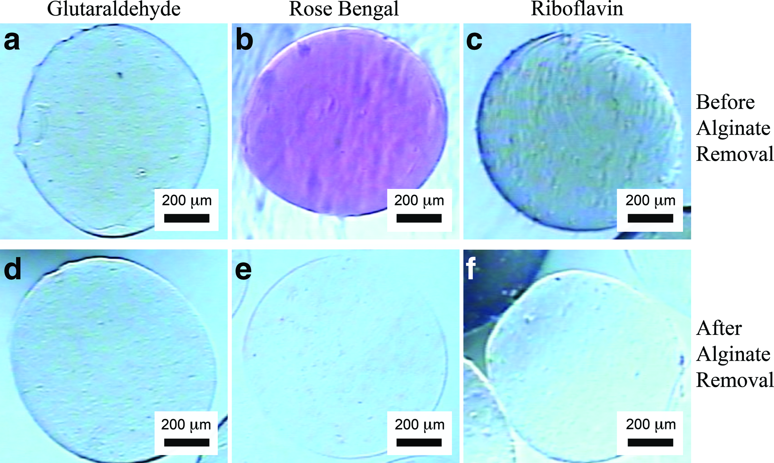
Representative crosslinked DAT microcarriers prepared using a 3:2 DATsol to alginate ratio before and after alginate extraction. Composite DAT/alginate crosslinked with
DAT crosslinking and alginate extraction
Initial stabilization of 3:2 and 4:3 DAT/alginate microcarriers by the crosslinking agents RB, Rib, and GTA was confirmed by the persistence of the microcarriers after alginate extraction, yielding DAT microcarriers with minimal residual alginate (Fig. 2). Qualitatively, the DAT microcarriers were spherical, colorless, and translucent. Composite microcarrier SEM images (Fig. 3a, b) were obtained before alginate extraction to permit visual evaluation of alginate removal efficacy. Optical microscopy confirmed alginate removal from all formulations, with no alginate core or residual fluorescence detected after sodium citrate treatments of at least 15 min (Fig. 3c inset, 3d–f). Under SEM, the spherical morphology of the microcarriers was conserved after alginate extraction (Fig. 3d), with a microporous surface topography generally observed (Fig. 3e). SEM imaging of the freeze-fractured microcarriers confirmed a porous inner structure (Fig. 3f).
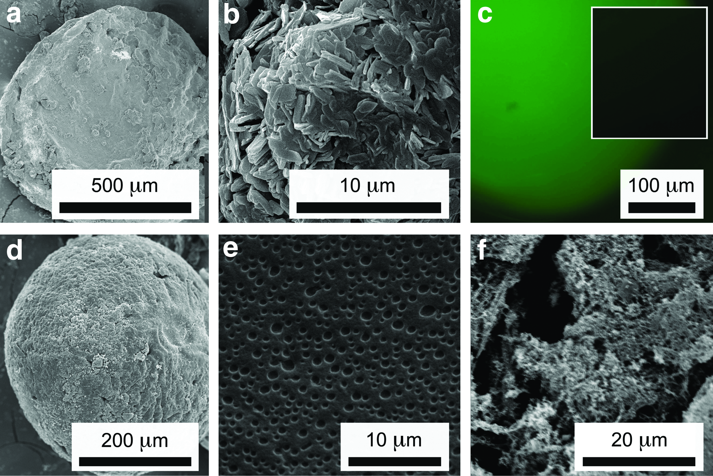
Representative images of DAT microcarriers by scanning electron microscopy and fluorescence microscopy.
Microcarrier characterization: Phase I
When transferred from deionized water to Ringer's solution, the microcarriers demonstrated an increase in diameter at early time points, consistent with equilibrium swelling in the ionic solution. The overall equilibrium mean diameter for the hydrated DAT microcarriers in Ringer's physiological fluid was calculated as 934±51 μm. The formulations crosslinked with RB were found to have greater initial mean diameter values than those formulations crosslinked with Rib or GTA. For the DAT microcarriers, the mean hydrated diameter of the 3:2 RB-crosslinked microcarriers was found to be 954±41 μm, while the mean hydrated diameters of the DAT microcarriers crosslinked by GTA and Rib were 851±58 and 915±44 μm, respectively. DAT microcarriers formulated using a 4:3 volumetric ratio of DAT to alginate were found to have mean hydrated diameters of 1096±59, 912±44, and 882±57 μm, for microcarriers crosslinked with RB, GTA, and Rib, respectively. Statistically, all mean initial diameters for each DAT microcarrier formulation were significantly different (p<0.05). Analysis of the RB-crosslinked 3:2 DAT microcarrier size distribution indicated that 78% of the microcarriers (n=100) ranged between 850 and 950 μm in diameter, and 69% of the microcarrier diameters were within±1 SD of the mean, without sieving (Fig. 4).

Representative microcarrier size distribution of one hundred RB-crosslinked 3:2 DAT/alginate microcarriers (n=100) from one fabrication batch.
RB-crosslinked DAT microcarriers, with a 3:2 formulation of solubilized DAT to 3% (w/v) alginate demonstrated fewer significant changes in mean diameter over time than the Rib-crosslinked microcarriers, and comparable stability to the control microcarriers crosslinked with GTA (Fig. 5). Differences in protein release for each formulation over time, as well as between different microcarrier formulations at each time point, were not found to be statistically significant. Protein release from each DAT microcarrier formulation was minimal, remaining below 15 μg/mL at each time point. Protein release from the 3:2 RB-crosslinked DAT microcarriers remained below 10 μg/mL at all time points (results not shown). Similar results were observed for the gelatin microcarriers in terms of diameter and stability (Supplementary Fig. S1; Supplementary Data are available online at

DAT microcarrier stability over 28 days within a simulated physiological environment, as a function of diameter. At each time point, one hundred microcarriers were selected from each formulation (n=100) and used to determine the sample means. Data are expressed as the mean±standard deviation (SD). Dashed lines indicate the overall mean diameter for each microcarrier formulation. DATsol:alginate ratio and crosslinker:
DAT microcarriers dehydrated before equilibration in Ringer's fluid demonstrated no significant changes in the mean microcarrier diameters beyond 72 h (Supplementary Fig. S2). Therefore, the weight of each microcarrier formulation at 72 h was used to calculate the swelling ratio (Table 1), with the 3:2 RB-crosslinked formulation demonstrating the lowest swelling ratio. Predrying the DAT microcarriers did not significantly impact the final mean microcarrier diameters (Supplementary Fig. S2), or protein release from microcarriers observed over 28 days in a simulated physiological environment. Similar trends were observed with the dried gelatin microcarriers after rehydration (results not shown).
Microcarrier characterization: Phase II
Since the RB 3:2 DAT/alginate microcarriers were more stable than the Rib-crosslinked microcarriers, they were selected to undergo further characterization. Gelatin microcarriers (RB 3:2 formulation) were included as a control. As measured by liquid displacement, the RB 3:2 DAT microcarriers were estimated to be 29%±4% porous, while the RB 3:2 gelatin microcarriers were found to be 28%±9% porous.
During injectability testing, the soft and compressible DAT and gelatin microcarriers were successfully passed through 18-gauge needles, without any observable impact on microcarrier integrity, size, or shape (Fig. 6), pointing to the potential for the microcarriers to be used not only as a cell culture substrate, but also as an injectable cell delivery vehicle. Further, the microcarriers retained their structural integrity when dynamically cultured with human ASCs. The seeded and unseeded microcarriers could be extruded to form monolayers or larger-volume aggregates, ∼1 cm3 in volume (Fig. 6).

DAT microcarrier architecture and injectability.
Cell attachment and proliferation was greater on the DAT microcarriers as compared to the gelatin microcarriers (Fig. 7). For both types of microcarriers, the attached ASCs appeared to have a more rounded cell morphology, relative to cells grown on TCPS, over the 14-day study. Individual ASCs were distributed over the surface of the microcarriers at early time points, with increased cell densities observed at 14 days.
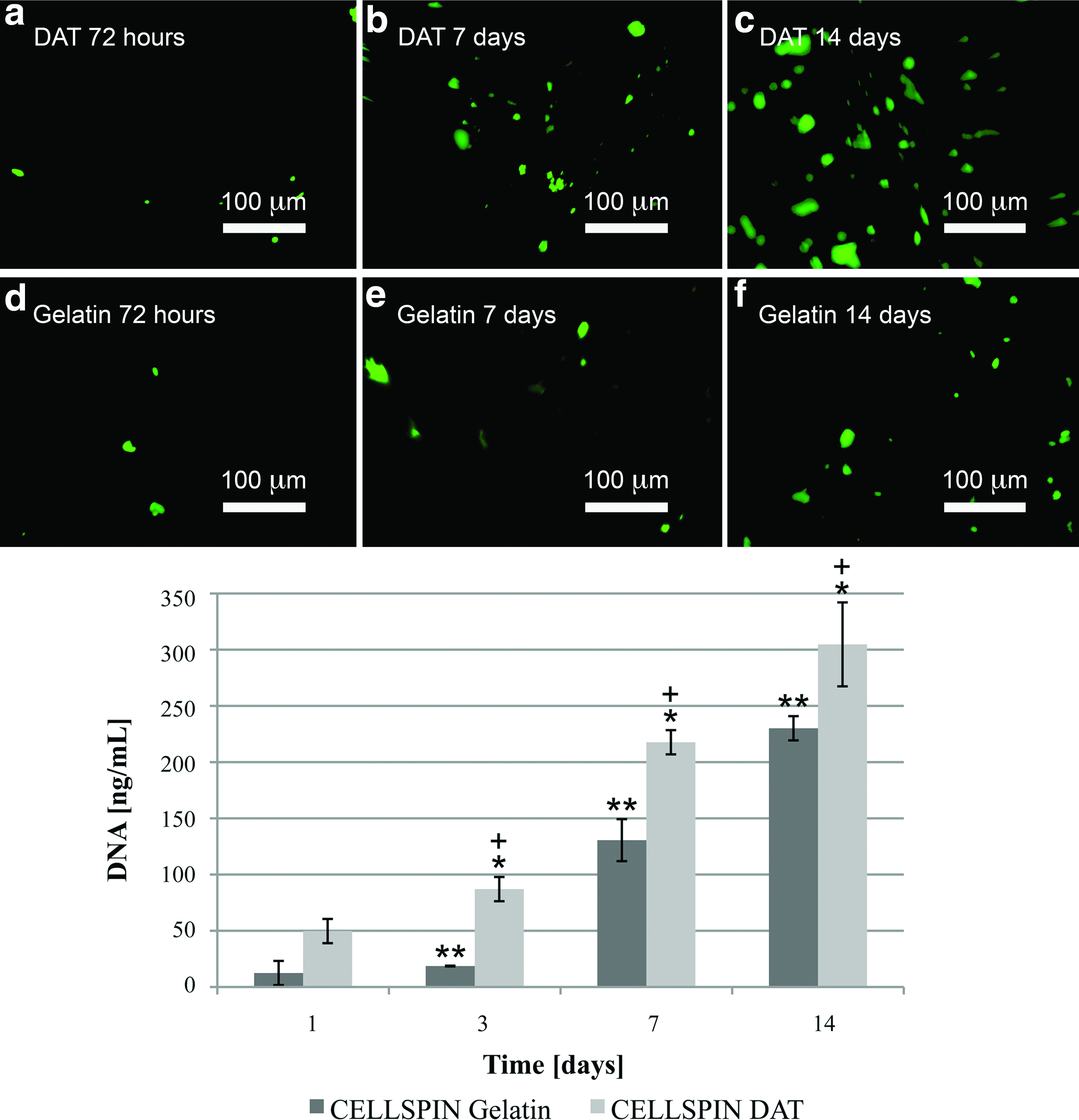
Human ASC distribution and proliferation on DAT and gelatin microcarriers under dynamic culture conditions in the CELLSPIN system. ASCs were labeled before seeding with CellTracker™ Green (Invitrogen), and imaged under fluorescence microscopy at
The DNA content on the ASC-seeded DAT microcarriers was significantly greater than the DNA content on the ASC-seeded gelatin microcarriers at 72 h, 7 days, and 14 days (Fig. 7). The highest mean DNA content was found at 14 days for the seeded DAT microcarriers, measured to be 304.73±37.31 ng/mL. Further, the mean DNA content measured on the DAT microcarriers after 14 days of culture was also found to be significantly greater than the mean DNA content measured for ASCs grown in 2D on TCPS (Fig. 8), with repeatable results obtained with cells from different donors. Overall, the trends were indicative of enhanced cellular attachment and proliferation on the DAT microcarriers.

Total dsDNA content measured using the PicoGreen® assay on DAT microcarriers, gelatin microcarriers, and tissue culture polystyrene (TCPS), following 14 days of culture. Proliferation Study 1 and 2 were conducted under identical conditions. Data are expressed as means±SD.
Discussion
The microcarrier fabrication protocols developed in this work produced stable, photochemically crosslinked, porous microcarriers derived from the ECM. The specific application of DAT as a decellularized base scaffold material yielded injectable adipose tissue-engineering scaffolds that were highly supportive of ASC attachment and proliferation.
In previous work, microcarriers fabricated from a variety of naturally derived materials, such as collagen and gelatin, have shown promise in short-term cell culture studies.5,23 However, the fact that these materials are often derived from xenogenic tissue sources raises potential clinical safety concerns. Further, only varying degrees of success have been achieved with the injectable cell-adhesive substrates developed to date. Moving toward the development of optimized strategies for 3D cell culture and injectable cell delivery, emerging data supports the implementation of a more tissue-specific approach, in terms of matching the scaffold properties to the native ECM of the cells or tissues of interest.24,48,49
In vivo, the ECM is dynamic and multifunctional, not only imparting structural integrity, but also modulating cell migration, morphology, apoptosis, and cytokine secretion, amongst other responses. 50 Adhesion of cells to the ECM is essential for normal growth and development, as well as for maintaining tissue homeostasis. 51 Variations in the relative concentrations of the various ECM macromolecules, as well as their 3D organization, give rise to tissue-specific characteristics. 52 As such, the incorporation of naturally derived ECM components within tissue-engineering scaffolds has the potential to significantly contribute to the overall regenerative response. 53 The methods developed in this work could be extended to produce microcarriers derived from other matrix sources, such as any type of decellularized tissue or cell-secreted ECM, to engineer an array of customized microcarriers that tailor the 3D cellular microenvironment, with the goal of promoting more normal cellular behavior and tissue-specific responses in cultured cell populations, to enhance tissue regeneration.
Our initial studies investigating microcarriers and films fabricated from solubilized DAT indicated that uncrosslinked materials completely dissolved within 72 h in a simulated physiological environment, emphasizing the need for stable crosslinking. While GTA has been commonly employed in the past as a chemical crosslinking agent for collagen, it is cytotoxic and has been associated with the in vivo calcification of decellularized biomaterials, both of which raise concerns with regard to its broad clinical applicability. 54 As a result, the effective stabilization of DAT microcarriers via photochemical crosslinking was a primary focus of this work. Previous work has demonstrated that RB and Rib can be implemented at relatively low concentrations as photo-crosslinkers for collagen-based biomaterials.44,45 Further, the results in the literature indicate that the cytotoxicity of both RB and Rib at the concentrations used in the current study would be quite low. In addition, our methods include extensive rinsing to eliminate any unreacted compounds, which would further reduce the potential risk.
In this study, the stability of DAT and gelatin microcarriers photo-crosslinked with RB and Rib was confirmed over a 28-day period, in comparison to more traditional GTA crosslinking. During the testing, the microcarriers were subjected to agitation speeds greater than 45 RPM, to confirm that they could withstand long-term culture under dynamic operating conditions, as found in spinner flasks or other mixed culturing systems.21,55,56 In addition, the stability testing was conducted in simulated physiological fluid at 37°C, to model short-term in vivo stability. Under these conditions, the DAT microcarriers crosslinked with RB demonstrated comparable stability to those crosslinked with GTA. When stored at 4°C in Ringer's solution, the crosslinked DAT microcarriers were qualitatively stable for more than 6 months. Swelling studies indicated that drying the microcarriers had no significant impact on their long-term stability. The capacity to store the DAT microcarriers sterilely in a dehydrated form, and then to easily rehydrate them on demand, has significant logistical advantages for employing these scaffolds in a clinical setting. More specifically, DAT microcarriers could be fabricated at a centralized location and dehydrated before distribution, to facilitate storage as an allogenic off-the-shelf biomaterial.
Confirmation of effective alginate removal after crosslinking was another focus, as residual alginate could influence cell attachment, 57 and consequently, cell proliferation and differentiation. Our results confirmed the efficacy of the finalized alginate extraction protocols involving treatment in sodium citrate buffer. Extracting the alginate from the composite microcarriers produced a microporous structure, as observed by SEM imaging and estimated by liquid displacement. The surface topography of the DAT microcarriers may have contributed to the favorable ASC attachment observed in culture. Initial sterility testing confirmed that the fabricated microcarriers demonstrated no detectable signs of contamination when maintained in culture medium that was not supplemented with antibiotics or antimycotics.
Over the 14-day proliferation study, DNA quantification confirmed a statistically significant increase in DNA content over time on the microcarriers, indicative of cell proliferation. The fluorescent cell imaging studies supported this trend, demonstrating cell adhesion and proliferation on the surface of the DAT and gelatin microcarriers. Based on the SEM imaging, the surface pores of the DAT microcarriers were not sufficiently sized to permit the penetration of the seeded ASCs into the interior regions of the microcarriers. While the DAT is a biodegradable scaffold, there was no evidence of microcarrier degradation to facilitate cell infiltration over the time course of the current study. Furthermore, the confocal imaging studies did not demonstrate any evidence of ASC infiltration into the microcarriers, and preliminary histological staining results corroborated this data (results not shown).
The enhanced cell proliferation on the DAT microcarriers, as compared with the gelatin controls, points to a compositional benefit of using DAT as a tissue-specific base scaffolding material for human ASC culture. Control testing of cell proliferation under static conditions indicated that the dynamic culture environment promoted cell proliferation, with dynamic culturing in the spinner culture system increasing the mean DNA content on the DAT and gelatin microcarriers by approximately 1.5 times (results not shown). The more rounded appearance of the dynamically cultured ASCs relative to 2D static culture controls is consistent with previous results obtained in dynamic culture systems with other cell types, and could be related in part to the low-shear mechanical forces exerted in the stirred culture environment. 22 Since cell shape has been shown to play a key role in the lineage commitment process in cultured mesenchymal stem cells,58,59 the flexibility of this type of 3D dynamic culture system may allow for better control and tuning of the cellular microenvironment, to facilitate future optimization of the desired cellular responses.
Longer-term cell culture experiments demonstrated that the DAT microcarriers were structurally robust and able to withstand dynamic culturing with human ASCs for at least 28 days with no detectable signs of microcarrier degradation. For clinical applications, freshly isolated human ASCs from small autologous tissue biopsies could be directly seeded on hydrated DAT microcarriers, fabricated from allogenic DAT sourced from donor adipose tissue. In general, human adipose tissue represents a particularly abundant and expendable source of both human regenerative cells and ECM that is routinely discarded as medical waste. If desired, adipose tissue could also be collected from liposuction procedures to be used as a source of solubilized ECM for fabricating DAT microcarriers. The approach could also be applied to develop autologous microcarriers from excess fat in an accessible donor region. ASCs could then be expanded on the DAT microcarriers within spinner flasks to achieve a desired cell density, and then the cell-seeded microcarriers could be injected into the appropriate tissue target.
The DAT microcarriers have the potential to be incorporated as biodegradable and biocompatible injectable cell delivery vehicles, either independently or in combination with other scaffolding materials, such as hydrogels or porous sponges. Promisingly, the microcarriers in the current study passed easily through 18-gauge needles, within the size range commonly used in the clinic. 60 For regenerative strategies based on subcutaneous or intramuscular stem cell delivery, the cell-adhesive DAT microcarriers may enhance ASC retention and survival after injection or implantation. Ongoing studies in our lab are probing the cell response to the DAT microcarriers in detail, with a focus on human ASC differentiation along the adipogenic lineage. In particular, the DAT microcarriers hold promise as clinically relevant, tissue-specific injectables for the treatment of small-volume soft tissue defects in plastic and reconstructive surgery, such as for cosmetic volume augmentation of wrinkles or folds in the face. Similarly, DAT microcarriers could be applied as a cell-adhesive delivery vehicle within a larger composite construct, for use in a wide range of soft tissue repair strategies, including the treatment of traumatic injuries, burns, congenital birth defects, or for contour deformities resulting from tumor resection.
After in vitro expansion, the ASCs can also be readily isolated from the microcarriers through enzymatic treatment. Collagenase is routinely used in established methods to isolate cells from adipose tissue and lipoaspirates, and can be used to digest the DAT microcarriers to separate purified populations of culture-expanded ASCs. Spherical microcarriers possess favorable surface area-to-volume ratios for achieving high cell densities. Relative to traditional monolayer culturing, microcarriers offer advantages in terms of reducing space and material costs, and can also allow for better control over the cellular microenvironment. Overall, our results support the potential of DAT microcarriers as both cell-adhesive substrates for ASC cell expansion, and in vivo cell delivery vehicles.
In summary, methods were developed to fabricate injectable microcarriers from decellularized ECM, and in particular, human adipose tissue, using minimally cytotoxic synthesis protocols. The DAT microcarriers showed enhanced cell attachment and proliferation of seeded human ASCs relative to gelatin controls, indicating that the DAT composition is favorable for ASC culture. The developed methods could be applied to other decellularized tissues or matrix sources, to fabricate cell- or tissue-specific strategies, which may promote more normal cellular organization and behavior in cultured cell populations. Overall, DAT microcarriers hold great potential for clinical use in a wide range of reconstructive and cosmetic applications.
Footnotes
Acknowledgments
Funding for this work was provided by the Natural Sciences and Engineering Research Council (NSERC) of Canada, the Canadian Foundation for Innovation (CFI) Leader's Opportunity Fund, and the Ontario Ministry of Research and Innovation (MRI) ORF-RI grant program, with scholarship funding from the R. Samuel McLaughlin Fellowship. The authors would like to thank Dr. J.F. Watkins, Dr. M. Harrison, Dr. K. Meathrel, Dr. J. Davidson, Dr. C. Watters, and Mrs. K. Martin for clinical collaborations, Mr. C. Cooney for assistance with SEM, and Dr. X. Yan for assistance with supercritical drying and SEM. They would also like to thank Dr. B. Amsden for the use of his light sources, Dr. S. Waldman for the use of the microplate reader, and Mr. J. Hayami for technical assistance.
Disclosure Statement
No competing financial interests exist.
References
Supplementary Material
Please find the following supplemental material available below.
For Open Access articles published under a Creative Commons License, all supplemental material carries the same license as the article it is associated with.
For non-Open Access articles published, all supplemental material carries a non-exclusive license, and permission requests for re-use of supplemental material or any part of supplemental material shall be sent directly to the copyright owner as specified in the copyright notice associated with the article.