
Abstract
Tissue engineering strategies are often limited by in vitro culture techniques of three dimensional scaffolds. Here we develop a method to form an aggregated cell-containing construct in vitro in a bioreactor system. Human mesenchymal stem cells (hMSCs) are cultured in individual alginate beads in a tubular perfusion system (TPS) bioreactor and then aggregated to form a single large construct. Mechanical evaluation of this construct demonstrated that aggregated alginate constructs (AACs) made from beads with 2.15 mm diameters had a Young's modulus of 85.6±15.8 kPa, a tensile strength of 3.24±0.55 kPa and a yield strength of 1.44±0.27 kPa. These mechanical properties were shown to be dependent on the bead size used to fabricate the AACs with smaller bead sizes resulting in stronger constructs. Analysis of metabolic activity revealed that hMSCs encapsulated in alginate exposed to AAC treatment sustained metabolic activity while live dead staining indicated cells remain viable. These results demonstrate the formation of AACs in the TPS bioreactor as an elegant method to create tissue engineering constructs in vitro.
Introduction
In a bottom up tissue engineering approach small-scale tissue engineering building blocks are created. These tissue building blocks are then assembled into a large tissue engineering construct. 2 For example, using endothelial cell coated collagen gel constructs a modular tissue engineering approach has been used to create a multicell microvascularized tissue engineering construct. 3 These endothelial coated gels contained an encapsulated hepatoma cell line and after perfusion culture in a bioreactor system the individual modules assembled to form a construct with interconnected channels.4,5
The goal of this work is to create an engineered tissue aggregated from many small alginate beads that are cultured individually before aggregation. This will allow for the in vitro development of tissue engineering constructs on size scales not easily possible with previous culture methods. The system involves the culture of human mesenchymal stem cells (hMSCs) in alginate beads in the tubular perfusion system (TPS) bioreactor. The TPS uses an elegant bioreactor design in which alginate scaffolds are tightly packed in a tubular growth chamber and the medium is pumped through the growth chamber from a reservoir by a peristaltic pump. The TPS bioreactor has been shown to support the growth and osteoblastic differentiation of hMSCs as well as enhance late osteoblastic differentiation and calcium matrix production. 6 hMSCs are utilized in this study as a promising cell source for both bone and cartilage tissue engineering as they can be isolated from the bone marrow and readily differentiated into both osteoblasts and chondrocytes.6,7 The hMSCs are encapsulated in alginate, a natural biomaterial derived from algae that is frequently utilized in cartilage tissue engineering8–11 as well as some bone tissue engineering applications.12–15 Alginate is composed of mannuronic acid and guluronic acid chains. 8 When a divalent ion such as calcium is added to an alginate solution the calcium binds between guluronic acid blocks of the alginate chain ionically crosslinking the alginate chains and gelling the alginate solution. 16 Thus, cells can be easily encapsulated in an alginate hydrogel by mixing an alginate cell solution and adding the solution dropwise into a calcium chloride solution. The size of these beads can be controlled by changing the size of the needle. The beads can then be dissolved through the addition of a calcium chelating agent such as ethyldiaminetetraacetic acid (EDTA), which sequesters the crosslinking calcium ions. This makes alginate advantageous for in vitro experimentation as cells can easily be removed and analyzed.
In this work these properties are utilized to create an aggregated construct where cells are cultured in smaller beads, which are joined to form a single, mechanically intact large construct. The objectives of this study are to first develop this aggregated construct in the TPS bioreactor. After this the mechanical properties of the construct will be tested. Finally, hMSCs will be encapsulated in the construct and the viability of these cells will be demonstrated throughout the creation of this construct.
Methods
Bioreactor design
Dynamic culture is completed in the TPS bioreactor as previously described. 6 Briefly, the bioreactor system consists of a tubular growth chamber and the medium reservoir connected via a tubing circuit which consists of platinum cured silicone tubing (Cole Parmer) for all areas except the area that passes through the pump, which is composed of Pharmed BPT tubing (Cole Parmer) (Fig. 1). Medium flow was driven by an L/S Multichannel Pump System (Cole Parmer) at 3 mL/min. After loading with cell-containing beads the tubing was fully assembled inside a cell culture hood and then placed in a cell culture incubator at 37°C and 5% CO2. Fifty milliliters of the osteogenic medium was loaded into separate 125 mL Erlenmeyer flasks for each growth chamber topped with rubber stoppers. The medium was withdrawn and replaced from the reservoir through two tubes that penetrate the stopper and changed every 3 days.

Schematic of tubular perfusion system (TPS) bioreactor (left) and enhanced view of growth chamber (right). Aggregated alginate constructs (AACs) are formed in the growth chamber. Color images available online at
Fabrication of aggregated alginate constructs
Alginate solutions of 2.0% w/v were prepared as previously described by adding alginic acid sodium salt from brown algae (Sigma), into 0.15 M NaCl (Sigma), and 0.025 M HEPES (Sigma) in deionized water.10,11,17 Alginate beads were fabricated by dropwise addition of this solution into a stirred solution of 0.1 M calcium chloride (Sigma). The beads were stirred for 15 min using a magnetic stir bar and stir plate set to 60 rotations per minute. The beads were then removed from the calcium chloride solution and rinsed in a phosphate-buffered saline (PBS) solution for 15 min. Bead size was varied by changing the needle gauge size. Gauges of 16, 18, 20, 27, and 30 were used. To make aggregated constructs beads were loaded into a growth chamber to make an aggregated construct ∼2 cm in length. A 0.025 M solution of EDTA (Sigma) was flowed through the growth chamber at 1.8 mL/min for 5 min. Allowing the EDTA to flow through the growth chamber for 5 min permits all the beads to be in full contact with the EDTA solution, and causes the alginate beads to expand in size and overlap. After this step, the EDTA solution was replaced with a 0.5 M solution of calcium chloride. This solution was flowed into the TPS for 5 min at 1.8 mL/min to remove EDTA, and then perfused through the growth chamber at 10 mL/min for 20 min to form aggregated constructs by ionically crosslinking the alginate chains with the calcium ions. The 5 min calcium chloride step is important to wash all remaining EDTA, whereas the 20 min calcium chloride step distributes calcium ions to all beads. Aggregated alginate constructs (AACs) were then removed from the TPS bioreactor for experimentation.
Measurement of tensile mechanical strength
Before mechanical testing the dimensions of the AACs were measured using calipers and the mass of the sample measured using an Ohaus Analytic Plus analytical balance. Constructs were placed using forceps into custom fit clamps attached to the clamps provided by the manufacture and the tensile strength measured using a Tensilon RTF-1310 mechanical tester outfitted with a 50N load cell and MSAT0002 materials testing software. The AAC samples were stretched with a constant crosshead speed of 1.0 mm/min, with the software constantly recording the stress and strain. The test ended with sample fracture. Young's modulus, tensile strength, and yield strength of the AACs were calculated. The Young's modulus was calculated as the slope of the initial linear portion of the stress–strain curve. The ultimate tensile strength was identified as the maximum stress reached by each sample. The tensile strength at 0.2% yield was calculated by locating the intersection of the stress–strain curve and a line with the Young's modulus slope at 0.2% strain offset. 18
Measuring rate of bead dissolution
To determine the dissolution rates of beads in EDTA, beads were fabricated as described previously in the methods using an 18 gauge needle. Initial bead size was then measured by calculating the cross-sectional area using ImageJ software (NIH) of a bead based on a image taken with an Axiovert 40 CFL with filter set 23 (Zeiss) equipped with a digital camera (Diagnostic Instruments 11.2 Color Mosaic). Alginate beads were loaded into TPS growth chamber and perfused with EDTA with concentrations ranging from 0.008 to 0.1 M at 1.8 mL/min. At each time point, five beads were removed from the bioreactor and photographed. Cross-sectional areas were normalized to initial cross-sectional areas to determine bead dissolution at each time point.
hMSC culture
hMSCs (p≤5) from a single donor were purchased from Lonza. Single-donor cells were used to minimize variability associated with a primary cell population. Cells were cultured before the study in the control medium consisting of Dulbecco's modified Eagle's medium (Gibco) supplemented with 10% fetal bovine serum (FBS) (Gibco), 1.0% v/v penicillin/streptomycin (Gibco), 0.1 mM nonessential amino acids (Gibco), and 4 mM L-glutamine (Gibco) using protocols set forth by the manufacturer and previously described.6,19,20 Cells were cultured on tissue culture polystyrene flasks with medium changes every 3 days according to the manufacture's specifications. Cells were stored in a cell culture incubator at 37°C and 5% CO2 and passaged every 6–7 days using trypsin/EDTA (Lonza). The osteogenic medium was formulated as described in the literature by supplementing the control medium with 100 nM dexamethasone (Sigma), 10 mM β-glycerophosphate, and 173 μM ascorbic acid (Sigma).19–21
hMSC encapsulation in alginate
Alginate solutions were sterilized via sterile filtration. hMSCs were removed from tissue culture flasks using trypsin/EDTA and pelleted via centrifugation at 500 g for 5 min. The cell pellet was resuspended in the alginate solution at a density of 1.25–2.5×10⁶ cells/mL. The alginate cell solution was added drop wise through a 20 gauge needle into a stirred solution of 0.1 M calcium chloride (Sigma), which immediately crosslinked the alginate to form beads. Beads were allowed to stabilize for 15 min and were loaded into the bioreactor. Cells were cultured in the control medium for use in live dead staining and the osteogenic medium to differentiate the hMSCs into osteoblasts and determine if calcium is produced as measured using Von Kossa staining. The control medium was used for live dead and metabolic activity assays to observe the growth process when cells are not differentiating. The medium was changed every 3 days.
Metabolic activity
hMSCs were encapsulated in alginate beads at 100,000 cells per bead. After stabilizing for 24 h in the control medium in static culture, beads were exposed to either 35 min of the control medium with FBS (control group), 5 min of 0.1 M calcium chloride, 5 min of 0.025 M EDTA, and 25 min of 0.5 M calcium chloride (AAC treatment group), or 35 min of 70% methanol (Sigma) (dead control group). Metabolic activity was then assessed using a dimethylthiazolyldiphenyltetrazolium bromide (MTT) based in vitro toxicology kit (Sigma) as previously described. 22 Briefly 200 μL of 5 mg/mL of reconstituted MTT was added to each well with 2 mL of the control medium with 10% FBS. Beads were then incubated for 150 min to allow for the formation of formazan crystals. Crystals were dissolved in 2 mL of MTT solubilization solution (Sigma) and allowed to dissolve out of alginate beads overnight. Two hundred microliters of supernatant was then transferred to a 96-well plate to record the optical density in triplicate at 570 nm using an M5 SpectraMax microplate reader (Molecular Devices).
Live dead assay
Cell viability was assessed using a live dead assay following standard protocols as described previously. 22 Viability tests were completed on three groups. In the control group cells were cultured in static culture in the control medium. In the AAC group cells were cultured in the bioreactor at a 3 mL/min flow rate for 10 days. AACs were formed from bioreactor cultured beads and removed from the bioreactor. In the final group the AAC was cultured in a static culture plate to determine if the hMSCs could remain viable for 24 h. AACs were either soaked in PBS to remove FBS and the medium for 30 min or moved to six-well plate for 24 h culture. Control beads were also first soaked in PBS for 30 min to remove FBS and the medium. Beads and AACs were then placed in well plates and incubated in 2 μm ethidium homodimer and 4 μm calcein AM (Invitrogen) for 30 min. Fluorescent images were then taken using a fluorescent microscope (Axiovert 40 CFL with filter set 23; Zeiss) equipped with a digital camera (Diagnostic Instruments 11.2 Color Mosaic). AACs cultured in well plates were removed 24 h later and stained following the same procedures as other groups.
Histological analysis
Experimental hMSCs were cultured in individual alginate beads in the osteogenic medium in the TPS bioreactor for 21 days. On day 21, alginate beads were aggregated into AACs and were collected and fixed in 4% paraformaldehyde (Sigma) and 0.1 M sodium cacodylate (Sigma) buffer containing 10 mM calcium chloride at pH 7.4 at 4°C for 4 h. After fixation, the beads were placed in cassettes and washed with 0.1 M sodium cacodylate buffer and 10 mM calcium chloride at pH 7.4 at room temperature for 24 h. The beads were then dehydrated for histological processing by ethanol washes followed by two Citrisolv (Fisher Scientific) washes. The samples were then embedded in paraffin (Fisher Scientific) and sectioned to 5-μm-thick sections and placed on glass slides. Sections were oven dried at 64°C for 2 h, deparaffinized in Citrisolv, and rehydrated in ethanol. Von Kossa staining was performed using standard protocols to observe mineralization with a Nuclear Fast Red (Poly Scientific) counterstain.
Statistical analysis
All samples were completed in triplicate (n=3). Data were analyzed using single-factor analysis of variance followed by Tukey's Multiple Comparison Test assuming normal data distribution with a confidence of 95% (p<0.05). Mean values of triplicates and standard deviation error bars are reported on each figure as well as relevant statistical relationships.
Results
Formation and dissolution of alginate beads
Alginate beads were fabricated to consistently different sizes using needle gauges of 16, 18, 20, 27, and 30 (Table 1). By using a needle gauge of 30 an average bead diameter of 2.15±0.07 mm was obtained. Sixteen gauge needles resulted in average bead diameters of nearly twice this magnitude with an average diameter of 3.90±0.09 mm. Needles with gauges in between these two resulted in bead diameters inside this range with each gauge needle producing beads significantly different in diameter from all other gauges (p<0.05). Based on these results beads of discrete diameters can be fabricated using different needle sizes.
Data are reported as mean±standard deviation. All groups are statistically different (p<0.05).
Dissolution curves of beads were then generated using 0.1, 0.05, 0.025, and 0.008 M concentrations of EDTA. This analysis was performed with 2.97-mm-diameter beads. Results of bead dissolution experiments revealed that 0.008 M EDTA did not dissolve alginate beads over the 30 min experiment (Fig. 2). Cross-sectional area was shown to increase slightly over the time points. For groups dissolved in 0.025 and 0.050 M EDTA bead size initially increased but decreased over later time points until complete dissolution in the 0.05 M group and near-complete dissolution in the 0.025 M group. The 0.025 EDTA group increased to 127% of original bead diameter after 5 min before dissolving to 11% of original area after 30 min. The 0.05 M group increased to 108% its original diameter before completely dissolving after 30 min. The 0.100 M was shown to completely dissolve in 10 min without an increase at the 5 min timepoint. These observed results in 0.025 and 0.05 M EDTA likely occur as EDTA decreases the crosslink density of the alginate causing the bead to grow in size before the bead is dissolved.
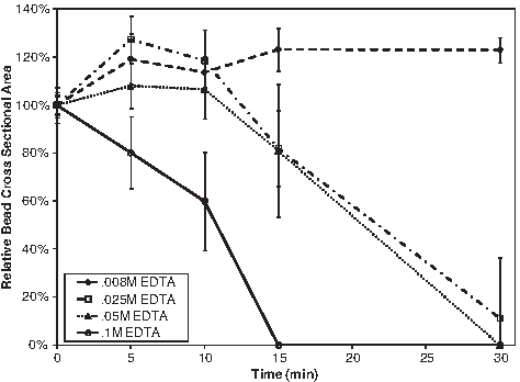
The dissolution of alginate beads at varying ethyldiaminetetraacetic acid (EDTA) concentrations at 1.8 mL/min flow rate. Bead sizes are reported as cross-sectional areas in relation to initial area.
Mechanical properties of AACs
The initial increase in size of beads in the 0.025 M EDTA group was used to develop a protocol to make AACs. Using this protocol these constructs were successfully created and shown to be easily transported and manipulated (Fig. 3). Mechanical testing of these constructs revealed that smaller diameter beads resulted in stronger aggregated constructs (Fig. 4). Aggregated constructs made from beads with 2.15 mm diameters had a Young's modulus of 85.6±15.8 kPa, a tensile strength of 3.24±0.55 kPa, and a yield strength of 1.44±0.27 kPa. These values were statistically similar (p>0.05) to constructs made from beads with 2.46 mm diameter. Increase of bead diameter slightly to 2.65 mm resulted in a relatively large and statistically significant change (p<0.05) in mechanical properties producing constructs with a Young's modulus of 39.2±10.6 kPa, a tensile strength of 0.85±0.25 kPa, and a yield strength of 0.52±0.15 kPa. These mechanical properties were statistically similar (p>0.05) to constructs made from beads with 2.97 and 3.90 mm diameters though beads with the largest diameter, 3.90 mm, exhibited the weakest mechanical properties. These samples had a Young's modulus of 18.2±5.6 kPa, a tensile strength of 0.45±0.04 kPa, and a yield strength of 0.27±0.03 kPa. AAC fracture was typically observed on the periphery of beads aggregated together; however, no other preferential breaking point was noted.
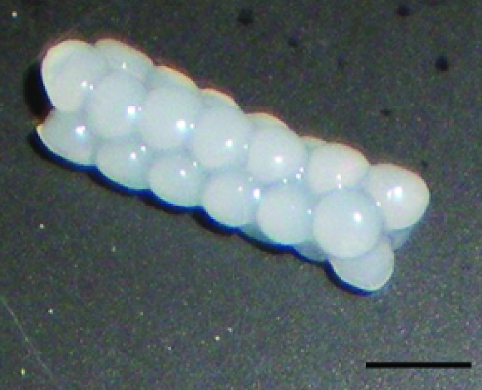
Image of AAC. Owing to the simplicity of the design, the construct can be easily moved and manipulated. Scale bar represents 5 mm. Color images available online at
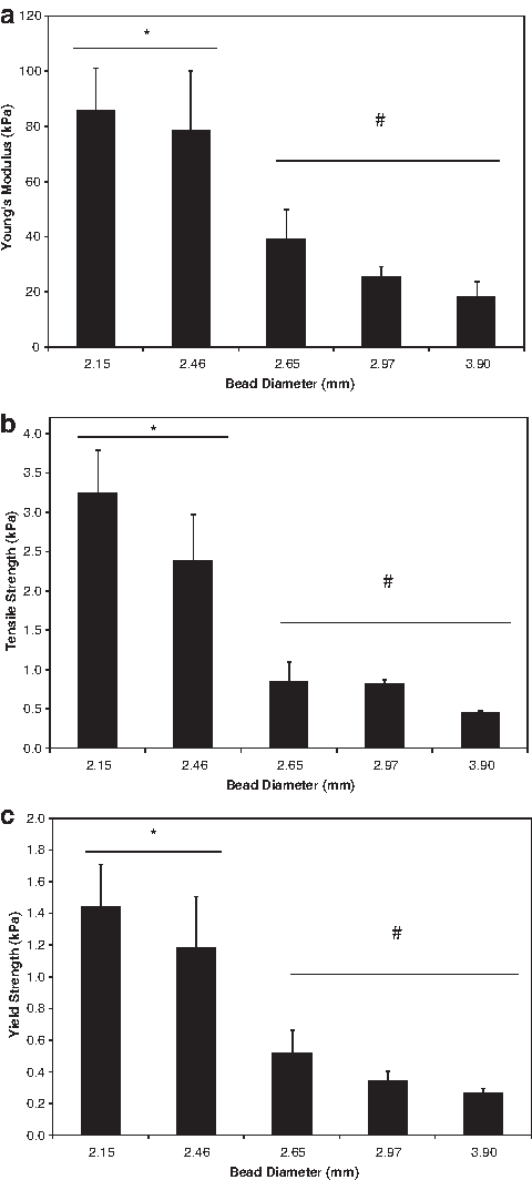
Mechanical properties of AAC including Young's modulus
hMSC viability and calcium deposition in AACs
Results of MTT assay indicate that AAC treatment has no effect on the metabolic activity of encapsulated hMSCs (Fig. 5). Cells in alginate beads exposed to AAC treatment had statistically similar metabolic activity to control hMSCs. Both these groups had significantly greater metabolic activity than hMSCs in beads exposed to methanol as a dead control. Microscopic images of the AAC reveal that hMSCs are homogenously distributed throughout the construct (Fig. 6). Upon live dead staining images reveal that the majority of these cells are viable after 10 days of culture and AAC formation (Fig. 7). Live dead images of cells 24 h after AAC treatment reveal that cells remain viable after the treatment. Following demonstration that hMSCs are viable in AACs, beads were cultured for 21 days in the osteogenic medium to demonstrate if calcium production was occurring as previously observed and if this calcium deposition would remain present throughout the AAC treatment. 6 Images of AAC sections stained using Von Kossa staining indicate that hMSCs produce calcium while being cultured before AAC formation and that AAC treatment does not eliminate these calcium deposits (Fig. 8). Calcium is stained black in these images and can be seen surrounding cells in AACs.

Metabolic activity of cells in alginate bead in the control medium, AAC treatment, and dead control. Dead control is significantly lower than control bead and AAC treatment, which are statistically similar. The symbol (*) indicates statistical significance (p<0.05).
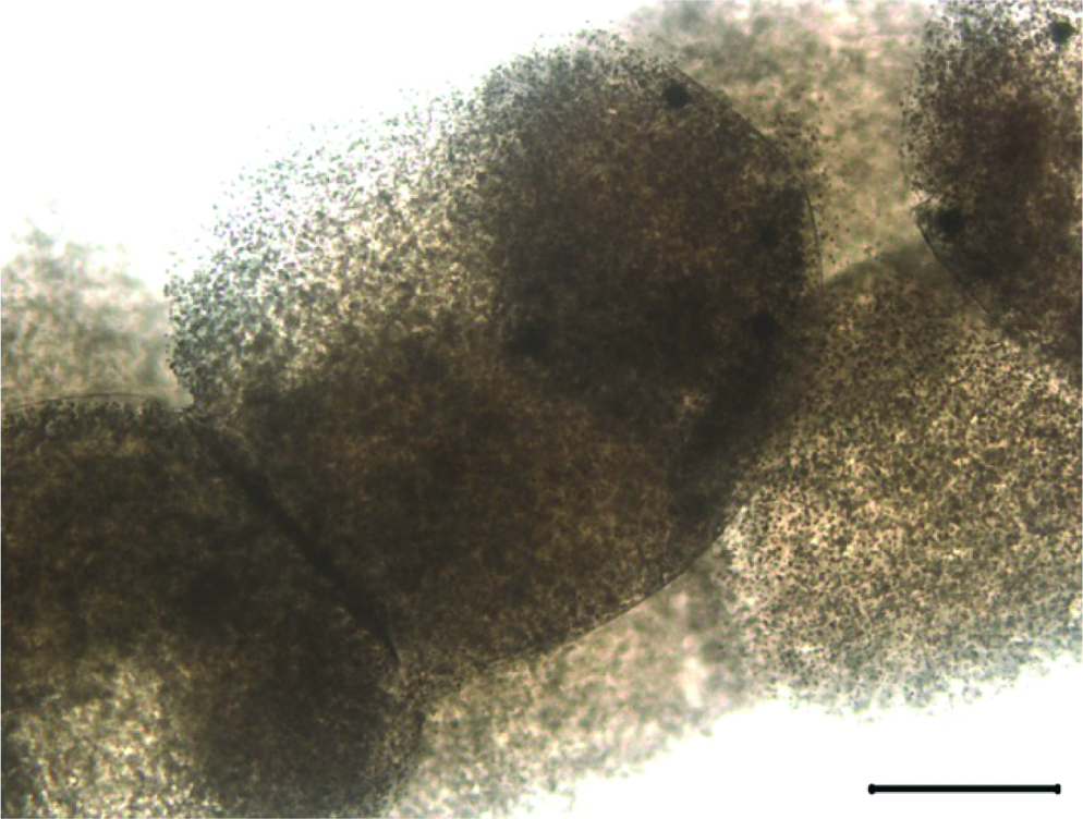
Image of cell-containing AAC. Cells can be observed throughout construct. Scale bar represents 1000 μm. Color images available online at

Live dead stain of AAC
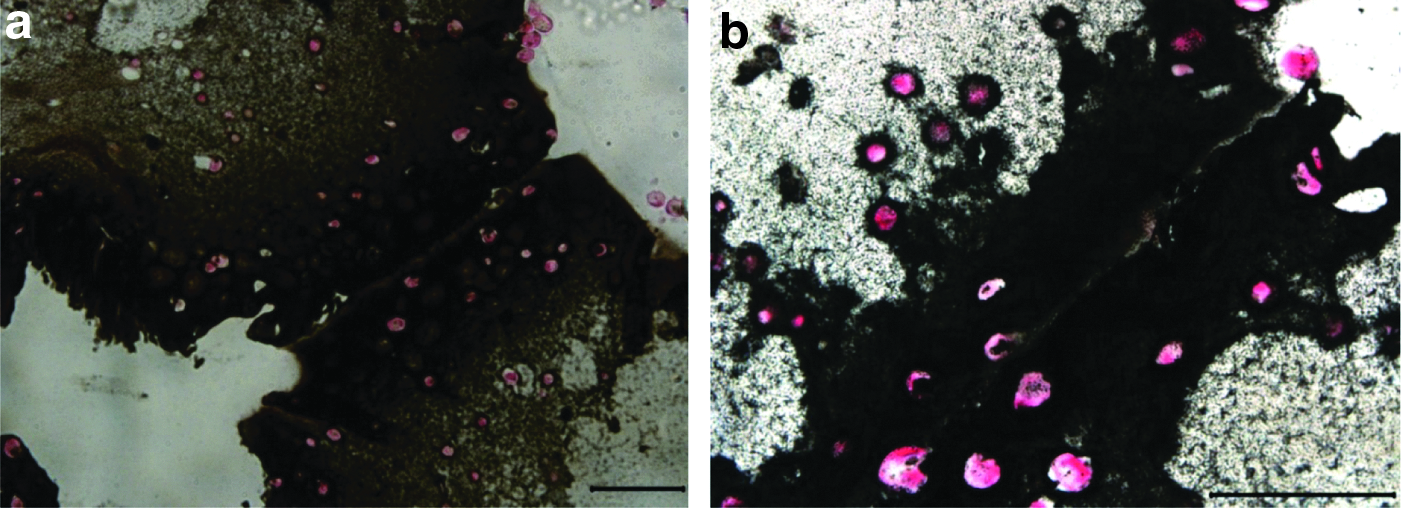
Von Kossa staining of AAC formed after 21 days of in vitro TPS culture of individual beads in the osteogenic medium at 20× objective
Discussion
A tissue engineering treatment option for bone, cartilage, and skeletal muscle represents a promising alternative to current clinical options. Despite the promise of tissue engineering, several significant hurdles exist. A central limitation is the culture of three-dimensional tissue engineering constructs in vitro. In static culture, nutrients and oxygen are replenished via diffusion. A nutrient gradient develops where cells on the exterior portions of scaffolds receive sufficient nutrients, whereas cells on the interior of scaffolds are deprived of nutrients and are exposed to hypoxic conditions.23,24 In a study analyzing oxygen concentration in three-dimensional scaffolds, a preosteoblast cell line was seeded on demineralized bone matrix scaffolds at 5×10⁴ cells/scaffold. 1 These scaffolds were 9 mm in diameter and 5 mm in height and cultured in static and dynamic conditions. In static culture central oxygen concentrations dropped quickly, below 10% in just 2 days and to 0% in just 5 days. Cell death was observed in areas where the central oxygen concentration was low. Dynamic culture in a bioreactor significantly improved oxygen transport and though central oxygen concentrations dropped to 4% cell death was not observed. Here we create aggregated constructs as large as 6 mm in diameter and 30 mm in height seeded at 3×10⁶ cells/scaffold. Before scaffold aggregation the individual alginate beads are cultured in the TPS bioreactor to enhance the growth of cells in the scaffolds. After aggregation the cells will no longer be cultured in the TPS bioreactor and are ready for implantation into a defect. By not attempting to culture such a large construct in vitro this method avoids nutrient transfer limitations, a major obstacle to three dimensional cell culture. To the best of our knowledge this is the first time an aggregated alginate scaffold has been created from many smaller cell-containing scaffolds in a bioreactor system. Cells were viable throughout the scaffold as these constructs were rapidly fabricated.
A modular tissue engineering approach previously has been used to create a perfusable cell-containing construct, 3 a cardiac sheet like construct, 25 and dermal equivalent tissue. 26 Previously used modular techniques have relied either on cell aggregation or a more complex approach such as tissue printing. Here we use a new approach where individual alginate beads are assembled together before implantation. This approach creates a controlled aggregated construct within a bioreactor system quickly with no major fabrication steps.
The method for fabricating these aggregated scaffolds was developed following analysis of alginate bead dissolution curves in EDTA, where it was discovered that alginate beads first increase in diameter before dissolution. When using EDTA concentrations ranging from 0.008 to 0.05 M this initial expansion occurs over a sufficiently long period to manipulate beads before dissolution. The goal of this experiment was to find an optimal EDTA concentration that could be used to form AACs. Results indicated that dissolution in 0.025 M EDTA would be optimal as this concentration resulted in the largest initial increase in bead diameter. These dissolution curves may not me applicable to dissolution of alginate beads in other systems as bead size can vary significantly due to swelling. 27 This swelling results from ion exchange between the calcium crosslinking ions and monovalent ions in the environment of the bead.28,29 To fabricate the AACs, the alginate beads were tightly packed in the tubular growth chamber and were perfused with 0.025 M EDTA for 5 min to allow for expansion. After this expansion, exterior edges of the beads overlapped with one another. Beads were then perfused with calcium chloride to ionically crosslink the overlapping edges, creating one aggregated construct from many beads.
Mechanical testing of this aggregated construct was then completed. Beads with different initial sizes were used in the study. AACs composed of beads with initial diameters of 2.15 and 2.46 mm had significantly higher Young's Moduli, Ultimate Tensile Strength, and Yield Strength than AACs composed of beads with initial diameters of 2.65, 2.97, and 3.90. Young's modulus, Ultimate Tensile Strength, and Yield Strength all decreased with increasing bead size. It is hypothesized that this occurs as beads with larger diameters are not able to pack as closely and create a less dense aggregated construct. Compression testing was not completed on the construct the primary goal was to measure the strength of bead aggregation. Alginate compression testing has been previously reported in the literature.30,31
Previous studies to determine the mechanical strength have shown large variations based upon the specific alginate polymer used; however, our results revealed that AACs have tensile properties in the lower range of alginates tested and Young's Moduli similar to previously described results. 32 This demonstrates AACs to be of sufficient strength for the engineering of tissue such as cartilage,10,11,33–35 non-load-bearing bone,13,36 skeletal muscle, 37 and other tissues that are commonly engineered using alginate. Specifically, AACs provide an advantage over typical alginate constructs, as individual alginate beads can be cultured in a bioreactor and then fabricated into one large construct. Thus, a large construct will be created without nutrient transfer problems that occur in statically cultured large constructs. Metabolic activity assays indicate that the AAC treatment does not have a negative effect on the metabolic activity of hMSCs encapsulated in alginate beads. This result is expected as the AAC treatment utilizes chemicals that are widely used in alginate bead fabrication and usage.10,11,33,34 This result was further confirmed by live dead staining, which indicated cells in AACs were viable immediately and 24 h after AAC fabrication.
In a prior study utilizing the TPS bioreactor hMSCs were shown to express much higher levels of osteogenic markers osteocalcin and osteopontin than static controls. 6 In addition to this mineralization was shown to be greatly increased throughout the bioreactor cultured beads. A goal of this study was to determine if this mineralization would remain after AAC treatment. Von Kossa staining for calcium was performed to examine this. This stain revealed that hMSCs in the AACs had deposited calcium and this calcium remained present throughout the AAC treatment. In vitro calcium deposition indicates that the hMSCs are able to differentiate into osteoblasts after 21 days and this calcium deposition remains present after AAC treatment.
The clinical strategy for use of this construct is to first extract bone marrow from the patient and isolate the MSCs. These stem cells will then be encapsulated in the alginate beads and cultured in the TPS bioreactor. When the tissue is ready to be implanted into the defect site the beads will be aggregated in the bioreactor, removed and the construct implanted into the patient. This follows the same strategy as traditional tissue engineering, but will allow for the production of larger cell-containing constructs than previously possible.
Conclusions
Through the course of this study a protocol has been developed and evaluated for the fabrication of a cell-containing tissue engineering construct from many smaller scaffolds in a bioreactor system. Results demonstrate this construct can be elegantly fabricated and has mechanical properties similar to traditionally fabricated alginate scaffolds. This AAC has many potential applications including non-load-bearing bone, cartilage, and skeletal muscle tissue engineering. By allowing cells to proliferate in smaller beads within the TPS bioreactor before aggregation, a large tissue engineering construct is created ready for implantation into a defect site.
Footnotes
Acknowledgments
Special thanks to Ms. Laura Hyland and Dr. Bruce Yu for use of the Tensilon RTF-1310, and Mr. David Hwang and Dr. Adam Hsieh for custom making testing clamps. This work was supported by the National Science Foundation (CAREER Award to J.P.F., #0448684) and the State of Maryland, Maryland Stem Cell Research Fund.
Disclosure Statement
No competing financial interests exist.