
Abstract
Scaffolds for tissue engineering applications must incorporate porosity for optimal cell seeding, tissue ingrowth, and vascularization, but common fabrication methods for achieving porosity are incompatible with a variety of polymers, limiting widespread use. In this study, porous scaffolds consisting of poly(1,8-octanediol-co-citrate) (POC) containing hydroxyapatite nanocrystals (HA) were fabricated using low-pressure foaming (LPF). LPF is a novel method of fabricating an interconnected, porous scaffold with relative ease. LPF takes advantage of air bubbles that act as pore nucleation sites during a polymer mixing step. Vacuum is applied to expand the nucleation sites into interconnected pores that are stabilized through cross-linking. POC was combined with 20%, 40%, and 60% by weight HA, and the effect of increasing HA particle content on porosity, mechanical properties, and alkaline phosphatase (ALP) activity of human mesenchymal stem cells (hMSC) was evaluated. The effect of the prepolymer viscosity on porosity and the mechanical properties of POC with 40% by weight HA (POC-40HA) were also assessed. POC-40HA scaffolds were also implanted in an osteochondral defect of a rabbit model, and the explants were assessed at 6 weeks using histology. With increasing HA content, the pore size of POC-HA scaffolds can be varied (85 to 1,003 μm) and controlled to mimic the pore size of native trabecular bone. The compression modulus increased with greater HA content under dry conditions and were retained to a greater extent than with porous scaffolds fabricated using salt-leaching under wet conditions. Furthermore, all POC-HA scaffolds prepared using LPF supported hMSC attachment, and an increase in ALP activity correlated with an increase in HA content. An increase in the prepolymer viscosity resulted in increased compression modulus, greater distance between pores, and less porosity. After 6 weeks in vivo, cell and tissue infiltration was present throughout the scaffold. This study describes a novel method of creating porous osteoconductive POC scaffolds without the need for porogen leaching and provides the groundwork for applying LPF to other elastomers and composites.
Introduction
Common methods of engineering porous scaffolds include solid freeform fabrication, thermally induced phase separation, gas foaming, and solvent casting followed by particulate leaching.23–28 Except for particulate leaching, these methods are not easily applicable to thermoset polyester elastomers because of their viscoelastic properties and the need to cross-link the scaffold in situ. 29 Although salt-leaching is often used to fabricate scaffolds for tissue engineering, it is a lengthy process that is performed in aqueous solutions and can cause premature polymer degradation through hydrolysis. Furthermore, because this method requires a large mass percentage of salt (>85%) to obtain pore interconnectivity throughout the scaffold, the fabrication of scaffolds with less porosity is limited. 30
Herein, we describe a novel method referred to as low-pressure foaming (LPF) that is used to fabricate porous thermoset nanocomposite scaffolds for bone tissue engineering applications. During LPF, air bubbles, nucleated within the prepolymer matrix during a mixing step, expand under vacuum, coalesce, and ultimately produce an interconnected pore network, addressing the disadvantages of the gas foaming and salt-leaching methods. The nanocomposites investigated consist of a poly(1,8 octanediol citrate) (POC) macrophase reinforced with hydroxyapatite (HA) (POC-HA) nanocrystals. The HA confers osteoconductivity and can mimic the mineral content of bone.3, 31, 32 The effect of the HA particle content and polymer viscosity on the scaffold porosity and mechanical properties are investigated. Furthermore, the biocompatibility of these novel nanocomposite scaffolds is investigated in vitro using human mesenchymal stem cells and in vivo in an osteochondral defect in a rabbit model.
Materials and Methods
Fabrication of POC and POC-HA scaffolds
Synthesis and characterization of the POC prepolymer is described elsewhere. 5 Briefly, equimolar amounts of citric acid and 1,8-octanediol (Sigma Aldrich, St. Louis, MO) were put into a round-bottom flask and melted together at 155°C for 9.5 minutes while being stirred at 140 rpm. The temperature was subsequently decreased to 140°C, and the mixture was stirred at 140 rpm for 45 minutes to obtain the POC prepolymer (pre-POC). These conditions were used unless otherwise specified. Ten g of pre-POC were dissolved in 18 g of ethanol in 80°C and stirred manually with a spatula for 30 seconds every 5 minutes for the first 2 hours in a Teflon dish. Next, pre-POC was mixed for 30 seconds every hour for 24 hours. Through the mixing process, the solvent was completely evaporated, and air bubbles were induced. POC was than postpolymerized at 80°C for 3 days under constant vacuum (2 Pa) followed by 120°C for 1 day also under vacuum.
To fabricate POC-HA scaffolds, pre-POC dissolved in ethanol (1:1 w/v) was mixed with various amounts of HA nanocrystals to obtain nanocomposites containing 20%, 40%, or 60% by weight HA. Medical-grade HA nanocrystals were 100 nm in size (100% HA, Berkeley Advanced Biomaterials, Berkeley, CA). 33 The POC-HA mixture was stirred manually as specified above and postpolymerized under the same conditions. Solid POC and POC-HA nanocomposites with 0%, 20%, and 40% by weight HA were also fabricated under the same temperature conditions but without the use of vacuum.
Effect of prepolymer viscosity on POC and POC-HA scaffolds
When specified, POC with 40% by weight HA was fabricated with pre-POC of varying viscosities and degrees of polymerization. Pre-POC was synthesized under one of the following synthesis conditions: 9.5 minutes at 155°C (melt only), 15 minutes at 155°C, or 15 minutes at 155°C followed by 35 minutes at 140°C. All three types of pre-POC were synthesized while being stirred at 140 rpm. The kinematic viscosities were measured at 25°C with 2% pre-POC solution in ethanol (w/v) using an Ubbelohde viscometer, following the manufacturer's instructions (Cannon Instrument Co., State College, PA). The POC-HA mixture was stirred manually as specified above and postpolymerized under the same conditions.
Fabrication of porous salt-leached POC and POC-HA scaffolds
Porous POC and POC-HA scaffolds were fabricated using the well-known salt-leaching technique.4,5,34–36 Pre-POC was dissolved in ethanol (1:1 w/v), followed by the addition of salt porogen (≤250 μm, 85% salt: 15% pre-POC by weight). For POC-HA, HA was added to the dissolved pre-POC before the addition of salt. The resulting slurry was placed into Teflon dishes, mixed, and placed in the oven for postpolymerization (80°C for 3 days, 120°C for 1 day). To avoid porosity induced by LPF, postpolymerization conditions did not include the use of vacuum. The salt in the resulting nanocomposites was leached out using successive incubations in Milli-Q water (Billerica, MA) every 12 hours for 6 days. Complete removal of salt was checked by adding silver nitrate to the water and looking for signs of silver chloride precipitation. The resulting scaffolds were dried at room temperature for 72 hours before testing.
Compression testing of POC and POC-HA scaffolds
The compression moduli under dry and wet conditions were measured according to Japanese industrial standard (JIS) 7208 using a Sintech mechanical tester model 20/G (Research Triangle Park, NC).3,37 At least three cubes with 10-mm dimensions were used for POC and POC-HA scaffolds fabricated using LPF and POC and POC-HA salt-leached scaffolds. Samples under wet conditions were soaked in Milli-Q water for 24 hours at room temperature before testing.
Porosity measurement
An Autopore IV (Micromeritics, Norcross, GA) mercury porosimeter was used to determine the incremental intrusion of mercury with pressure, from which a volume percentage for a particular pore diameter was calculated according to:
where I is the incremental intrusion, P is the porosity, and T is the total intrusion.
The cross-section and the surface of the scaffolds were sputter-coated with gold and observed using scanning electron microscopy (SEM; Hitachi 3500 N, EPIC, Northwestern University, Evanston, IL), and the average pore size and pore wall thickness were measured manually using the SEM images. At least four measurements were used to determine each parameter.
Cell culture
Human mesenchymal stem cells (hMSC) were (Lonza, Walkersville, MD) were cultured and expanded in growth medium containing low-glucose Dulbecco's modified Eagle medium (1 g/mL glucose) supplemented with 10% fetal bovine serum and 1% penicillin/streptomycin at 37°C in humidified air containing 5% carbon dioxide (CO2). Cells at passage 5 were used in this study.
Morphology, attachment, and effect of alkaline phosphatase activity of hMSC on POC and POC-HA scaffolds
Each sample was placed in a 24-well plate and sterilized using an AN74j/Anprolene ethylene oxide sterilization system that performed a 2-hour degassing step under vacuum after 12 hours of gas exposure (Anderson Sterilization, Inc., Haw River, NC). After the samples (10-mm cubes) had been conditioned in growth medium for 24 hours at 37°C, 40,000 cells were suspended in 15 μL of medium before seeding. Two hours after seeding, 2 mL of medium was aliquotted into each well.
To assess the attachment and morphology of hMSC after 24 hours, all samples were fixed with 2.5% glutaraldehyde, dehydrated in graded series of ethanol, and freeze-dried. The samples were then sputter-coated and observed using SEM. To quantify hMSC attachment, total DNA was quantified using Quant-iT Pico Green dsDNA Reagent (Invitrogen, Carlsbad, CA). Cells were lysed using 0.1% Triton-X 100 and sonicated for 20 minutes, and the lysate was used for the assay. At least three samples were used for each material.
The intracellular alkaline phosphatase (ALP) activity of hMSC was quantified by adding one aliquot of the cell lysate to an equal amount of reaction buffer containing 10mM p-nitrophenyl phosphate in 50mM glycine buffer, pH 10.5, supplemented with 0.5mM magnesium chloride (Sigma Aldrich). After 30 minutes at 37°C, the reaction was stopped by the addition of 0.05M sodium hydroxide and measured using spectrophotometry at 410 nm. At least three samples were used for each nanocomposite type, and results were normalized according to total DNA.
Animal experiments
POC-40HA scaffolds with a diameter of 3 mm and a length of 10 mm were sterilized using an AN74j/Anprolene ethylene oxide sterilization system (Anderson Sterilization). Three skeletally mature, male New Zealand rabbits were purchased (3.2-3.5 kg, 6-7 months, Covance, Kalamazoo, MI), and the surgical protocol followed National Institutes of Health guidelines for the care and use of laboratory animals and was approved by Northwestern University's Animal Care and Use Committee (Chicago, IL). Briefly, anesthesia was induced using an intramuscular injection of ketamine (40 mg/kg) and xylazine (5-7 mg/kg) and maintained with isoflurane (1-2% inhalation). After shaving and sterilely preparing the lower extremities, a 4- to 5-cm medial parapatellar arthrotomy was created in each knee, exposing the medial femoral condyle. A 3-mm-diameter, 10-mm-deep defect was drilled in the medial femoral condyles and filled with POC-40HA. 38 The wound was irrigated with sterile saline solution and an antibiotic flush (200 mg of cefazolin in 1 L of saline). The deep capsular layer was closed in an interrupted pattern using 3-0 and 4-0 Vicryl sutures (Ethicon, Somerville, NJ), and the skin was closed in a subcuticular pattern.
Buprenorphine (0.02-0.05 mg/kg) was administered subcutaneously before surgery and 8 hours postoperatively. A fentanyl transdermal patch was placed shortly after surgery (12 μg/h) and removed after 72 hours. All animals received a 5-day subcutaneous postoperative regimen of enrofloxacin (5 mg/kg). All rabbits were allowed to ambulate freely postoperatively.
Histology
The rabbits were killed 6 weeks postoperatively. Knee joints were collected, immediately fixed in 10% neutral-buffered formalin, dehydrated in graded series of ethanol, and embedded using JB-4 plus (Polysciences, Warrington, PA). All specimens were sectioned at 5 to 10 μm of longitudinal thickness for histological assessment using an HM 355 S Rotary Microtome (Richard-Allan Scientific, Kalamazoo, MI) and stained with Masson-Goldner trichrome. Sections were assessed under standard light microscopy (Nikon Eclipse TE2000-U). All histological assessment was determined in consultation with a pathologist, and representative images are shown.
Statistical analysis
The Student t-test was used to compare means of pairs. ANOVA with Newman-Keuls multiple comparison test post hoc analysis was used to determine significant differences between three or more means. A p-value of 0.05 or less was considered to be significant.
Results
Fabrication and effect of particle content on pore size, porosity, and mechanical properties of POC and POC-HA scaffolds
Solid or porous POC and POC-HA nanocomposites with 20% and 40% by weight HA (POC-20HA and POC-40HA) were fabricated (Fig. 1). The volume of the scaffolds fabricated using LPF was greater than that of the composites fabricated without added vacuum conditions, and the outer layer of the scaffolds fabricated using LPF was distinctly less porous (Fig. 1G).

Digital images of solid poly(1,8-octanediol-co-citrate) (POC), POC containing 20% by weight hydroxyapatite nanocrystals (POC-20HA), and POC-40HA
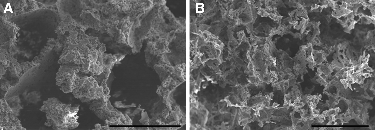
SEM images showed heterogeneous pore sizes for all scaffolds (Fig. 2), although smaller average pore size was demonstrated with greater HA content (Table 1). Pore sizes were 1,003±653 μm, 765±662 μm, 321±153 μm, and 85±57 μm for POC, POC-20HA, POC-40HA, and POC-60HA scaffolds, respectively (Table 1). Significant differences were determined between POC and POC-40HA, between POC and POC-60HA, between POC-20HA and POC-40HA, and between POC-20HA and POC-60HA (p<0.05). Mercury intrusion porosimetry confirmed the presence of interconnected pores with 47.2%, 66.9%, 69.7%, and 24.8% porosity for POC, POC-20HA, POC-40HA, and POC-60HA scaffolds, respectively (Table 1). The average pore size and porosity of scaffolds fabricated using LPF were in the range of human trabecular bone.39-44

Scanning electron microscopy images of
Values derived from Sikavitsas et al. 39
HA, hydroxyapatite.
The mechanical properties of POC, POC-20HA, and POC-40HA scaffolds prepared using LPF under dry and wet conditions were measured; values for porous scaffolds fabricated using the salt-leaching technique are also provided for comparison (85% salt by weight, <250 μm) (Table 2). Representative images of salt-leached scaffolds are shown in Fig. 3. Eighty-five percent salt content was chosen to ensure pore interconnectivity, and a heterogenous size range of salt crystals was incorporated because it parallels the heterogeneity of pore sizes within POC and POC-HA scaffolds fabricated using LPF (Fig. 2, Table 1). 45
Representative scanning electron microscopy images of salt-leached poly(1,8-octanediol-co-citrate) (POC) scaffolds. Scale bar:
Values derived from Athanasiou et al. 66
The compression modulus was greater with greater HA content under dry conditions for LPF scaffolds and salt-leached scaffolds. For POC and POC-HA scaffolds fabricated using LPF, significant differences were found between POC-20HA and POC-40HA and between POC and POC-40HA. For salt-leached scaffolds (POC salt, POC-20HA salt, and POC-40HA salt), significant differences were found between all groups (p<0.05). In the wet state, all compression moduli decreased from the dry state for both types of porous scaffolds with the exception of POC scaffolds. The compression modulus of POC-20HA scaffolds and POC-40HA scaffolds were 34.5% and 67% less, respectively. All salt-leached scaffolds had a greater loss in compression modulus than scaffolds fabricated using LPF (64%, 83%, and 97% less for POC salt, POC-20HA salt, and Poc-40HA salt, respectively). Differences between the dry and wet states, were found to be statistically significant only for POC-40HA scaffolds, POC-20HA salt, and POC-40HA salt (p<0.05). Under wet conditions, 40HA salt had the largest drop in compression modulus, and the stiffness was lower than that of POC-20HA salt and POC-40 HA scaffolds.
Effect of prepolymer viscosity on the porosity and mechanical properties of POC-40HA scaffolds
To investigate the effect of prepolymer viscosity on scaffold porosity and the mechanical properties, three different pre-POC were synthesized under varying polymerization conditions (Table 3, Fig. 4). Greater viscosity, which has been previously reported to correlate with greater cross-linking and molecular weight, was seen with longer polymerization time,. 46 These prepolymers were then used to fabricate POC-40HA scaffolds. The compression modulus was greater with greater pre-POC viscosity and was found to be statistically significantly different between all nanocomposite types (p<0.05). There were no statistically significant differences between pore sizes, which were heterogeneous (Table 3). The porosity was lower and the average distance between pores greater with greater pre-POC viscosity, although not statistically significantly so (Table 3, Fig. 4).

Cross section of poly(1,8-octanediol-co-citrate) containing 20% by weight hydroxyapatite nanocrystals (POC-40HA) scaffolds fabricated using POC prepolymer synthesized under one of the following synthesis conditions:
Biocompatibility in vitro using hMSC and in vivo at 6 weeks in a rabbit model
When hMSC were seeded onto POC, POC-20HA, and POC-40HA, cells attached and had spread after 1 day (Fig. 5). The amount of attachment was quantified, and no statistically significant differences were found between any scaffolds (Fig. 6), although greater HA content correlated with greater ALP activity, and significant differences were found between POC and POC-40HA and between POC-20HA and POC-40HA (p<0.05) (Fig. 6).
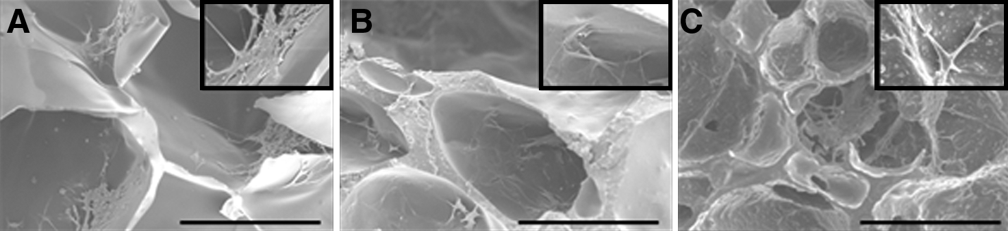
Human mesenchymal stem cells adhere and spread on
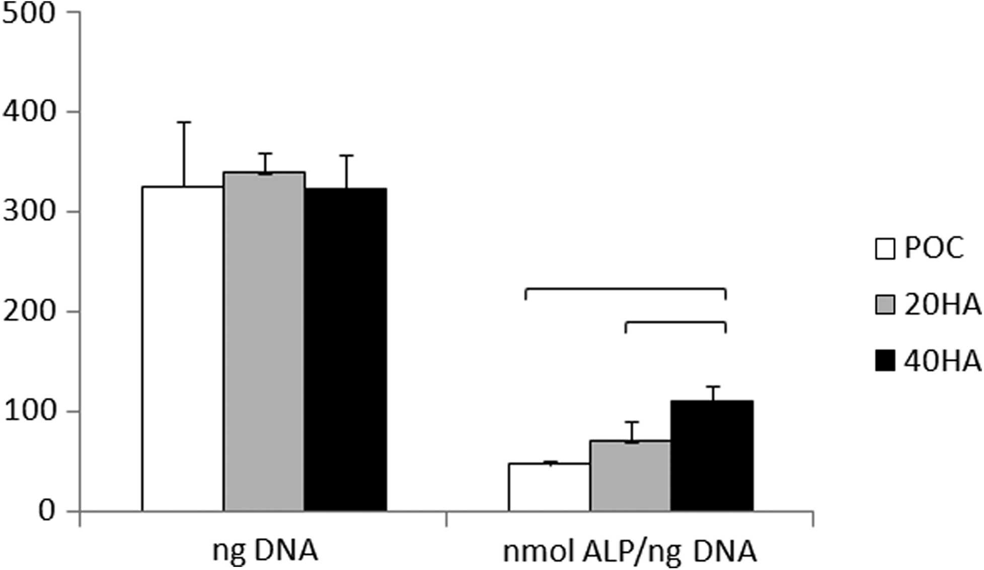
Quantification of human mesenchymal stem cell (hMSC) attachment and alkaline phosphatase (ALP) activity on all poly(1,8-octanediol-co-citrate) (POC)-based scaffolds at day 1. hMSC were seeded on 10-mm3 samples; bars represent significant difference between two groups.
After 6 weeks in vivo, there was no evidence of wound infection at the implant site, and all rabbits recovered well without any signs of erythema or sinus tract formation upon gross examination. Cell and tissue infiltrated throughout the pores of the POC-40HA scaffold (Fig. 7) and osteoblasts lining the osteoid were found at the implant–bone interface (Fig. 7C). Fibrous tissue was present within the scaffold and at the implant–bone interface.
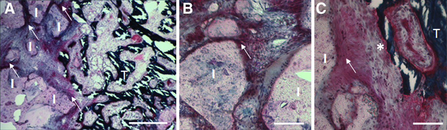
Representative images with Masson Goldner trichrome staining at 6 weeks of
Discussion
In the present study, interconnected, porous poly(diol citrate) scaffolds were fabricated using LPF. Through LPF, porosity can be incorporated within polymers that are not miscible in supercritical CO2, cytotoxicity due to trace organic solvents is eliminated, and there is no need for extensive salt-leaching in aqueous solution, eliminating concerns of premature polymer degradation through hydrolysis.29,30 Furthermore, HA nanocrystals can be incorporated to provide osteoconductivity and osteogenicity for bone applications.47,48
During LPF, pores are nucleated by trapping air bubbles created during the mixing process, and the number of pores can be controlled by regulating the stirring time.49,50 For example, POC samples that were not mixed did not result in porous scaffolds (Supplementary Fig. 1). Although performed manually in this initial study, future studies may automate the mixing speed and time for the most-consistent results and for large-scale applications.
The pressure is lower under vacuum conditions, causing the entrapped gases within the nucleation sites to exert pressure against the pore wall. The exerted pressure causes the scaffold to expand, increasing the total scaffold volume (Fig. 1). Expansion and coalescence of pores during LPF parallels the foaming process using supercritical CO2 (sCO2). During foaming using sCO2, polymers are dissolved in sCO2, and the solubility of the gas is reduced by decreasing the pressure.51,52 As a result, pores are nucleated and are able to grow and coalesce as gases diffuse throughout the material. The glass transition temperature of the polymer rises concurrently, and the porous network is stabilized in place. 51 Unlike foaming using sCO2, pore expansion and coalescence are feasible during LPF because of the elastomeric properties of POC. 5
The bubbles near the surface also expand and may rupture as the cell wall becomes thinned out. 53 This collapse causes a distinct outer membrane that resembles the less-porous, outer, cortical bone layer found throughout the skeleton (Fig. 1G),39,54 although to achieve tissue ingrowth, this outer layer can be removed easily by cutting or shaving it with a sharp tool. The porous structure is stabilized through complete cross-linking through postpolymerization (3 days at 80°C followed by 1 day at 120°C, at 2 Pa). When scaffolds are removed before complete polymerization, the scaffolds collapse, and porosity is lower (data not shown).
Pore size, porosity, and mechanical properties can be controlled according to particle content and extent of prepolymer polymerization. By increasing particle content or polymerization time of pre-POC, greater viscosity and rigidity is provided, which limits pore size and overall porosity while increasing the compression modulus (Fig. 2, Tables 1–3).3,55,56. Native trabecular bone is 50% to 90% porous, with pore diameters on the order of 1 mm, and scaffolds with pore sizes of 50 to 450 μm and pore volumes greater than 48% have displayed favorable bone regeneration,39–44 indicating that POC and POC-HA scaffolds can be engineered to meet the pore size range and pore volume criteria recommended for bone applications. The distance between pores can also be controlled to mimic the pore distance within bone, which has been reported to be on the order of 100 μm (Table 3, Fig. 4) 40 .
For LPF and salt-leached POC scaffolds, percentage mass loss from dry to wet states was greater with greater HA content (Table 2, Fig. 3). This is consistent with other reports that highlight water as an effective plasticizer through the disruption of polymer chain–chain hydrogen bonding and the lubrication effect of water. 57 All salt-leached scaffolds had greater percentage mass loss than HA-equivalent scaffolds fabricated using LPF (Table 2). Furthermore, POC-40HA salt had a lower modulus than POC-40HA when wet. In our previous studies, we reported that POC acted as the microphase binder and that 65% by weight HA was the upper limit of particle incorporation. 37 A high percentage of salt incorporation is necessary for interconnectivity, but salt particles may physically hinder cross-linking of the polymer, and salt-leaching under aqueous conditions can speed biodegradation. These actions can contribute to the lower compression modulus and highlights the disadvantages of the salt-leaching technique that are avoided during LPF. Although the mechanical properties of LPF scaffolds are inferior to those reported for bone (1–1,524 MPa, 211±78 MPa mean compression modulus),33,58,59 the mechanical properties are similar to those of other biodegradable, porous scaffolds reported for tissue engineering applications.60,61 The LPF scaffolds described in this study may be suitable for non-load-bearing bone defects, but future studies taking advantage of longer postpolymerization conditions will determine whether suitable mechanical properties for load-bearing defects can be fabricated as well. 5
To assess in vitro biocompatibility and the osteogenic potential of LPF scaffolds, hMSC were seeded and cultured in vitro for 1 day. hMSC adhered and spread on all LPF scaffolds, confirming biocompatibility (Fig. 5), and greater ALP activity was found with greater HA content (Fig. 6). This result is consistent with what is reported in the literature regarding the osteogenic potency of HA, confirming the utility of these scaffolds specifically for bone applications.48,62 In addition, after 6 weeks in vivo, cell and tissue infiltration was found throughout POC-40HA scaffolds, confirming their interconnectivity and ability to interlock with the surrounding tissue, an important requirement for implant stability and fixation (Fig. 7).39,40,54 The flexibility of adjusting the material and cellular properties of these novel scaffolds highlights their potential use for bone applications. Furthermore, these novel POC and POC-HA scaffolds may act as vehicles to deliver cells, growth factors, drugs, and genes, providing a wider range of applications and a basis for future directions.63–65
Conclusion
POC scaffolds were fabricated using a new technique (LPF). LPF does not use organic solvents, supercritical fluid, or particle leaching and is easily performed. In addition, it was demonstrated that HA content and prepolymer viscosity can affect the pore size, pore volume, and mechanical properties of scaffolds fabricated using LPF. The resulting porous scaffolds support cell migration in vivo. This study describes a simple method of creating porous scaffolds that may be adapted for other elastomers.
Footnotes
Acknowledgments
This work was supported by the National Science Foundation (NSF) CAREER award granted to Dr. Guillermo Ameer. This work made use of central facilities supported by the Materials Research Science and Engineering Center (MRSEC) of the NSF at Northwestern University. Electron microscopy was performed in the Electron Probe Instrumentation Center facility of the Northwestern University Atomic and Nanoscale Characterization Experimental Center at Northwestern University and is supported by the NSF Nanoscale Science and Engineering Center, NSF-MRSEC, Keck Foundation, the State of Illinois, and Northwestern University. We thank Matthew Johnson for assistance with mercury intrusion porosimetry and Mark Seniw for assistance with the mechanical tester.
Disclosure Statement
Northwestern University has applied for a patent for LPF and scaffolds fabricated using LPF.
References
Supplementary Material
Please find the following supplemental material available below.
For Open Access articles published under a Creative Commons License, all supplemental material carries the same license as the article it is associated with.
For non-Open Access articles published, all supplemental material carries a non-exclusive license, and permission requests for re-use of supplemental material or any part of supplemental material shall be sent directly to the copyright owner as specified in the copyright notice associated with the article.