
Abstract
Layer-by-layer biofabrication represents a novel strategy to create three-dimensional living structures with a controlled internal architecture, using cell micromanipulation technologies. Laser assisted bioprinting (LAB) is an effective printing method for patterning cells, biomolecules, and biomaterials in two dimensions. “Biopapers,” made of thin polymer scaffolds, may be appropriate to achieve three-dimensional constructs and to reinforce mechanical properties of printed materials. The aim of this work was to evaluate the effect of the tridimensional organization of cells and biomaterials on cell proliferation in vitro and in vivo. The experimental LAB setup was comprised of an infrared laser, focused onto a glass ribbon coated with an absorbing layer of gold. The cell bioink was made of MG63 cells (50 millions cells/mL in culture medium and 1% alginate), transduced with Luciferase gene for tracking and quantification. The printing substrate was a 100-μm-thick polycaprolacton (PCL) electrospun scaffold. The building sequence comprised sequential layers of cells and PCL scaffolds stacked using two different tridimensional arrangements, which were compared in this study (layer-by-layer vs. seeding on a single locus of the scaffolds). Then the cell-seeded materials were cultured in vitro or implanted in vivo in NOD-SCID mice. The qualitative follow-up involved scanning electron microscopy (SEM) observations, live-dead assays, and histology. The cell amount was quantified by photon imager during 21 days in vitro and 2 months in vivo. Live- dead assay and SEM revealed that the cells survived after printing and spread onto PCL membranes. Circle-shaped patterns were maintained in vitro during the first week but they were no longer observable after 2 weeks, due to cell proliferation. Luciferase tracking displayed that the cell amount was increased in vitro and in vivo when the materials and the cells where stacked layer by layer. Histological sections of the in vivo samples revealed a thicker fibrous tissue in the layer-by-layer samples. We have demonstrated in this study that PCL electrospun biopapers can act as a shock-absorbing mattress for cell printing and could further support cell proliferation. The layer-by-layer printing provided an appropriate 3D environment for cell survival and enhanced cell proliferation in vitro and in vivo.
Introduction
An alternative method for the assembly of cells and biomaterials is defined by the bottom-up approach 5 that “relies on the assembly of building blocks mimicking native functional units into larger tissue constructs, using layer stacking, random packing and 3D bioprinting.” 6 This paradigm shift should lead to the fabrication of more realistic and functional tissue constructs by creating patterns of cells and biomolecules that would favor tissue regeneration.
Among bioprinting methods, laser assisted bioprinting (LAB) is efficient for cell printing at high resolution 7 without alteration of cell viability 8 or phenotype. 9 We have previously demonstrated that high-throughput printing by LAB was achievable with a dedicated experimental set-up. 10 However, LAB is not completely suited for building by itself tissue constructs with cm3 size and handling capacity. The same issue can be observed using droplet-based bioprinting technologies (ink-jet, acoustic droplet ejection). Indeed, the characteristic droplet volume is around 1 pL, 10 and the printed bioinks lack sufficient mechanical properties to maintain the shape of three-dimensional hybrid materials. 11 As similar issues occurred with other bioprinting methods (ink jet printing, microsyringe printing, or spheroid extrusion), it was proposed to support the layer-by-layer assembly of biological elements with “biopapers,” made of hydrogels sprayed between two stacks of cells. This concept was applied for layer-by-layer inkjet printing of cells using fibrin12,13 or collagen biopapers. 14 Sprays of collagen were also used as biopapers for layer-by-layer cell printing using microsyringe,15,16 or cell spheroids technologies.17–19 More recently, a 3D structure was achieved with stem cells printed by laser and collagen sprayed between three stacks. 20
From these studies, we have learned that several hydrogels can be successfully used for layer-by-layer printing of cells, leading to relevant in vitro models to study cell–cell and cell–environment interactions. 20 However, there are currently no in vivo applications of layer-by-layer bioprinting using hydrogel biopapers, probably because the mechanical properties of these materials are not adapted to in vivo implantation. The latter would require more spatial stability for handling and grafting. It is noteworthy that most of the potential applications of bioprinting are today restricted to soft tissues, such as skin, 15 vascular, 17 or neural 16 tissue engineering. For wider in vivo applications of bioprinting, it is necessary to develop new materials and methods to fabricate three-dimensional layer-by-layer printed scaffolds that allow manipulation and implantation.
Electrospinning is commonly used to build scaffolds made of intermingled fibers of polymers, and results in materials with a highly controlled three-dimensional architecture. 21 The typical fiber diameter ranges from 0,1 μm to a few micrometers and the scaffold properties can be tuned by modifying processing parameters (voltage, flow rate, time of deposition). The pore size is directly correlated to the fibers diameter, 22 which may be a limit for cell invasion inside electrospun scaffolds.23,24 Several strategies have been developed to improve cell seeding efficiency into electrospun scaffold such as modification of the polymer solution to make the scaffold bioactive 25 and concomitant spraying of cells during electrospinning. 26
For in vitro applications of bioprinting, the layer-by-layer biofabrication using hydrogels sprayed alternatively between the stacks of cells is efficient to keep the cell pattern shape and to maintain cell viability.14–16,20 For in vivo applications of bioprinting, the biopapers should possess specific properties including handling for surgical implantation. Biopapers made of chromatography paper have been used for layer-by-layer stacking and in vivo implantation of tumor cells lines and study of oxygen diffusion. 27 However, the chromatography paper cannot be used for tissue regeneration. However, the electrospun PCL is a widely studied biomaterial, suitable for in vivo implantation for bone 28 or vascular26,29 tissue engineering.
The roles of the biopapers for the layer-by layer bioprinting are multiple and encompass microscopic to macroscopic aspects. The biopapers are receiving substrate for cell printing and should (1) be appropriate for a smooth deposition of cells to maintain their viability, 8 (2) provide a stabilization of printed patterns, 30 and (3) bring an appropriate environment for cell nutrition and function. 31 Then, the biopapers should also bring stability and relevant mechanical properties to the three-dimensional assembly. This second aspect is essential for in vivo applications, whatever the engineered tissue considered.
We hypothesize that a combination of LAB and electrospinning will result in adding up the respective advantages of these methods and eliminate their specific drawbacks: toward this aim, these technologies could be combined in a layer-by-layer bottom-up approach, using LAB to deposit cells containing droplets onto alternate layers of electrospun scaffold biopapers, to fabricate a tridimensional microtissue.5,6 This combination could provide efficient cell seeding of electrospun scaffolds and cohesion to hybrid materials fabricated layer by layer with LAB. In this study, we have evaluated for the first time the behavior of three-dimensional hybrid materials built layer by layer, using electrospun scaffolds of polycaprolacton (PCL) (biopapers) and cell printed by LAB. More specifically, the objective of this work was to evaluate the influence of the three-dimensional organization of MG63 cells and PCL electrospun scaffolds on cell proliferation in vitro and in vivo, using a layer-by-layer sandwich model of assembly, and to compare it to a control hybrid material made of the same amount of material with an alternative 3D arrangement.
Materials and Methods
Cell culture and cell bioink preparation
A human osteosarcoma cell line MG63 (ATCC: CRL 1427™) was used for in vitro and in vivo experiments. Cells were cultured in Iscove modified Dulbecco's medium (IMDM, Invitrogen, Carlsbad, CA), supplemented with 10% (v/v) bovine serum albumin (BSA) at 37°C in a humid atmosphere composed of 5% CO2.
Cells were transduced with lentiviruses harboring the firefly luciferase gene, under the control of the MND promoter (myeloproliferative sarcoma virus enhancer, negative control region deleted, dl587rev primer-binding site substituted) 32 using a lentiviral vector. For viral transduction, 2×10⁵ freshly trypsinized MG63 cells were mixed with 4×10⁶ viral particles (multiplicity of infection=20). After 24 h in culture, virus-containing medium was replaced by fresh medium and cells were allowed to grow. Cells were then amplified and used for laser printing.
The cell bioink for laser printing experiments was prepared as follows. Cells were detached from the plastic dish with a solution of 0.125% (w/v) trypsin and 2 mM EDTA, then the clot was suspended in an appropriate volume of IMDM at a cell concentration of 50×106 cells/mL. The cell suspension was finally supplemented with 1% (w/v) sodium alginate solution (Protanal 10/60–FMC BioPolymer®).
Fabrication of PCL biopapers by electrospinning
PCL (Mn=42,500) was purchased from Aldrich® and a PCL solution (20% w/v in CHCl3) was loaded into a syringe and a desired flow rate was set using a syringe pump (KDS 100, KD Scientific). The other end of the syringe was connected to a needle, which acted as the positive pole when a high-voltage generator (NCE 30000, Heinzinger Electronic GmbH, Germany) was turned on. A metallic sheet of stainless steel was the collector (ground). An electrostatic field was formed between the needle and the collector when high voltage was applied. The polymer solution was pushed through the syringe to the tip of the needle at a flow rate of 1 mL/h. When the electrostatic field strength overcame the surface tension of the liquid drop at the tip of the needle, the drop was stretched and deposited onto the collector. A voltage of 12 kV was applied and the distance between the needle and collector was 20 cm. The processing parameters were adjusted to obtain membranes of 100 μm thick. For the in vivo and the in vitro experiments, disc-shaped PCL membranes of 5 or 10 mm diameter were punched using a die cutter and then they were sterilized by UV exposure (2×10 min).
LAB workstation and cell printing conditions
The LAB workstation has been described elsewhere. 10 Briefly, it was composed of an Nd:YAG infrared laser (SpectraPhysics, λ=1064 nm, 30 ns) driven with galvanometric mirrors (scanning system) and focused onto the ribbon. The LAB workstation was equipped with dedicated software, allowing each printing parameter to be adjusted individually. The experiments were performed using a 5 kHz laser pulse rate, a 18 μJ laser energy, a 200 mm/s scanning speed, and a 500 μm space between the ribbon and the substrate. Ribbons were made of a quartz slide transparent to laser wavelength, and were coated with a laser-absorbing interlayer of gold (50 nm) using a sputter coater (Emscope SC500). Ribbons and substrates were sterilized in an autoclave before cell printing experiments.
Feasibility and viability of cell printing on electrospun PCL membranes
This first experiment was done to evaluate the fate of cells printed onto PCL scaffolds. A 20 μm-thick film of cell bioink was spread 1 min before printing on the ribbon and a 100-μm-thick Matrigel® layer was placed beneath the PCL membrane to stabilize it onto the quartz receiving substrate. The cells were printed in the shape of 2-mm-wide circles (band width=500 μm) and the receiving substrates were placed in a controlled humid atmosphere (5% CO2, 100% humidity, 37°C) for 5 min. A set of four circles was printed on each PCL membrane, which measured 10 mm in diameter. Then the receiving substrates were placed in a six-well plate and 2 ml of complete culture medium was added over cell-PCL hybrid materials before returning in the incubator.
The cells were stained 4 days after the printing experiment on PCL biopapers using Live/Dead assay kit (Invitrogen) according to the manufacturer's instructions. The samples were incubated in IMDM supplemented with 10% FBS and 2 μM calcein-AM and 4 μM ethidium homodimer (EthD-1) for 20 min in a controlled atmosphere (100% humidity, 37°C, 5% CO2). The observations were done with Nikon Eclipse 80i microscope equipped with a digital camera Dxm1200C. Finally, the hybrid materials were fixed in paraformaldehyde (4% w/v, 10 min), dehydrated in graded increasing ethanol solution, and sputtered with gold for scanning electron microscopy (SEM). The observations were done with a Hitachi® S-2500 microscope (Tokyo, Japan), working at 10 keV.
Three-dimensional assembly of PCL membranes and cells
The layer-by-layer assembly of PCL biopapers and MG63 cells transfected with luciferase was done with the same protocol for in vitro and in vivo experiments.
The PCL membranes were cut with a 5-mm-diameter die cutter for these experiments. The receiving substrate (quartz) was covered with a 100-μm-thick Matrigel® layer to stabilize the first PCL membrane. Then a 20-μm-thick film of cell bioink was spread 1 min before printing on the ribbon and the cells were printed in the shape of a 4-mm-wide disc centered onto the PCL scaffolds.
The layer-by-layer sandwich assembly (LBL) of cells and PCL membranes consisted of three membranes of PCL and nine layers of cells stacked alternatively 3 by 3. The cell seeded scaffold (CSS) of PCL membranes by cells consisted in nine layers of cells printed onto three membranes of PCL stacked (Fig. 1). In both conditions (LBL and CSS), the same amounts of cells and PCL membranes were used but their three-dimensional arrangement was modified.
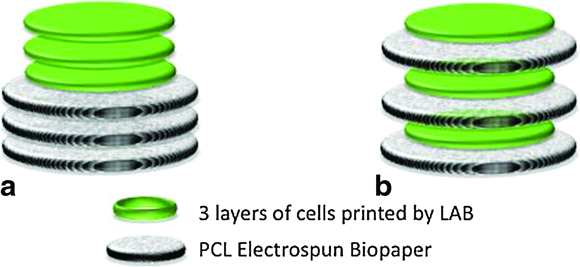
Design of the experiment.
Finally, the tridimensional PCL/cell constructs were designed to measure 5 mm diameter and an average of 300 μm thick (three sheets of 100 μm each; the thickness of cells printed being negligible). The same tridimensional constructs were used for in vitro and in vivo experiments.
Study of in vitro cell proliferation into LBL and CSS hybrid materials
Both types of cells-PCL assemblies (LBL and CSS) were cultured in static conditions (96-well culture dish) during 21 days and culture medium was changed every third day. Each replicate was placed in a single well with 200 μl of medium. The wells of the plates have been coated with a 2% (w/v) agarose solution to prevent cell attachment to the tissue culture plastic. The cell number in each construct at each time point was deducted from photon-imager analyses performed every week, using a standard curve built before the experiments. Emission was recorded in real time with a Biospace® Imaging System (BIOSPACE Lab., Paris, France). Prior to the observations, 150 μg/ml of a Beetle Luciferin (Promega) solution in PBS 1X was added in each well; the measurements then started immediately. The results corresponded to the values measured when the signal was optimal and stable for at least 3 min. The signal was integrated for 100 s and values were expressed as p/s/sr (photons/second/steradian) (maximum and minimum values are fixed, respectively, at 100 and 10, smoothing=2.5). Each experiment was repeated five times.
Study of in vivo cell proliferation into LBL and CSS hybrid materials
Twenty NOD-SCID mice (female, 8 weeks old) were used in this study to compare the effect of biofabrication methods on cell proliferation using bioluminescence quantitative imaging and histology.
The animals were anesthetized with isofluorane and skin antisepsis was done with povidone-iodine (Betadine, France). An incision was performed in the skull midline and the scalp was dissected to expose the calvaria. Then the periosteum was carefully peeled off and two lateral calvaria bone defects (4 mm wide) were performed in each animal with a 4-mm-diameter trephine (TBR, Toulouse, France). The surgical procedures were performed under constant saline irrigation and care was taken to prevent dura mater injury. The material “LBL” was inserted in the right defect and the material “CSS” was inserted in the left defect. Each animal was its own control and all the 20 animals were treated using the same procedure. The soft tissues were repositioned and sutured using 3:0 Vicryl® at the end of the surgery. Animals recovered in a warm environment before being returned to animal facilities and received 1 μg/10 g animal weight of buprenorphine intraperitoneally. The in vivo experiments have been performed after the protocol was approved by the ethics committee of the Aquitaine Region and Bordeaux University (France).
The in vivo follow-up of cell number was done using a photon imager, two times per week during 4 weeks (n=20) or 8 weeks (n=10). The mice were anesthetized with isofluorane and injected intraperitoneally with 150 mg/kg of firefly luciferin (Promega), dissolved in PBS 1X from a solution concentrated at 30 mg/mL. Light emission was recorded in real time with a Biospace® Imaging System (BIOSPACE Lab., Paris, France). Measurements started immediately after substrate injection and the results corresponded to the values measured when the signal was optimal and stable for at least 3 min. The signal was integrated for 100 s and values were expressed as p/s/sr (photons/second/steradian) (maximum and minimum values are fixed respectively at 100 and 10, smoothing=2.5). Signal quantification on the regions of interest (right or left side of calvarias) was done using the M3 Vision® software.
The animals were sacrificed after 4 weeks (n=10) or 8 weeks (n=10), then the skulls were harvested and fixed in PFA (4% v/v) for 3 days. The samples were demineralized 12 h (Decalcifiant osseux BAYER, France), dehydrated in ethanol (70%, 80%, 95%, 100%), and then embedded in paraffin. Coronal sections were obtained (6–8 μm), stained with hematoxylin–eosin–saffron (HES), and observed under a photomicroscope (Nikon eclipse 80i).
Comparisons of the independent groups (unpaired samples) were done with the nonparametric Mann–Whitney U-test. The paired groups (LBL vs. CSS) were compared using the Wilcoxon test for in vivo and in vitro experiments. Differences were considered significant when p<0.05.
Results
Fabrication of electrospun sheets
PCL fiber meshes to be used as biopapers were successfully fabricated using electrospinning. Figure 2 shows an overview of an electrospun PCL sheet along with a high magnification image of the fibers observed by SEM. The fiber diameters were measured to be 5.96±1.9 μm and the average thickness of the PCL sheets was 100 μm.

SEM observations of electrospun membranes of PCL.
MG63 cells printed onto PCL biopapers
The MG63 cells printed on a receiving substrate made of PCL maintained their viability after printing. Indeed, the live-dead assay was strongly positive after 4 days of culture and revealed that the initial circle-shaped pattern was maintained at this time (Fig. 3a). Higher magnification displays cell spreading on the PCL fibers (Fig. 3b) and extracellular matrix secretion between the fibers of PCL (Fig. 3c).

MG63 cell printing on electrospun PCL biopapers.
In vitro cell proliferation
One day after printing, the quantification of luminescence corresponded to an average of 20,000 printed cells and was similar in the LBL and in the CSS three-dimensional hybrid materials (Fig. 4).
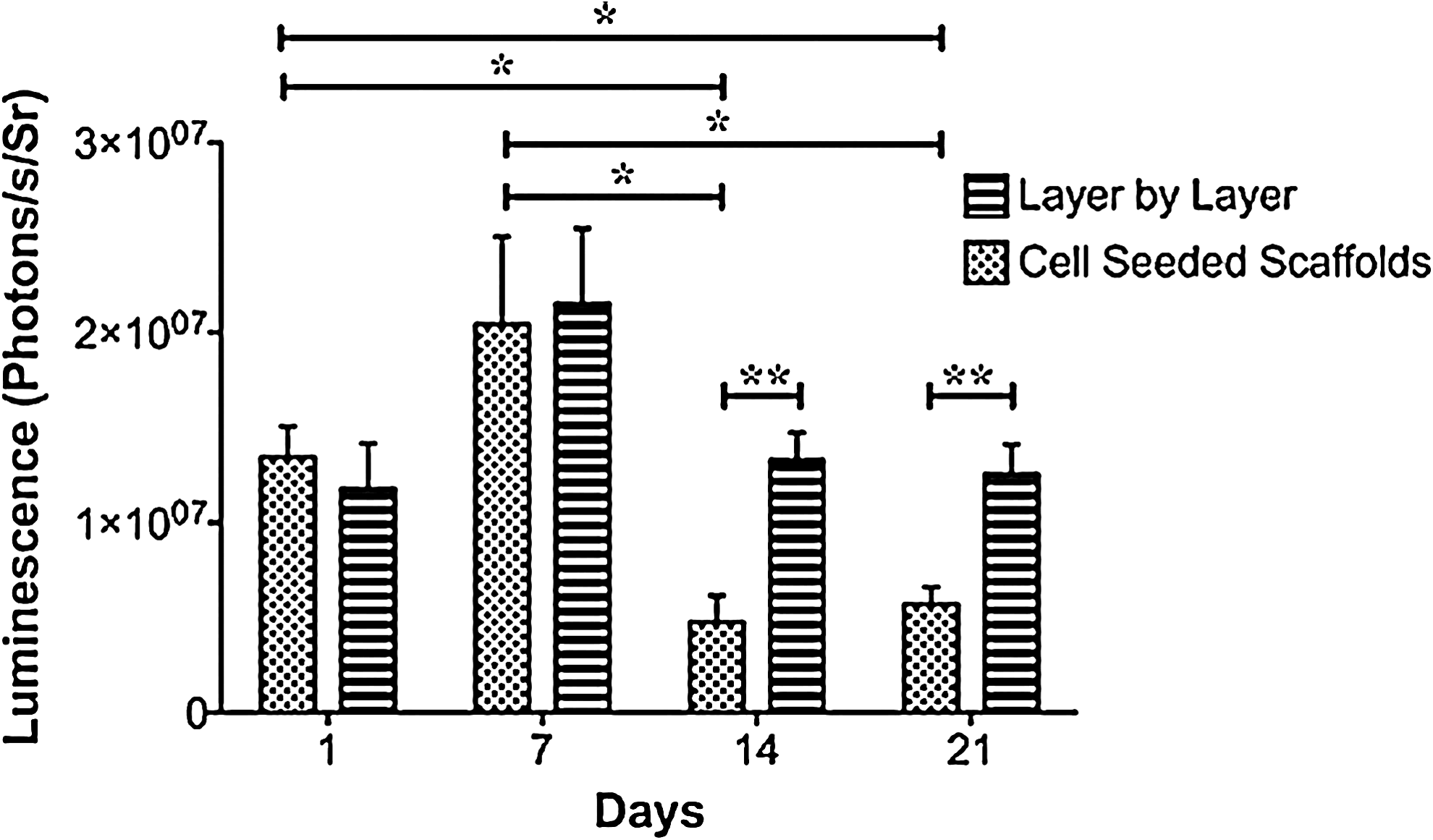
In vitro cell number assessed by photon imager. The layer-by-layer (LBL) sandwiches and the cell seeded scaffolds (CSS) displayed similar cell amounts until 7 days. After 2 and 3 weeks, the LBL arrangement enhanced cell proliferation when compared to CSS. *p<0.05; **p<0.01.
The LBL experiment displayed that the amount of cells was not statistically different at all the time points of the experiment (p>0.05). In the CSS experiment we observed that the cell number was stable during the first week, then it decreased significantly at day 14 and day 21 versus day 1 or day 7 (p=0.028) (Fig. 4).
The comparison of the two conditions at each time point did not reveal statistical differences at day 1 or day 7, whereas the amount of cells in the LBL sandwiches revealed by photon-imager quantification was significantly higher than in CSS at day 14 (p=0.008) and day 21 (p=0.008) (Fig. 4).
In vivo cell proliferation
All the animals survived the experimental procedure and they were all included for the in vivo follow-up of cell proliferation. The cell number was observed by photon-imager at each time point on both sides of the calvaria and the results confirmed the previous in vitro observations (Fig. 5). Light emission was always significantly higher in the LBL sites when compared to the CSS sites (Fig. 6). Concerning the LBL sites, there were significantly less cells between day 1 and day 10 and all the other time points. Values observed at day 47 were higher than at all the other time points. Concerning the CSS sites, a significant increase between day 1 and 10 and all the other time points was observed. There was also a significant increase between day 15 and day 30 and between day 47 and day 15 and 23.

In vivo follow-up illustration of cell number after implantation of PCL/MG63-Luc hybrid materials in mouse calvarial bone defects. The follow-up of the five mice depicted was done by placing them in the same order for each photon-imager analysis session. This picture is representative of the luciferase expression observed in the 20 mice used in this study. LBL were implanted on the right side and CSS were implanted on the left side. Follow-up images were obtained at 7 days

Quantification of in vivo cell proliferation using light emission of MG63-Luc cells. Light emission was significantly higher in test vs. control sites at all the time points of the experiment (*p<0.05).
Histological analyses of the decalcified samples observed after 8 weeks of healing revealed that a dense and thick fibrous tissue was observed in the LBL sites, whereas a thinner fibrous tissue was present in the CSS samples (Fig. 7a–c). Similar histological observations were done after 4 weeks of healing.

Histological pictures of the materials implanted in the calvaria defects after 8 weeks.
Discussion
One of the major goals of layer-by-layer biofabrication is to enhance the control on the three-dimensional position of cells seeded inside the support material. Several studies have achieved this goal using inkjet printing14–16 or laser printing. 20 However, the influence of the three-dimensional layer-by-layer organization on cell number has not been evaluated in these experiments. We have hypothesized that the layer-by-layer bioprinting would make the seeding more efficient than the conventional seeding of porous materials, and we have proposed using a combination of laser assisted bioprinting and electrospinning. We have demonstrated in this study that the position of the cells in a three-dimensional tissue engineered product had a significant effect on cell number. Indeed, cell number was increased in vivo using a layer-by-layer biofabrication model, combining PCL electrospun scaffolds and MG63 cells printed by LAB. The conventional seeding of the same amount of scaffolds and cells was used as a control and led to a significant lower cell number in vivo. The positive effect of LBL constructs on cell proliferation was not observed in vitro and the cell number in this condition remained stable after 21 days of culture. However, the LBL constructs provided a suitable environment to maintain cell viability over time, while the CSS constructs did not. This study demonstrates the superiority of LBL constructs to maintain cell viability in vitro and to enhance cell proliferation in vivo.
The bioprinting assisted by laser has been used in several studies to print cells with different set-ups, 33 and the protocols to maintain cell viability, phenotype, proliferation, and differentiation after printing have been studied with different cell types. 34 The critical parameters are related to the bioink composition, the droplet generation laser process, and the nature of the receiving substrate. In this study, the bioink composition and the printing set-up were similar to those of previous studies, which have both shown their efficacy concerning the printing resolution and maintenance of biological materials activity.8,35 More specifically in this study, the cell bioink was prepared using the MG63 osteoblastic cell line. These cells are widely used as a model for in vitro biocompatibility studies of biomaterials, and they have also been successfully printed by Matrix-Assisted Pulsed Laser Evaporation–Direct Write (MAPLE-DW).36,37 The bioink contained a high cell concentration and was supplemented with alginate 8 and the printing parameters were similar to previous studies with the same set-up. 35
The main original points of our study concern the nature of the receiving substrate and the layer-by-layer printing. The maintenance of cell viability after LAB requires a shock-absorbing mattress, which is usually made of hydrogels such as Matrigel 8 or Gelatin. 38 We have also recently demonstrated that human osteoprogenitors could be printed onto a nanohydroxyapatite receiving substrate printed by LAB. 35 In the present study, we have shown that the PCL biopapers can be used as a receiving substrate for layer-by-layer bioprinting. The PCL biopapers have provided a shock-absorbing mattress for the cells printed and they have supported the cell viability (Fig. 3) and proliferation (Figs. 4–7). The LAB printing conditions were compatible with the preservation of the viability of printed cells. The integrity of the printed patterns was maintained by the use of PCL biopapers, which retained the circular shape of MG63 cells after 4 days (Fig. 3).
Growth of a cell population is a critical step for engineered tissue growth and maturation, and this parameter was evaluated to characterize the efficiency of three-dimensional cell organization of the hybrid materials. Thus, the MG63 cells were used for the in vitro and the in vivo experiments because of their high proliferation rate. The cell transduction by luciferase and photon imager analysis allowed quantification of the amount of cells in the hybrid materials in vitro and in vivo (Figs. 4 and 6). Photon imager was chosen as a noninvasive method to follow the cell number in the mice after implantation of the hybrid materials in calvarial defects, and both conditions were evaluated simultaneously in each animal. The calvarial model allowed repeated observations of the light emitted in each animal with a precise localization of cells during the entire experiment (Fig. 5).
Cell quantification for the in vitro study has shown that after 1 day, a similar amount of printed cells was present in the two types of hybrid materials (Fig. 4), which confirms that laser-assisted bioprinting allows printing controlled amounts of cells. 7 The intensity of light quantified by photon imager analysis was correlated to a standard amount of MG63 cells. We have estimated that an average of 18,000 cells were printed inside each CSS or LBL hybrid material, i.e., 2000 cells were printed in each individual layer. Concerning the in vitro follow-up of the hybrid materials, the amount of cells was stable during the entire experiment using the LBL organization, which means that cell number had reach a threshold, probably due to the limited renewing of culture medium in the wells (every 3 days, static culture conditions). The reduced pore size of the PCL biopapers (40 μm: Fig. 2) could also explain why the cell number reached a threshold, due to a limited diffusion of nutrients and oxygen inside the constructs.
In contrast, the quantity of cells in the CSS scaffolds decreased significantly after 14 and 21 days of culture: in this case, the cells were not trapped initially inside the PCL biopapers and were probably partially washed out when the culture medium was changed (Fig. 4).
The same hybrid materials have been implanted in mice calvarial defects bilaterally and each animal was its own control (Fig. 5). The LBL organization displayed a significantly higher cell number than the CSS organization at each time point of the experiment (Fig. 6), suggesting that this configuration favors cell proliferation. The signal was not visible at the first time points because the threshold applied to the picture was higher than the quantity of light emitted at that time. The discrepancy between the in vitro (Fig. 4) and the in vivo (Fig. 6) results could be explained by more nutrients transferring in vivo than in vitro. The histological analyses of the materials implanted in the mice are consistent with the photon imager data: a thick fibrous tissue with fibroblastic cells was observed in the LBL sites, whereas a thin tissue with a poor cell content was observed in the CSS site (Fig. 7).
Increase of cell number in vivo was not observed during the first week, whereas the presence of cells was detected in vitro after 1 day of culture (Fig. 4). The luciferin was added directly in the culture medium for in vitro experiments, and the cells in culture have metabolized it immediately. Concerning the in vivo quantification, the luciferin was injected intraperitoneally and was transported by blood flow before reaching the MG63-Luc cells, which required the development of an efficient vasculature in the surgical site around the implanted materials: this could explain the delay observed between the implantation and the first observation of the signal after 10 days (Fig. 6).
In this model, the cell number was increased in the LBL hybrid materials due to (1) the three-dimensional environment developed for cell proliferation and (2) the trapping of the cells inside the PCL scaffolds. The LBL biofabrication of 300-μm-thick hybrid materials provided a 3D environment that enhanced cell proliferation. For thicker hybrid materials, and depending on the nature of the tissue, it may be necessary to develop a vasculature inside the stacked material, 6 because the low oxygen and nutrient supply inside a thick material fabricated layer by layer can prevent cell proliferation. 27 The second advantage of the LBL biofabrication is related to the trapping of the printed cells between the PCL biopapers, which reduced their dispersion in the surrounding tissues after in vivo implantation. It is likely that a significant amount of cells printed in the CSS scaffold migrated away from the implanted materials. In a previous study, we have shown that nHA particles printed in situ and in vivo without stabilization were partially dispersed in the surrounding tissues after 1 week of healing. 11 This study confirms that postprocessing of printed elements is mandatory to maintain cell position and viability. The stabilization of cells on their substrate is usually achieved by the spraying of a hydrogel after cell printing, but the final mechanical properties of these hybrid materials are generally very limited and may hinder their postprinting manipulation. 30 Here we show that cell immobilization is also possible with electrospun meshes, which allow an easy handling and surgical implantation of the printed materials.
Conclusions and Perspectives
We have established a model for microtissue fabrication combining laser assisted bioprinting and electrospinning and we have shown that layer-by-layer biofabrication supports cell viability in vitro and cell proliferation in vivo. This layer-by-layer model for biofabrication is consistent for cell proliferation, but the following steps leading to the establishment of a functional tissue require further explorations. Following this proof of concept, the influence of the LBL organization should be evaluated for cell proliferation, differentiation, and extracellular matrix secretion using cells more relevant for tissue regeneration applications. The diffusion of nutrients and oxygen inside the construct as well as the elimination of metabolic wastes should be addressed to achieve clinical-size tridimensional cellularized constructs. This model could be easily modified using different cell types printed alternatively during the LBL fabrication and different scaffolding biopapers with specific bioactive properties, to achieve further biomimicry for more complex tissue fabrication.
Footnotes
Acknowledgments
The authors would like to thank the “Fondation de la Recherche Médicale (FRM),” the Institut Français de la Recherche Médicale (IFRO),” the “Région Aquitaine,” and the GIS “Advanced Materials in Aquitaine” for their financial support.
Disclosure Statement
The authors have no financial affiliations that would have biased this article.