
Abstract
Commercially available permeable supports with microporous membranes have led to significant improvements in the culture of polarized cells because they permit them to feed basolaterally and thus carry out metabolism in a more in vivo-like setting. The porous nature of these membranes enables permeability measurements of drugs or biomolecules across the cellular barrier. However, current porous membranes have a high flow resistance due to great thickness (20–40 μm), low porosity, and a wide pore size distribution with tortuous diffusion paths, which make them low-throughput for permeability studies. Here we describe an alternate platform that is more flexible, allows for more control over physical parameters of the membranes, and is high-throughput. This study reports on the synthesis, nanofabrication, and surface characterization of a 3-μm-thick transparent membrane based on poly(4-hydroxy styrene) (PHOST). The membranes are nanofabricated using electron beam lithography and deep ion plasma etching to achieve an organized array of straight pores from 50 to 800 nm in diameter, with at least 23 times less flow resistance. It also shows for the first time the potential utility of PHOST as a cell culture substrate without cytotoxicity, and suitability for nanofabrication processes due to temperature stability.
Introduction
Microporous membranes in commercially available permeable supports are made of either polyester (partially transparent) or polycarbonate (translucent). They are 20–40 μm thick depending on the manufacturer, and their pore diameter is 0.4, 1.0, or 3.0 μm with their nominal pore density being 2×10⁶ (polyester) and 1×10⁸ (polycarbonate) pores/cm2. Even though polycarbonate membranes possess higher porosity, they are not commonly used in cell culture applications due to the difficulty in performing microscopy on them. The process of fabrication of these membranes results in a random distribution in the size and placement of pores, and more importantly the resulting diffusion path is tortuous causing entrapment of biomolecules.5–12 Thus, key drawbacks of current commercial permeable supports are their high flow resistance due to a wide pore size distribution with tortuous paths, low total porosity, and relatively thick membranes which make them low-throughput for permeability studies.8–12
Here we describe an alternate platform that is more flexible, versatile in its physical characteristics, and high-throughput. Conventional cell culture dishes and inserts are generally made of polystyrene, and these surfaces are further plasma treated to make them negatively charged for better cell attachment. For this reason, poly(4-hydroxy styrene) (PHOST) was our polymer of choice for this study because it is already negatively charged due to the presence of the hydroxyl group, and it has also proven suitable for nanofabrication purposes due to high thermal resistance.13,14 A 3-μm-thick transparent polymeric membrane based on PHOST was synthesized and nanofabricated using a combination of electron beam lithography with deep ion plasma etching to achieve an organized array of straight pores with a narrow size distribution (50 to 800 nm in diameter). The porosity of the membrane is 8×10⁶ pores/cm2, which is four times higher than commercial membranes. These membranes have at least 23 times less flow resistance than their commercial counterparts, making them ideal for high-throughput permeability studies. Moreover, we report on the preliminary cell culture studies, showing the successful attachment and growth of several cell lines commonly used in in vitro models of the blood-brain barrier and the intestinal barrier, with focus on the viability, rates of metabolism, and differentiation of the cells on the nanofabricated membranes.
Materials and Methods
Chemicals and supplies
Solvents and reagents for the polymer synthesis were obtained from Sigma (St. Louis, MO) and used without further purification unless otherwise stated. Twelve-well transwell permeable supports (polyester; pore size, 0.4 μm; pore density, 4×10⁶ pores/cm2) were purchased from Corning (Lowell, MA). Poly-D-lysine and collagen type I were purchased from Fisher Scientific (Pittsburg, PA). Human fibronectin was purchased from Millipore (Billerica, MA). EGM® MV and trypsin-EDTA were purchased from Lonza (Basel, Switserland). Alamar Blue (AB), fetal bovine serum (FBS), and Dulbecco's modified Eagle's medium (DMEM, with L-glutamin, sodium pyruvate, and low glucose), polyclonal rabbit anti-glial fibrillary acidic protein (GFAP) antibody, rabbit anti-occludin antibody, Alexa Fluor® 488 goat anti-rabbit IgG antibody, and 4′,6-diamidino-2-phenylindole (DAPI) dihydrochloride nuclear stain were purchased from Invitrogen (Carlsbad, CA). Dulbecco's phosphate buffered saline (DPBS, without CaCl2 and MgCl2), HEPES sodium salt, Triton X-100, goat serum, fluorescein sodium salt, and gentamicin were purchased from Sigma (St. Louis, MO). Paraformaldehyde (16%) was purchased from Electron Microscopy Sciences (Ft. Washington, PA). Acetylated low-density lipoprotein labeled with 1,1′-dioctadecyl-3,3,3′,3′-tetramethylindo-carbocyanine percholorate (DiI-Ac-LDL) was purchased from Biomedical Technologies (Stoughton, MA).
Polymer synthesis
The reactions used for the preparation of PHOST is based on a modified reaction scheme (Fig. 1).13,14 Poly(tert-butoxy styrene) was synthesized by free radical polymerization. Briefly, 7.5 mL of 4-tert-butoxy styrene (99%) was polymerized in 5 mL of anhydrous toluene at 65°C with 10 mg of recrystallized α,α′-Azoisobutyronitrile (AIBN) as the initiator. All glassware was flame dried before use and the reaction mixture was purged with dry argon for 10 min. Polymerization was carried out for 19 h and then terminated in 800 mL of 4°C methanol. Due to the high molecular weight of poly(tert-butoxy styrene), it was sticky and self-folded, so no further washing was performed. Poly(tert-butoxy styrene) was converted to PHOST by a hydrolysis reaction. First, it was dissolved in 20 mL of 1,4-dioxane, and then 50 mL of 4 M hydrochloric acid in dioxane solution (fivefold) was added. The mixture was reacted at 80°C under reflux for 24 h and then precipitated in 800 mL 4°C water. After neutralization with NaOH solution to a pH value of 7–7.5, the resulting polymer was filtered and washed several times with 4°C water and lyophilized for 48 h.
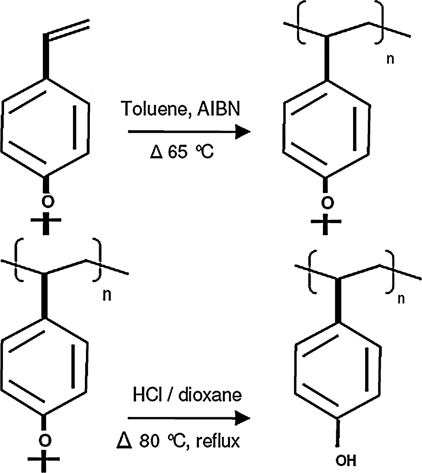
Synthesis of poly(4-hydroxy styrene) (PHOST).
Polymer characterization
Gel permeation chromatography (GPC) measurements were performed using THF as elution solvent at a flow rate of 1 mL/min in a Waters size exclusion chromatography system. 1 H-NMR spectroscopy was performed on 300 MHz Mercury 300 NMR instrument using CDCl3 and DMSO-d6 as solvents. Fourier transform infrared spectroscopy (FTIR) measurements were carried out on a Mattson 2020 Galaxy series instrument. Thermal analysis was carried out on a TA Differential scanning calorimeter (DSC, DSCQ1000) and a TA thermogravimetric analyzer (TGAQ500).
Nanofabrication process
Figure 2 illustrates the fabrication process that was applied to obtain spatially controlled nanopores. Briefly, a silicon substrate was treated with oxygen plasma (standard recipe) in an Oxford Plasmalab 80+Reactive Ion Etching (RIE) system for 2 min. A solution of poly(acrylic acid) (PAA, 20 wt% in water) was spin-coated at 2000 rpm for 45 s on the substrate followed by baking at 130°C for 5 s. The layer thickness was 200 nm by profilometry. Following the deposition of the PAA layer, a solution of PHOST (15 wt% in propylene glycol methyl ether acetate) was spin-coated at 1100 rpm for 30 s followed by baking at 130°C for 1 min. The layer thickness was 3 μm by profilometry (at least four scratches were made on the film at different locations using a razorblade and the depth of each scratch was measured at three different locations). Low stress thermally grown SiO2 was deposited using the IPE 1000 plasma enhanced chemical vapor deposition system at 115°C for 5 min. The layer thickness was 200–300 nm. ZEP-520A (positive electron beam resist) was spin-coated at 4000 rpm for 1 min and baked at 115°C for 2 min. A Leica VB6-HR electron beam lithography system (100 kV) was used to write a hexagonal pattern of octagons with the current at 1 nA and the exposure dose at 1500 μC/cm2. The pore diameters were 50, 200, 400, 600, or 800 nm with 5 μm center to center spacing. The pattern area was a square with an area of 0.25 cm2. The exposed samples were developed at 4°C in ZED-N50 for 1 min, followed by methyl isobutyl ketone for 30 s, and finally rinsed with isopropyl alcohol for 1 min. The SiO2 mask was etched with CHF3/O2 plasma (50 mTorr, 50 sccm CHF3, 2 sccm O2, 200W) in an Oxford Plasmalab 80+ RIE system for 6 min, and the PHOST plus the remaining ZEP-520A were etched with an oxygen plasma (5 mTorr, 50 sccm O2, 50W) in an Oxford Plasmalab System 100 for 2 min. Finally, the SiO2 mask was removed with the CHF3/O2 plasma for 5 min. Pore dimensions were characterized using Zeiss Ultra scanning electron microscope at 3 kV.

Nanofabrication scheme for obtaining spatially controlled nanopores. Poly(acrylic acid) (PAA) was spin-coated on a silicon wafer, followed by spin coating of PHOST, deposition of low stress SiO2 layer, and electron beam resist. After electron beam exposure, samples were developed at 4°C. A three-step etch process was used to etch through the SiO2 layer followed by the PHOST. The final oxide etch removes the SiO2 layer. Porous membranes are detached from the Si wafer by placing them in water and thus dissolving the PAA layer.
Cell culture maintenance
Astrocytes isolated from 3-day-old Wistar rat pups were provided by Dr. W. Shain (Wadsworth Center, Albany, NY) following established techniques. 15 Cells were maintained in DMEM (low glucose, with L-glutamine and sodium pyruvate) supplemented with 5% FBS and gentamicin at the supplier's recommended concentration. Cells were grown on 75 cm2 tissue culture flasks at passage 0 and were fed every 3 days by completely replacing the medium. Cryo-preserved, single-donor, bovine aortic endothelial cells (BAEC) were purchased from Lonza (Basel, Switzerland) at passage 1. Cells were thawed, maintained, and sub-cultured according to the supplier's recommendations. In summary, cells were maintained in the endothelial basal medium supplemented with bovine brain extract, heparin, human epidermal growth factor, hydrocortisone, gentamicin-amphotericin, and 10% FBS (EGM MV). Cells were grown on 25 cm2 tissue culture flasks and used at passage 6 for experiments. Human colon carcinoma cells (Caco-2) were maintained in DMEM with 4.5 g/L glucose, 25 mM HEPES buffer, 4 mM L-glutamine, and 10% FBS. Cells were used at passage 34. All cultures were maintained in a 37°C humidified cell culture incubator with 5% CO2.
Cell culture on nanofabricated membranes
The PHOST membrane was detached from the silicon substrate after the fabrication process by placing it in water. This step dissolved the PAA layer and ensured detachment of the membrane from the substrate. An in-house support device was designed and made from polycarbonate (Fig. 3). The membrane was chemically attached to the device using methylene chloride. After membrane attachment, the device was placed in a 12-well culture dish and incubated in an antibiotic solution (100 U/mL penicillin, 100 μg/mL streptomycin, and 0.25 μg/mL amphotericin) for 4 h in a culture incubator for sterilization. The membrane was washed with DPBS before cell culture. For astrocyte attachment, both the membrane and a control commercially available permeable support were treated with 5 μg/cm2 of poly-D-lysine in DMEM without serum for 4 h in the culture incubator. Before use, poly-D-lysine was aspirated and the surface was rinsed once with DPBS. Astrocytes (passage 1) were seeded at 50,000 cells/cm2 on both surfaces. For BAEC, the membrane and the control surface were treated with 10 μg/cm2 of human plasma fibronectin in DMEM without serum for 20 min in the culture incubator. Before use, fibronectin was aspirated and the surface was rinsed once with DPBS. BAEC (passage 6) were seeded at 20,000 cells/cm2 on both surfaces. For Caco-2 attachment, the membrane and the control surface were treated with 8 μg/cm2 of collagen type I in water for 20 min in the culture incubator. Before use, collagen was aspirated and the surface was rinsed once with DPBS. Caco-2 (passage 34) was seeded at 100,000 cells/cm2 on both surfaces. Cells were observed with an Olympus inverted phase contrast microscope and images were captured with a Retiga 1300 CCD camera 1 day and 4 days after culture establishment.
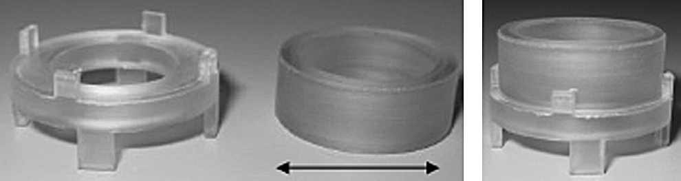
Cell culture support device. After detachment of fabricated membranes from the silicon wafer, they are chemically attached to the top cylindrical segment of the device using methylene chloride. The top segment will then sit firmly inside the bottom segment with the membrane dividing the chambers. The device fits inside a 12-well culture dish. The inner diameter (arrow) is 1.12 cm.
Immunocytochemistry
DiI-Ac-LDL was diluted to 10 μg/mL in EGM MV and added to BAEC. The cells were then incubated in a 37°C humidified cell culture incubator with 5% CO2 for 4 h. The cells were rinsed three times with DPBS and then fixed in 4% (w/v) paraformaldehyde at room temperature for 20 min. Astrocyte and Caco-2 cultures were rinsed twice with DPBS followed by fixation in 4% paraformaldehyde for 20 min (all steps performed at room temperature unless stated otherwise). Following three 5 min rinses in DPBS, astrocytes were permeabilized with 0.1% Triton X-100 in DPBS for 15 min, and Caco-2 cells were permeabilized with 0.05% Triton X-100 for 5 min on ice. Caco-2 cells were also prepermeabilized with 0.2% Triton X-100 for 2 min on ice for occludin staining. After three 5-min rinses in DPBS following permeabilization, astrocytes and Caco-2 cells were blocked in 10% goat serum in DPBS for 15 min. All subsequent steps were performed on a shaker. The following concentrations in 10% goat serum were used for primary antibodies: rabbit anti-GFAP, 4 μg/mL; rabbit anti-occludin, 15 μg/mL. Primary antibodies were added to the appropriate compartment for 1 h followed by three 10-min rinses in 1% goat serum (occludin incubation was done at 4°C). Cells were incubated for 1 h in 1:500 dilution of Alexa Fluor 488 goat anti-rabbit IgG antibody followed by three 10-min rinses in 1% goat serum. DAPI was added at 300 nM for 3 min before mounting the membrane on a glass slide with a cover slip in Vectashield mounting medium. Samples were observed in a Leica SP2 scanning confocal microscope (Leica Microsystems, Inc., Bannockburn, IL). Control samples were treated exactly as stated above, except that the primary antibodies were replaced with a rabbit IgG fraction negative control. A secondary antibody-only control was also included.
Cell metabolism and viability assays
AB assay, a fluorometric indicator of cell metabolic activity, was used to determine the cell metabolic rate.16,17 Before the experiment and according to the manufacturer's protocol, the seeding density and incubation period consistent with linear reduction of AB was determined by seeding cells at various densities and monitoring AB reduction over 72 h. This initial study indicated that for the seeding densities reported for all three cell lines in the former section, the maximum incubation time in which the cells turn AB from the oxidized form (blue) to the fully reduced form (red) is 24 h, with the signal from the first 6 h being linear. Thus, 24 h postseeding, all cultures were fed with a 10% (v/v) solution of AB in the culture medium and allowed to incubate for 6 h in the incubator. The fluorescence was measured at room temperature every hour in a fluorescence plate reader (Molecular Devices SpectraMax Gemini EM) using an excitation and emission wavelength of 540 and 590 nm, respectively. The fluorescence of an empty culture dish and the culture medium were also measured as controls. At day 4, all cultures were washed with DPBS and cells were trypsinized from the surface. Cell viability was determined with a hemocytometer using 50% Trypan Blue. Three independent experiments were performed with n=4 in each experiment. The statistical analysis was carried out using JMP® 7.0 (SAS Institute, Inc., Cary, NC). Statistical significance was determined using one-way analysis of variance with the global risk fixed at p<0.05. All results are mean±standard deviation.
X-ray photoelectron spectroscopy
Protein adsorption on PHOST was evaluated with X-ray photoelectron spectroscopy (XPS). XPS measurements were performed using a Surface Science Instruments SSX-100 system with a monochromatic Al Kα X-ray source at 1486.6 eV with operating pressure of less than 2×10⁻⁹ Torr. Samples were tilted to obtain emission angles of 0° and 75° from the sample normal. Charge compensation was carried out by a low-energy electron flood gun. The hemispherical analyzer utilized pass energies of 150 eV for survey scans, and 50 eV for high resolution scans. The spectra were analyzed using Casa XPS v.2.3.12Dev4 software. The C-C peak at 285 eV was used as the reference for binding energy calibration.
Flux measurements
The top and bottom compartments of the commercial and nanofabricated membranes were pre-equilibrated with transport buffer (10 mM HEPES). A solution of sodium fluorescein (376 Da, water soluble) in transport buffer was then added to the upper compartments to yield a final concentration of 1 μM. Aliquots (100 μL) were removed from the bottom compartment for later analysis in a plate reader and the volume replaced with transport buffer every 15 min over 60 min. The rate of influx of sodium fluorescein into the bottom compartment divided by the total porous area equals the flux. 18
Results and Discussion
Although several attempts have been made to create high flux membranes,8–12 they are usually targeted toward separation and microfiltration processes by creating silicon nitride or silicon oxide nano-films or nano-sieves with limited pore sizes (<100 nm) and random pore arrangements. Due to the proposed application as well as the extreme fragility of the above membranes, these studies fall short in showing the absence of cytotoxicity and appropriateness for cell culture and tissue engineering. In this study, selection of the appropriate polymer involved consideration of two factors. First, the material should lack cytotoxicity and require minimum postfabrication modifications for cell attachment, mainly due to the inherent fragility of a 3-μm-thick membrane and preference for minimal manipulations. Second, the polymeric membrane should have enough thermal resistance to withstand high temperatures needed at various nanofabrication steps. This study proposes the potential utility of PHOST as a cell culture substrate with suitability for nanofabrication processes for the first time. PHOST was chosen over poly(styrene) (material of preference for cell culture applications) because the presence of the negatively charged hydroxyl group in PHOST makes it a better candidate for cell attachment. More importantly, since the hydroxyl group was incorporated into the polymer during the synthesis, the need for postfabrication treatment for better cell attachment was eliminated.
Synthesis and characterization of PHOST
PHOST was prepared in a novel reaction scheme by free radical polymerization and subsequent hydrolytic de-protection (Fig. 1). The molecular weight before de-protection was 186,000 g/mol. GPC results indicated that the molecular weight distribution (PDI) is 1.7. The high molecular weight of PHOST is necessary to ensure proper mechanical stability at 3-μm thickness. While Higashimura et al. (1989) proposed a living cationic polymerization scheme for PHOST synthesis, our free radical polymerization scheme is superior because it is faster, does not require in-lab preparation of reactants, is capable of creating high-molecular-weight PHOST, and involves safer chemicals.
The chemical composition of both polymers was determined by 1 H-NMR spectroscopy (Fig. 4). A chemical shift of 1.2 ppm corresponds to the tert-butyl group of poly(tert-butoxy styrene) in CDCl3. The spectrum of PHOST in DMSO-d6 shows a peak at 8.9 ppm, which corresponds to the hydroxyl group after the de-protection reaction. The polymer backbone protons appear in the chemical shift region of 1–2 ppm with the signal due to the tert-butyl group being eliminated. The change in water's chemical shift in both 1 H-NMRs is due to the use of different solvents for each reaction. 19 The FTIR spectrum of PHOST shows a peak at 3380 cm−1, which corresponds to the presence of the hydroxyl group after de-protection (Fig. 5). The disappearance of the peak at 1132 cm−1 indicates complete removal of the tert-butyl ether group during hydrolysis.
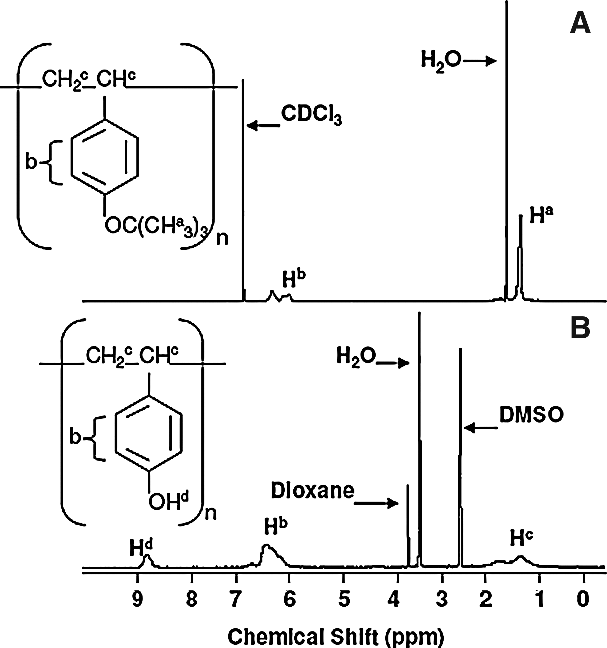
1H-NMR spectrum of poly(tert-butoxy styrene) in CDCl3
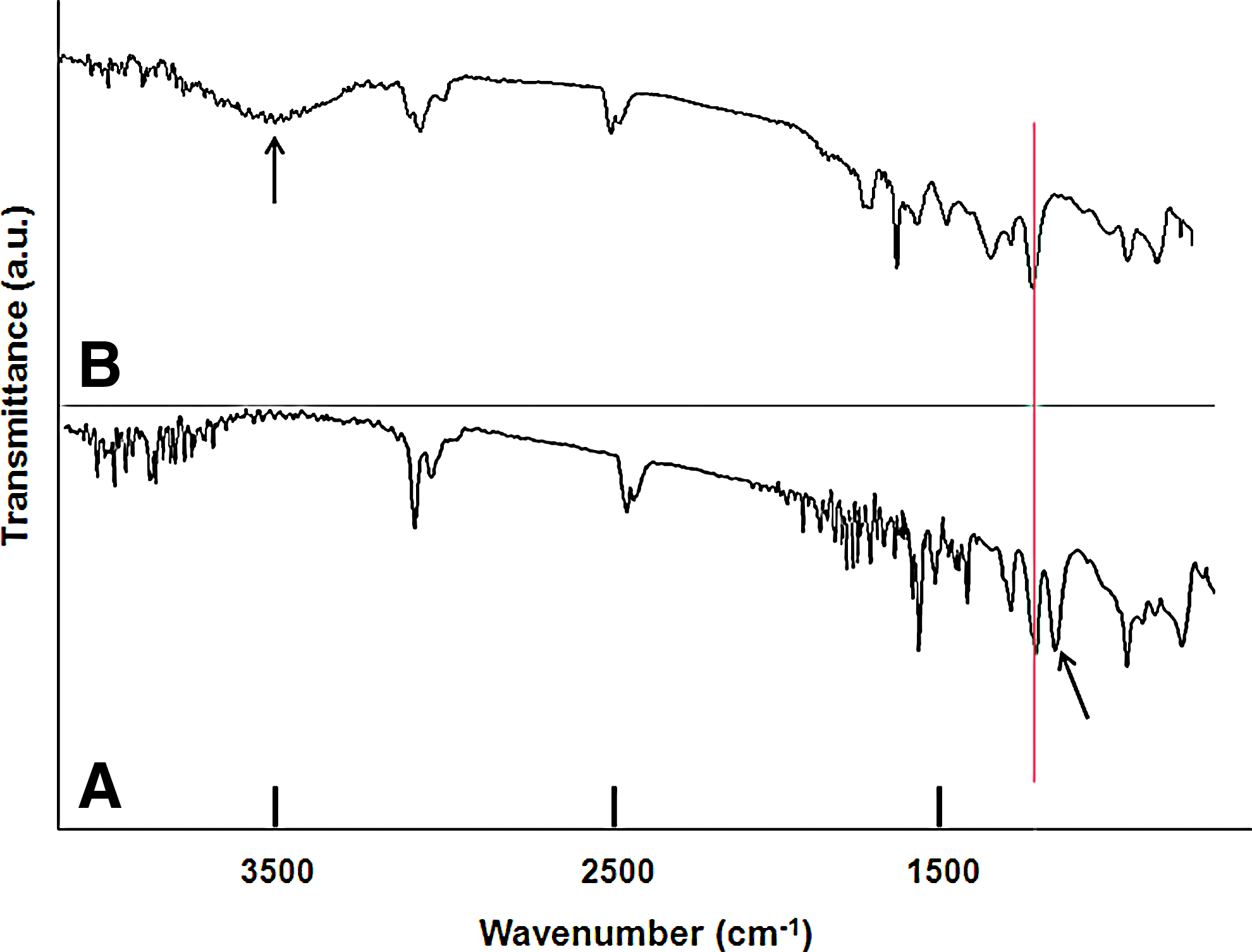
FTIR spectra of poly(tert-butoxy styrene)
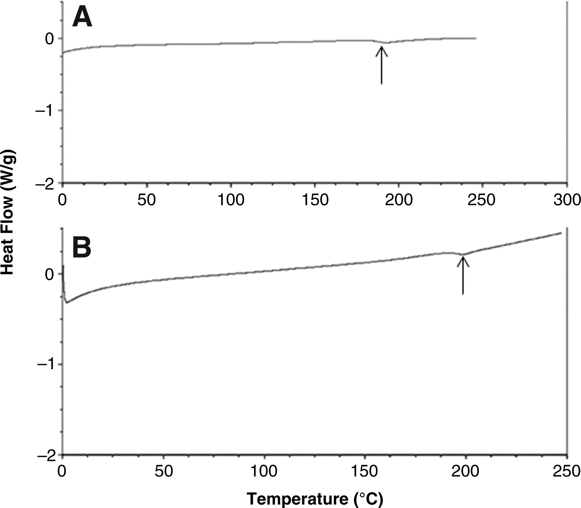
DSC was carried out on both polymers (Fig. 6). The Tg of both poly(tert-butoxy styrene) and PHOST is ∼180°C. Thermogravimetric analysis (Fig. 7) indicated no significant weight loss in PHOST below 361°C. The significant weight loss at 220°C in poly(tert-butoxy styrene) is due to decomposition of the tert-butyl group.
Differential scanning calorimetry analysis of poly(tert-butoxy styrene)
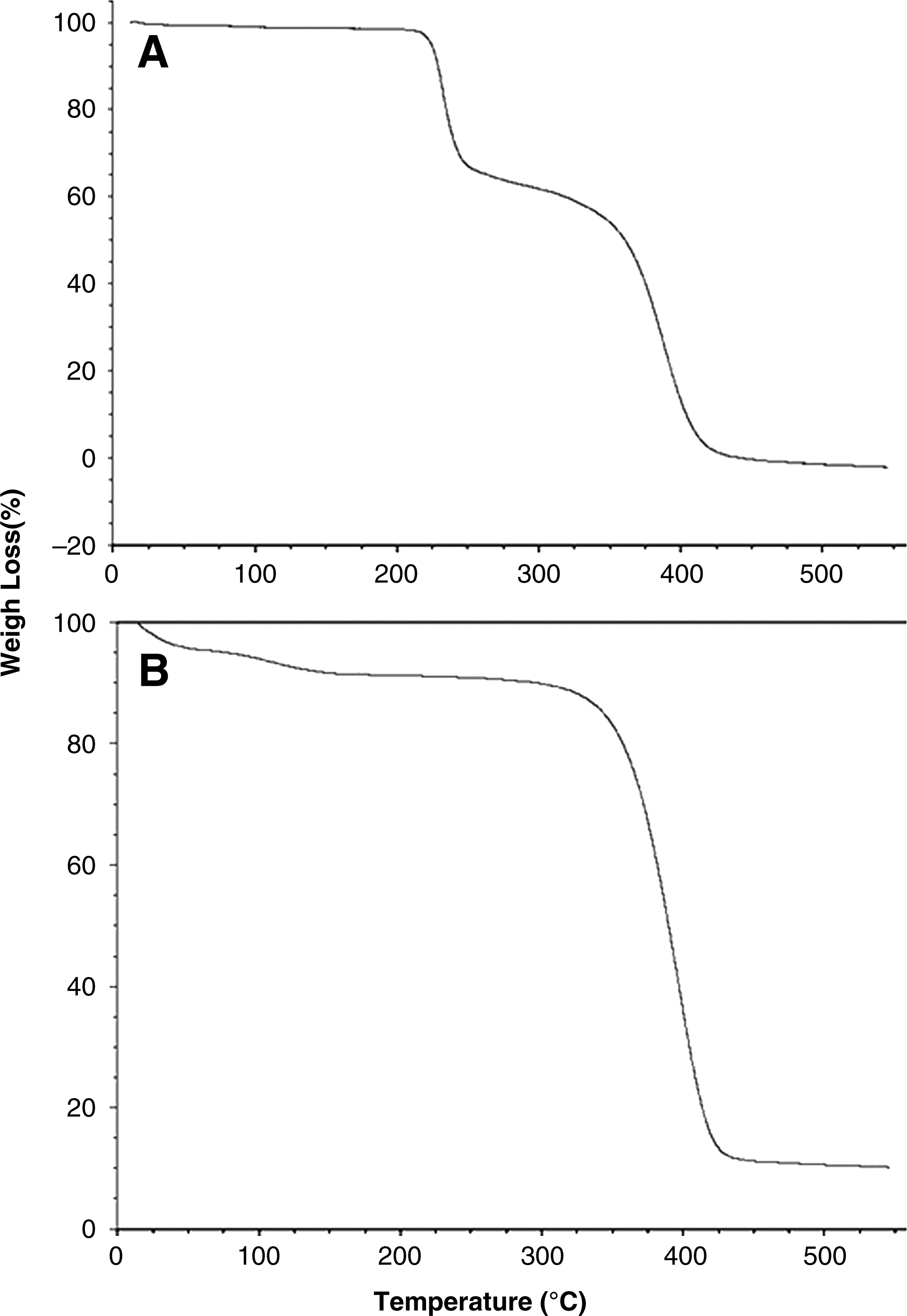
TGA analysis of poly(tert-butoxy styrene)
Characterization of nanofabricated membranes
Here we describe a nanofabrication scheme that is used for the first time to create high-resolution porous structures directly on the membranes. Although traditional photolithography approaches have long been used in creating micro-patterned surfaces, electron beam introduces a higher level of precision and resolution. Furthermore, the proposed fabrication scheme provides flexibility over the choice of polymer. Also, the process is not limited to thin (<500 nm) conducting films (as seen in semiconductor/mask nano-patterning using electron beam) due to the presence of an internal mask (SiO2). SiO2 is resistant to oxygen etch, and thus can be selectively used as an internal mask to transfer the e-beam pattern to PHOST or other underlying polymers with thicknesses in the micron or nanometer range. Susceptibility of the SiO2 layer to oxide etch, however, makes its removal easy.
The PAA layer ensures the complete detachment of fabricated PHOST from the Si wafer once the sample is placed in water. Due to the extreme hygroscopic properties of PAA, it makes a uniform film on the Si wafer only after O2 plasma treatment of the wafer. 20 The thickness of the PAA layer and PHOST were 200 nm and 3 μm, respectively, by profilometry. The thickness of PHOST was chosen based on its mechanical stability and ease of handling in cell culture.
ZEP-520A (positive electron beam resist) was specifically chosen over more traditional PMMA due its high resolution and more importantly high etch resistance. 21 The etch resistance ensured the complete transfer of pattern from the ZEP layer all the way down to the SiO2 layer during the CHF3/O2 etch process. Cold development temperature was also used to minimize increases in the pattern dimensions.22,23 After the O2 plasma etching of PHOST, pores with sloped sidewalls measuring 150–250 nm larger than the target size on the patterned side (top side) were typically observed. Figure 8 shows scanning electron microscopy images of the top view and cross section of membranes and compares them to a commercially available polyester membrane with random pore arrangement and size distribution. The process is capable of producing pores with diameters in the range of 50–800 nm.

Top and cross-sectional views of the porous membrane. Top views of 400-nm pores
After detachment of a fabricated membrane from the Si wafer, it was attached to the top cylindrical segment of the in-house support device (Fig. 3) using methylene chloride. Even though this may change the thickness and chemistry of the membrane at the attachment site (the cross-sectional thickness of the ring is 0.2 cm), the chemical attachment is fully controllable with respect to creating a leak-proof chamber. Moreover, the inner diameter of the support device (and hence the membrane) is 1.12 cm, with the nanofabricated portion being a square located at the center of the support device (the area of the nanofabricated square is 0.25 cm2). Thus, the nanofabricated region is 0.31 cm away from the attachment site in every direction.
Cell viability, rate of metabolism, and differentiation
To test the lack of cytotoxicity and the cell culture handling ability of very thin nanofabricated membranes, astrocytes, BAEC, and Caco-2 cells were cultured on PHOST with pores of 400 nm in diameter. This pore size was selected because the smallest available pore size in commercial permeable supports is 400 nm. The three cell lines were chosen to show that the membrane is generally applicable to the culture of any cell type, and more importantly because these cells are commonly used in constructing in vitro models of barrier tissues, the blood-brain barrier (BAEC and astrocytes) and the intestinal barrier (Caco-2).
AB was used to determine cell metabolic rate.16,17 AB is a water-soluble dye that contains a fluorometric/colorimetric redox indicator that changes from the oxidized to the reduced form in response to the chemical reduction of the growth medium resulting from cellular metabolism. AB is extremely stable and most importantly nontoxic to the cells and thus cultures can be monitored continuously over time. The maximum amount of time it takes for AB to be completely reduced depends on the cell seeding density. Before testing the rate of metabolism of cells cultured on the membranes, the desired seeding densities were used to determine the incubation period consistent with linear reduction of AB over 72 h. In this experiment, the maximum incubation time in which the cells turn AB from the oxidized form to the fully reduced form was 24 h, with the signal from the first 6 h being linear.
Figure 9 shows the fluorescence over time for astrocytes, BAEC, and Caco-2 cells cultured on the membrane and the control culture dish. According to the slopes, the rate of metabolism of astrocytes was 36% lower on the membrane compared to the control. For BAEC grown on the membrane the rate of metabolism was 19% lower than the control and for the Caco-2 cells the rate was statistically the same on both surfaces. The Trypan Blue test indicated that cell viability is the same on membranes compared to control surfaces for all three cell lines (94%, 96.7%, and 96.9% for astrocytes, BAEC, and Caco-2 cells, respectively).
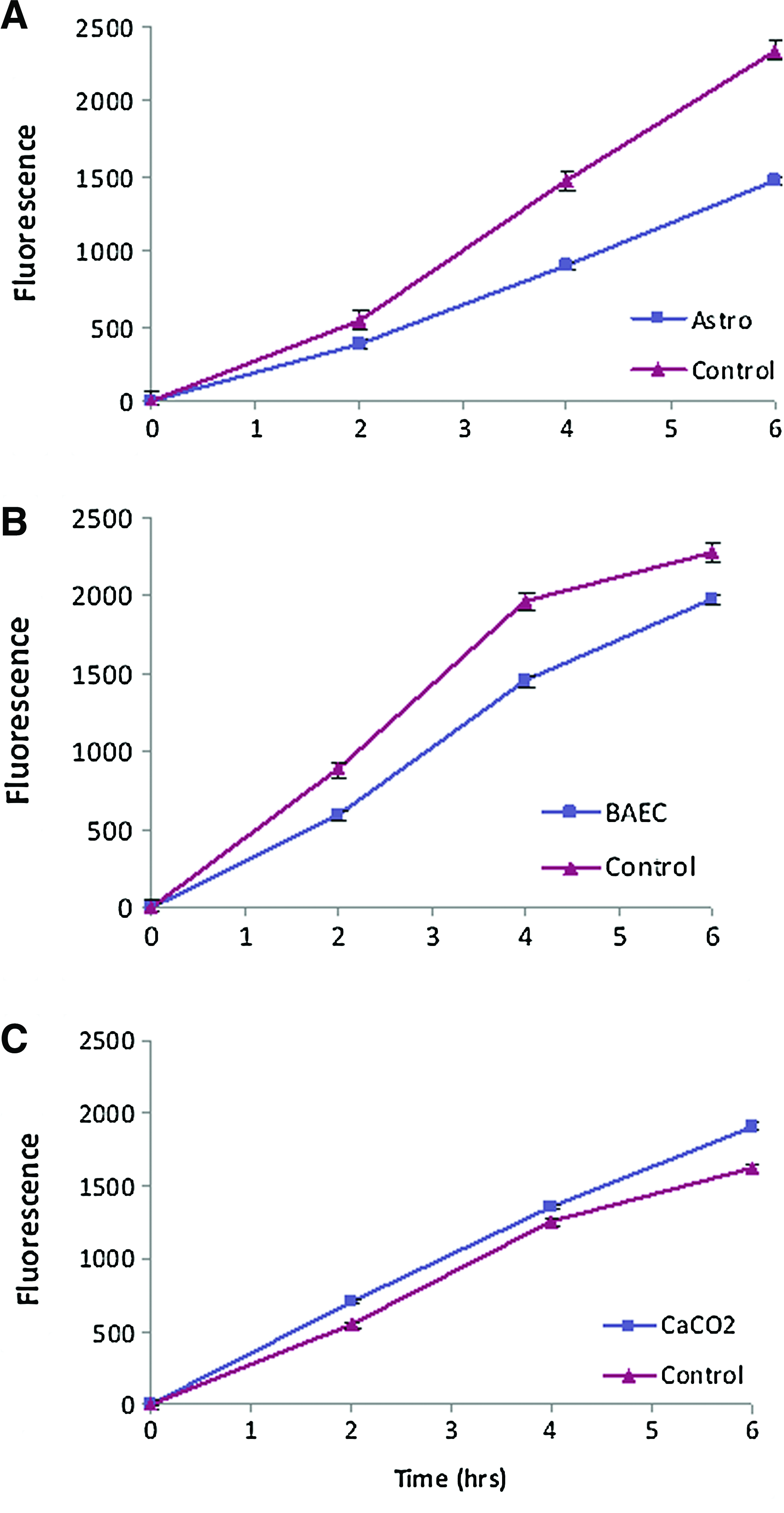
Alamar Blue assay showing cellular rate of metabolism on PHOST with 400-nm pores versus the control surface (tissue culture treated petri dish).
Even though the rate of metabolism of astrocytes and BAEC is lower on PHOST, their viability is the same as those cultured on the control surfaces. The geometry and consequently the thickness of the liquid film above the cells differ in these two constructs (PHOST vs. commercial inserts) and the different responses may be due to differences in surface properties or gas transfer. We believe that Caco-2 cells grow as fast on PHOST compared to control surfaces because they are transformed (i.e., cancer cell line) and thus are robust and properties of the culture substrate may not play a significant role in their metabolism. However, in the case of astrocytes, which are primary cells and thus extremely sensitive, the rate of metabolism is lowest. BAEC are also primary cells, but they can be passaged multiple times before they lose their properties and thus they are more robust than astrocytes.
Figure 10 shows the phase-contrast images of cells 1 day after culture establishment on both surfaces (A–F). This shows the ability of the cells to adhere to porous PHOST with their morphology being indistinguishable from that on the control surface. Phase-contrast image of the cells 4 days after culture establishment shows the ability of the cells to grow and proliferate on PHOST (Fig. 10 G–I). Immunocytochemical characterization of BAEC for uptake of Ac-LDL (a marker of endothelial cells), astrocytes for the expression of GFAP, and Caco-2 cells for the expression of occludin junctional protein shows that the differentiation of these cell types is maintained when cultured on porous PHOST (Fig. 10 J–L).
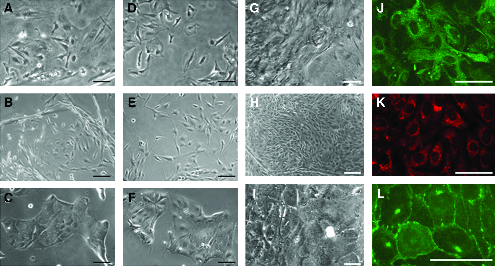
Cell attachment, growth, and differentiation on PHOST.
Membrane surface characterization with XPS
XPS is an effective analytic method that can be used to determine the chemical composition in the near-surface region of a sample. Figure 11 shows the XPS survey scan of the non-protein-treated PHOST (A and B) and protein-treated PHOST (C and D) taken at two different incident angles (0° and 75°). The spectra are normalized so that the total area under the carbon peaks is equal to unity. PHOST shows a strong intensity peak from C=C and C–C near 285 eV. The surface is dominated by the peaks associated with C 1s and O 1s, located at ∼285 eV and ∼535 eV, respectively. The atomic composition of the PHOST was determined to be 80.33% C and 19.67% O at 0°. Oxygen concentration increases 27.64% as the incident angle is increased from 0° to 75°. This indicates that the hydroxyl group of the PHOST is hidden underneath the surface. The protein-treated surface is dominated by the peaks associated with C 1s at ∼285 eV, O 1s at ∼535 eV, and N 1s at ∼400 eV. The atomic composition of the protein-treated PHOST was determined to be 73.14% C, 20.40% O, and 6.46% N at 0°. The peak associated with N 1s is indicative of protein adsorption onto the surface.
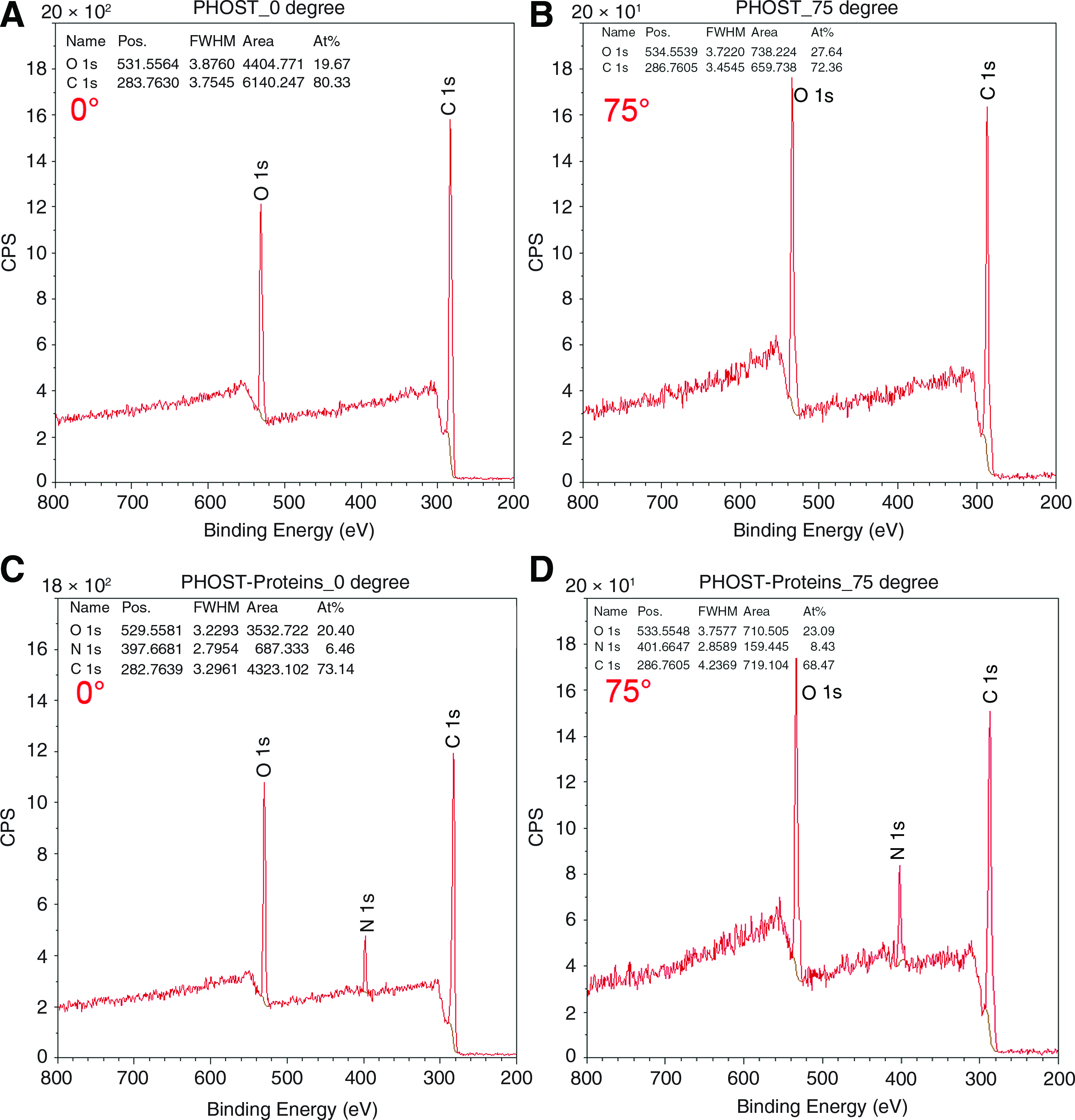
X-ray photoelectron spectroscopy (XPS) spectra of the PHOST surface
Flow resistance across nanofabricated membranes
As mentioned previously, a key drawback of current commercial permeable supports is their high flow resistance due to a wide pore size distribution with tortuous paths, low total porosity, and relatively thick membrane, which makes them low-throughput for permeability studies.8–12 Sodium fluorescein was used to assess the flux across the nanofabricated PHOST and commercially available permeable supports. The rate of transport of sodium fluorescein divided by the porous area is equal to the flux. 18 We evaluated the flux over two nanofabricated PHOST membranes both with 400 nm pore sizes: one with the same total porosity as a commercial counterpart, and the second one with twice the porosity. Figure 12 shows the corresponding rates of transport across the membranes. These results indicate that the flux over PHOST is 23 times higher compared to commercial counterparts due to the reduced thickness and the straight pore profiles. When the porosity is doubled, flux is 53 times higher compared to commercial membranes. To our knowledge, this is the first study that establishes a direct comparison between the porosity of commercial membranes versus nanofabricated PHOST and reports a minimum of 23 times higher flux.
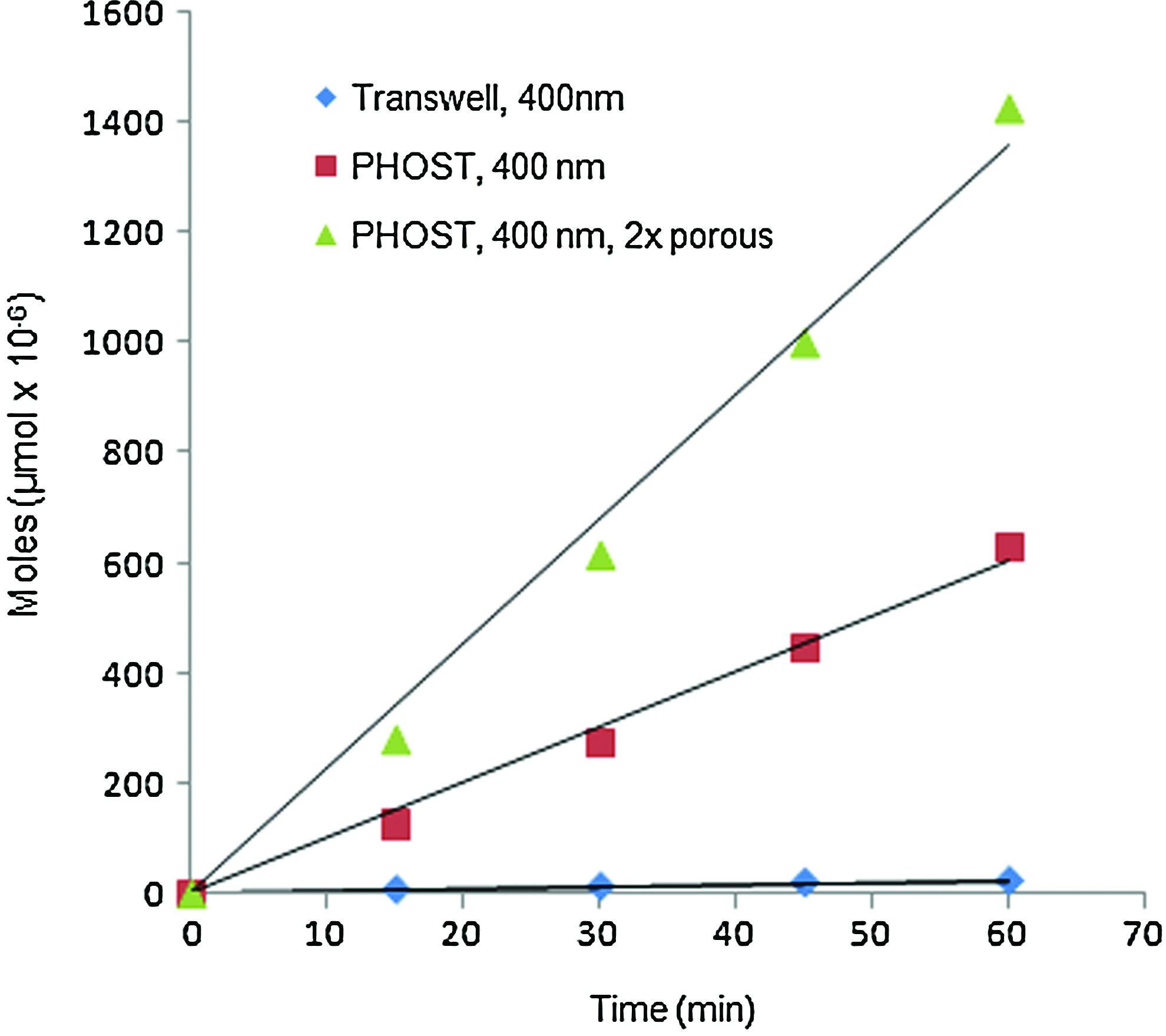
Rate of transport across PHOST with 400 nm pores, its commercial counterpart, and PHOST with twice the porosity of the commercial membrane. Color images available online at
Conclusion
In summary, 3-μm-thick, transparent, porous polymeric membranes based on PHOST with defined pore sizes and pore density are nanofabricated. The membrane is attached to a support device to facilitate its use for cell culture and/or permeability studies. Moreover, the membranes have low flow resistance, which contributes to their application for high-throughput permeability and/or separation assays. Cell culture studies show the successful attachment, proliferation, and differentiation of several cell lines that are commonly used in constructing in vitro models of the blood-brain barrier, and intestinal barrier. We believe that this is the first study that proposes the suitability of nanoporous PHOST as a high-throughput polymeric membrane without cytotoxicity, and has appropriate characteristics for use in nanofabrication processes.
Footnotes
Acknowledgments
This work was funded in part by the New York State Office for Science, Technology, and Academic Research (NYSTAR) and the National Science Foundation through the Nanobiotechnology Center (NBTC). We specially thank Cornell NanoScale Science and Technology Facility (CNF) for their continuous support on material fabrication as well as Cornell Center for Materials Research for polymer characterization. We thank Professor William Shain at the Center of Neural Communication Technology, Wadsworth Center (Albany, NY), for providing us with astrocytes. We thank Professor David Putnam at Biomedical Engineering, Cornell University (Ithaca, NY), and Sara Yazdi for their guidance on 1 H-NMR and PHOST synthesis. We also thank Jaewook Seok for his support on chemistry and fabrication.
Disclosure Statement
No competing financial interests exist.