




Abstract
The study of the mechanical behavior of soft biological materials presents many challenges due to the materials' time-dependent mechanical response as well as inherent size and shape limitations. In this study, by using agar as a surrogate material for soft tissues, the effects of these limitations upon standardized macroscale dynamic compression protocols are compared to dynamic nanoindentation procedures. Both techniques are then applied to dynamically test porcine sclera tissue, showing a significant difference in recorded loss and storage modulus values between the two methodologies. Additionally, the tissues of the porcine eye wall are characterized with macrocompression in their layered in vivo arrangement. The overall constraints of standardized macroscale tests for dynamic mechanical characterization of thin and soft biological tissues are discussed.
Introduction
Reliably characterizing the mechanical properties of soft tissues, like those found in the eye, is a difficult challenge, as biological tissues differ greatly in their structure and composition and have complex hierarchical structures with elements interacting across length-scales, which span several orders of magnitude from nano- to millimeters.12,13 Furthermore, these tissues have dimensional limits, which may not allow sample sizes that conform to testing protocols.14,15 It has been well documented that part of the challenge of characterizing biological tissues in general lies with the fact that most biological tissues are highly sensitive to environmental conditions, and exhibit anisotropic behaviors.6,8,16–18 Additionally, their mechanical behavior is viscoelastic, dependent upon both the strain and the strain rate. 19 Previously cited studies on eye tissues5–11 have not taken all of these factors into account, and to our best knowledge, no cohesive protocol has been developed for the macroscale studies that evaluated similar soft (E<100 kPa) and thin (thickness <2 mm) tissues. The closest commonly practiced characterization procedures, such as dynamic macrocompression, require large standardized sample shapes and sizes to measure storage and loss modulus,20,21 and their applicability toward thin biological tissues remains unknown. Some have investigated the use of instrumented nanoindentation to quasistatically and dynamically characterize soft biological materials which could minimize length-scale related issues. 19 Earlier nanoindentation studies have characterized hard (E>1 GPa), 22 soft (1 GPa >E>100 kPa), 19 and ultrasoft biological tissues (E<100 kPa),6,23,24 and the published procedures accommodate for the required hydration and size constraints.24,25
To assess the limitations of standardized mechanical characterization techniques on soft biological materials, we compare the storage and loss modulus measurements obtained with dynamic macrocompression and nanoindentation methodologies for both agar and porcine eye tissue samples. This study is the first to analyze the effects of sample size and stiffness upon the dynamic mechanical characterization of low-modulus and thin biological materials using surrogate samples and tissues under identical testing conditions. The macroscale compression tests employ equipment previously used to mechanically characterize the soft biological tissues3,26 as adapted from current standards.20,21 Following the standardized protocol for macroscale dynamic characterization of viscoelastic materials, the effects of sample shape and stiffness can be distinguished by using agar, a biological tissue surrogate that can be made to the concentrations and dimensions required for the standard protocols.23,24,27 Both methodologies, macrocompression and nanoindentation, are also used to measure the mechanical properties of porcine sclera tissue, which is used as a comparative base.
Procedure
Sample selection and preparation
The dynamic macrocompression protocols specified a minimum stiffness and proportional thickness to ensure valid results,20,21 while nanoindentation only requires a maximum sample displacement of less than 10% of the total sample thickness. Previous tests showed that 5.0% agar samples met the minimum stiffness requirement (E >100 kPa) for macrocompression and can be prepared for a wide range of thicknesses. 0.5% agar concentration samples were also selected, since they closely aligned with the low stiffness properties of soft eye tissues, which are outside the standards stiffness specifications for macrocompression. Therefore, by using both nanoindentation and macrocompression protocols, 5.0% and 0.5% agar samples were tested to evaluate sample shape effects, while the 0.5% agar samples were used to study effects due to testing low stiffness materials. For the shape studies, three different thickness agar samples were characterized for both agar concentrations. Medium samples most closely matched standard protocol-defined shape characteristics, taller samples helped to evaluate macrocompression sensitivity to these shape parameters, and short samples were made to match the thickness of the eye tissues as closely as possible.
For these experiments, agar samples were prepared from agar pellets (Bioline USA, Inc., Taunton, MA) and phosphate buffered saline (Dulbecco's Sigma-Aldrich Company, Ltd, Irvine, Ayrshire, United Kingdom) as per the manufacturer's instructions. The 0.5-g agar pellets contain molecular grade, DNase/RNase-free, sterilized, and purified polysaccharides, which are first dissolved at room temperature into phosphate buffered saline (PBS), rapidly heated to boiling using a microwave, mixed for up to 15 s, and poured into a petri dish with a diameter of 53 mm. After solidification, the samples cool overnight in a refrigerator at 15°C±1°C. Agar samples were made at 0.5% and 5.0% concentrations as determined by weight before heating. For all samples, the final water loss after heating and cooling remained consistently below 1% of the total PBS volume used. Samples prepared for the nanoindentation experiments followed the protocol described previously, and were 11±1 mm thick. 24 Macrocompression samples were cut from this larger petri dish sample using a 14-mm diameter sharp plastic trephine. For these macroscale tests, petri dish samples were produced using three different average thicknesses, 2 mm short, 6 mm medium, and 12 mm tall. Macroscale tests used at least 10 trials per condition and nanoindentation tests used at least 20 trials per condition. All samples from both methodologies were kept hydrated through bathing in PBS, and all tests were completed within 4 h at room temperature to ensure sample integrity.
Eye tissue samples were obtained from healthy mixed breed, porcine specimens between 5 and 7 months of age from Sierra Medical (Sylmar, CA). Before harvesting, the skin surface was sterilized in hot water at 64°C for all animals by submerging their heads and necks for 4 min postsacrifice. Prior studies have shown that this is believed to have no effect upon the integrity of deeper tissues due to the superficial meat remaining raw and the harvest site lying underneath 3 cm of skin, muscle, and bone. 8 Porcine eyes are harvested and delivered in refrigerated containers the same day, and all tests are conducted within 24 h of harvesting. Once delivered, eye preparation begins by clearing orbital fat and muscle tissue. From there, the eyes are sectioned coronally, and square 1.0±0.2 cm on side segments are taken from the posterior region of the eye, adjacent and lateral to the ocular nerve, to find segments of consistent thickness.3,8 SCR samples had the sclera, choroid, and retina layers in their original arrangement, while sclera-only SO samples had the choroid and retina layers removed. For the nanoindentation experiments, tissue samples have their outside surface lightly patted dry, and then glued using a single drop of cyanoacrylate (All-Purpose Instant Krazy Glue, Elmer's Products, Inc., Columbus, OH) to the bottom of a shallow and rigid plastic well with an inner diameter of 1.50 cm and a height of 0.50 cm. This sample mounting procedure has been shown not to affect the modulus of sclera-only samples. 6 The wells are then partially filled with PBS to maintain tissue hydration during testing at room temperature. Nanoindentation of the SCR samples was not possible due to a machine-limited maximum displacement of only 5 μm, which is not deep enough to test the properties of the SCR sample. Macroscale test samples were not glued and instead placed in a petri dish with a diameter of 53 mm lined with self-adhesive sand paper of 300grit as adapted from similar standardized protocols for the viscoelastic characterization of elastomers. 20
Testing protocol
Nanoscale dynamic (nanoDMA) characterization of the agar and eye tissue samples was obtained using a Triboindenter (Hysistron, Inc., Minneapolis, MN).
24
Following the calibration procedures and equations outlined elsewhere,19,23,28,29 a cylindrical, 80 μm diameter, flat-punch, sapphire probe indents the fluid-covered samples at room temperature. Dynamic characterization of the storage and loss modulus was performed using a frequency sweep test where an initial static load is applied to the sample with a superimposed dynamic oscillation of fixed amplitude, but varied frequency. This procedure for viscoelastic characterization of soft biological materials has been used previously with consistent results.24,25 The dynamic oscillation frequencies are at 25 equally spaced intervals between 10 and 250 Hz, in line with previous dynamic studies on biological materials.
25
Every dynamic indent for both agar and sclera-only samples is repeated 20 times at each combination of loading depth and frequency with at least 200 μm distance between indents; previous work has not shown this distance between indents to have any effects upon the agar or porcine sclera samples since the indents are fairly shallow (<2 μm).6,24 In this paper, the dynamic indentation characteristics are reported in storage (E′) and loss (E′′) modulus (kPa).18,20,21 Together, they model the time-dependent viscoelastic behavior in the form of the complex reduced modulus E* as follows:
Equations 1–3 derive from the Sneddon equations and the Kelvin–Voigt material model for time-dependent strain-related elastic and viscoelastic behavior. The reduced storage modulus describes the elastic in-phase response of a material at a specific frequency, defined as:
while the reduced loss modulus measures the phase delay between the applied signal and the material's response at that frequency calculated by:
Where S is the measured contact stiffness, Ac the projected contact area(5.0×103 μm2 for the flat punch used in this study), Dc the measured contact damping, and ω the frequency. The ratio of the phase-delayed loss modulus to the in-phase storage modulus reflects the overall damping response of the material, termed tan(δ) and defined as follows18,25:
Macroscale dynamic compression tests were performed using a Bose Electroforce 3100 (Bose Corp., Eden Prairie, MN), following the calibration procedures and equations listed in the user manual and primarily based off of existing viscoelastic characterization protocols.20,21,30 The resolution of the system includes a load cell sensitivity (<0.1 g) across the load cell's entire range (1000 g) in addition to the sensitivity (<1 μm) throughout the actuator's range (4 mm). Electrostatic forces are accounted for before the experiment and overall machine dynamic compliance is calibrated throughout the entire frequency range (0.01–100 Hz) as per the manufacturer's instructions.
30
The sample rests in a 300 grit sandpaper-lined petri dish while the sample remains hydrated in PBS. The petri dish is adhered with double-stick tape (3M, St. Paul, MN) on top of a 40-mm metal platen, which is connected to the load transducer below. An inverted, but otherwise identical platen setup is attached to the electromagnetic actuator above the sample (Fig. 1b). The upper platen is lowered into the sample, while the recorded load and displacement are exported. Current length, L, and force, F, were recorded independently from the actuator and load cell, respectively. Dynamic tests used a variable preload designed to maintain sample contact throughout the 0.02-mm peak-to-peak oscillations. Agar samples were tested in the short, medium, and tall shapes, and eye tissues were tested with the SO and SCR configurations. Calculation of the storage and loss modulus used the Fourier analysis of the reference (displacement) and measured (load) channels with a variable sample rate taking a minimum of 512 data points per oscillatory cycle. The Kelvin–Voigt parallel spring and dashpot model is also used here to represent the sample, and relates the proportionality factor between the peak-to-peak displacement and load as well as the phase delay between the recorded and reference signal to measure the complex modulus. As with the nanoindentation experiments, the storage and loss modulus are calculated from Equations 2 and 3. The relevant protocols for macroscale studies recommend that samples used in these tests have an ideal shape factor equal to 0.5, as defined by Equation 520,21:

A summary of the tests can be seen in Table 1 with the respective loading parameters in Table 2.
Results and Discussion
Nanoindentation
Nanoindentation, which has been used previously to dynamically characterize soft biological materials and is less affected by the sample shape constraints, was used as a reference for the macroscale tests. Dynamic nanoindentation tests were performed on the 0.5% agar, 5.0% agar, and porcine SO samples between the frequencies of 10 and 250 Hz (Fig. 2a, b). Storage modulus values of all samples generally increased with frequency, while loss modulus values displayed a less obvious trend. 5.0% agar samples produced a storage modulus 1–2 orders of magnitude higher than the 0.5% agar or SO samples. Previous tests have shown that quasistatically, porcine sclera was expected to have mechanical properties similar to the 0.5% agar samples,6,24 but here the storage and loss modulus values for the sclera-only samples were almost twice that of the 0.5% agar samples depending upon the oscillatory frequency. The 0.5% agar and porcine SO samples showed standard deviations, which exceeded their mean modulus values when tested outside of the 90–200 Hz range and consequently are not included here for an analysis. Overall, the nanoindentation results produced a larger storage modulus relative to the loss modulus, with differences between the two measurements varying 1–2 orders of magnitude. The low ratio of loss to storage modulus, tan(δ), shows that the material stores more energy than it dissipates.24,31–36 For all the nanoindentation tests presented here, indentation depth is far below the 10% displacement limit and therefore, no substrate effects are expected.

Dynamic nanoindentation results of agar and porcine sclera-only (SO) samples for the
Macrocompression
Dynamic macrocompression tests were performed using the Bose system on the short, medium, and tall 0.5% agar samples between 0.1 and 100 Hz (Fig. 3a, b) to analyze the effects due to sample shape. The medium sized agar samples are close to the protocol-specified shape factor of 0.5, while the tall and short samples are used to explore the shape factor effects. Storage modulus generally increases with frequency for all sample thicknesses. The short size agar samples also showed the greatest values for storage and loss modulus as well as the greatest standard deviation, while the medium and tall samples closely aligned with each other. As the preloads produced a consistent displacement for all 0.5% agar samples, the relative compression is greatest with the short samples. For compressive displacements greater than 10% of the sample's thickness, a significant portion of the recorded force is due to the contribution of the substrate underneath the sample.37,38 Similar sample thickness effects were seen in the 5.0% agar samples, as the short samples also showed an increased storage and loss modulus.
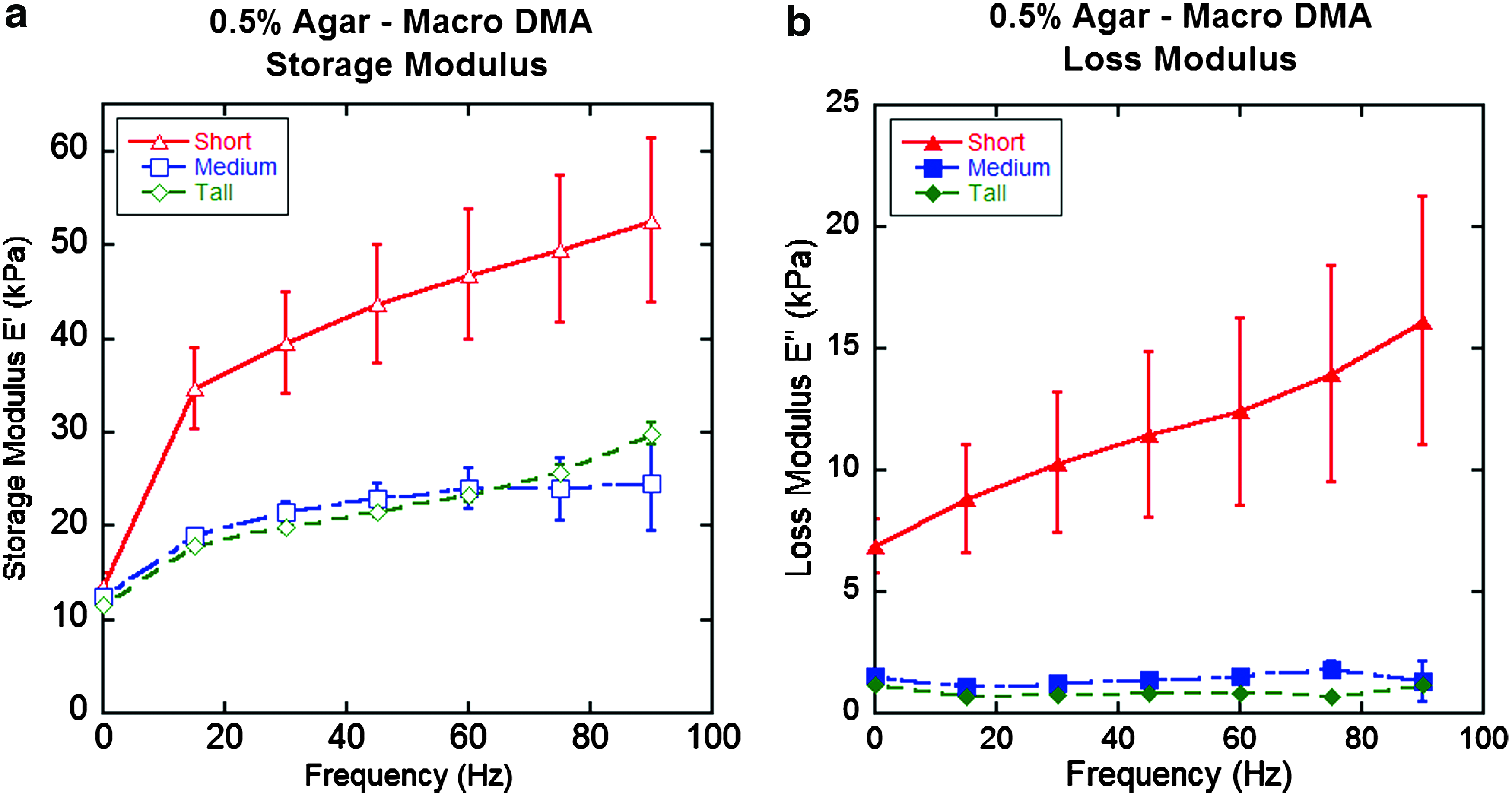
Macroscale compression of 0.5% agar with three different sample heights noted as tall, medium, and short. The plot shows
Macroscale tests on medium size agar and the two different porcine tissue sample preparations, SO and SCR, are shown in Figure 4a and b. It should be noted that SO and SCR samples fall outside the minimum modulus range specified in the dynamic macroscale standards,6–8,39 as well as the shape factor requirements.20,21 As the 0.5% agar samples are also expected to be in this low modulus range, effects due to sample stiffness can be separated from shape effects by comparing samples of different thicknesses. Only macroscale tests of the short 0.5% agar samples show a similar qualitative trend for the storage and loss modulus to the SO and SCR samples, indicating that substrate or sample size effects result from the sample shape and not low modulus. This substrate effect has not been previously expanded to dynamic macroscale compression tests to the authors' knowledge, but the low modulus thin materials appear to have an increased modulus under large displacements relative to thicker samples. This increased storage and modulus effect is seen in both the agar and tissue samples. Furthermore, the effect increases with frequency, as deeper displacements were required to prevent loss of contact during higher frequency dynamic oscillations at the same amplitude.

Macroscale compression of 0.5% and 5.0% agar and porcine tissue samples showing
Protocol comparisons
Dynamic characterization results are summarized in Table 3 at 90 Hz, the only common frequency between the two methodologies for all samples. It should be noted that previous nanoindentation studies have shown 5.0% agar samples to have elastic modulus values close to 700 kPa,23,24,40 which meets the minimum 100-kPa limit specified for protocol applicability.20,21 Therefore, the medium sized 5.0% agar samples were the only samples that met the stiffness and shape requirements of the macroscale protocols and have a region of significant frequency overlap, 10–100 Hz, between the two testing methodologies. In this window, the 5.0% agar samples closely agree in storage modulus when tested using macrocompression (2079.5 kPa) and nanoindentation (1970.3 kPa), but loss modulus values for macrocompression (157.0 kPa) disagree with measurements taken with nanoindentation (13.1 kPa). From this, dynamic nanoindentation of 5.0% agar shows the general material response to be more energetically conservative than the macroscale tests, even when closely adhering to the standardized protocols. Dynamic nanoindentation data were not available for frequencies lower than 90 Hz for the 0.5% agar 24 and SO samples, and hardware limitations prevented acquisition of macroscale dynamic compression data above 100 Hz.
Macro results refer to the medium size agar samples.
The effects of machine sensitivity and material stiffness are shown when comparing the two protocols on the 0.5% medium agar samples. The storage modulus results between macrocompression (24.6 kPa) and nanoindentation (10.9 kPa) tests are within the same order of magnitude, and loss modulus values obtained through macrocompression (1.3 kPa) and nanoindentation (1.9 kPa) agree more closely. When comparing SO samples, the macroscale storage modulus results (405.4 kPa) are much greater than those values obtained through nanoindentation (53.4 kPa). Similar results were found with the loss modulus with macrocompression (280.4 kPa) measuring much higher than nanoindentation (6.1 kPa). This difference between nanoindentation and macrocompression results was also seen with the 0.5% short agar samples, but not with the medium or tall agar samples, indicating that the difference in the SO tests most likely arises from possible sample size and substrate effects. Macrocompression of the full eye wall thickness SCR samples, comprised of the sclera, choroid, and retina, measured a storage modulus of 665.3 kPa and a loss modulus of 548.2 kPa.
These results can be due to two possibly simultaneous factors. First, thickness-related substrate effects would show an increased modulus on thinner specimens. However, the tissue samples of the thicker SCR samples (∼1.6 mm) showed a higher storage and loss modulus when compared to the thinner SO samples (∼1.2 mm). The second factor arises from the strain in the sample, since previous macroscale tensile tests have shown a great increase in the choroid and retina modulus under high strain.8,35,39 High and low strain elastic effects in eye tissues are explained elsewhere, 8 but can be summarized as follows: biological materials exhibit a low modulus behavior under low strain (<10%), and high modulus behavior under higher strain (>10%) possibly due to structural protein and polysaccharide strain limits or movement of water throughout the sample. 26
Efforts were made to mitigate preload-induced effects as the preload was increased for each group of samples just enough to ensure constant contact with the sample throughout the 0.02-mm peak-to-peak dynamic oscillations. The dynamic amplitude was minimized as much as possible in consideration of the system sensitivity and resolution of both force and displacement. If a significant preload was needed to ensure contact during oscillation, then high prestrains can be expected. This prestrain could induce the tissue to exhibit its high-strain modulus behavior, which would show as an increased modulus of the SCR samples. The modulus of these layered tissues can be calculated by using models based on composite materials for mixtures following Equation 5
41
:
where V represents the volume fraction of the individual layers a through z, and E the modulus for the entire composite and the respective layers. From this equation, an increased modulus associated with high-strain effects would explain the higher modulus from the SCR compared to the SO samples.
To predict Ecomposite, quasistatic high and low strain modulus measurements were taken from available sources from static data: for the sclera (Ehigh=5050 kPa, Elow=20 kPa), choroid (Ehigh=2380 kPa, Elow=15 kPa), and retina (Ehigh=30 kPa, Elow=0.5 kPa). 8 If only low-strain or high-strain modulus numbers are used, the model predicts a composite modulus for SCR samples of ∼16 kPa and ∼4050 kPa, respectively. Although, these comparisons are limited given that previous studies have shown that quasistatic elastic modulus values are not directly comparable to storage loss modulus values, 24 the effect of low and high strain modulus values are shown to be significant.
In general, nanoindentation tests minimize the effects due to high-strain densification, and substrate effects related to sample size because of the low deformation relative to the overall sample thickness. It should be noted that nanoindentation data were not acquired on the SCR samples due the limited maximum displacement of the nanoindenter. However, by comparing the SCR tests to the SO tests at the macroscale, the effects due to contact adjustment and high strain can be evaluated. Additionally, the differences between in vivo and in vitro samples can only be speculated, since there are active metabolic mechanisms that promote adhesion between the tissue layers that terminate shortly after harvesting.42,43 Finally, macroscale protocols cite temperature as a possible variable in dynamic tests,20,21 but the temperature change in the sample was measured through a noncontact infrared thermometer and remained less than 1°C throughout the tests. Previous studies on the effects of temperature on biological materials focused only on elastic characterization, and effects remained statistically insignificant. 8
Conclusions
This study focuses on size effects in the mechanical characterization of soft (E <100 kPa) and thin (thickness <2 mm) biological tissues and surrogates. Comparative testing of agar samples presents inherent differences between macrocompression and nanoindentation. With agar acting as a mechanical surrogate for tissues, the size of the samples can be controlled and its effects separated from other variables. Macroscale compression tests show a greater average and standard deviation for storage and loss modulus on materials outside the protocol-designated size and stiffness specifications. Tests on the eye tissue samples using dynamic macrocompression must aim to minimize substrate and high-strain effects. The results demonstrate how substrate effects can dominate the measured material response under standardized dynamic macrocompression and that these protocols may not be applicable for soft and thin biological materials and tissues. These effects are important to understand when characterizing biological materials that are often limited in their size and shape, highlighting when nanoindentation is a viable alternative. As demonstrated here, mechanical characterization of soft viscoelastic biological materials and tissues must take into account effects due to high strain, sample size, and low stiffness to yield reliable values.
Footnotes
Disclosure Statement
No competing financial interests exist.