




Abstract
Current protocols for the scalable suspension culture of human pluripotent stem cells (hPSCs) are limited by multiple biological and technical challenges that need to be addressed before their use in clinical trials. To overcome these challenges, we have developed a novel bioprocess platform for large-scale expansion of human embryonic and induced pluripotent stem cell lines as three-dimensional size-controlled aggregates. This novel bioprocess utilizes the stepwise optimization of both static and dynamic suspension culture conditions. After screening eight xeno-free media in static suspension culture and optimizing single-cell passaging in dynamic conditions, the scale-up from a static to a dynamic suspension culture in the stirred bioreactor resulted in a two- to threefold improvement in expansion rates, as measured by cell counts and metabolic activity. We successfully produced size-specific aggregates through optimization of bioreactor hydrodynamic conditions by using combinations of different agitation rates and shear protectant concentrations. The expansion rates were further improved by controlling oxygen concentration at normoxic conditions, and reached a maximum eightfold increase for both types of hPSCs. Subsequently, we demonstrated a simple and rapid scale-up strategy that produced clinically relevant numbers of hPSCs (∼2×10⁹ cells) over a 1-month period by the direct transfer of “suspension-adapted frozen cells” to a stirred suspension bioreactor. We omitted the required preadaptation passages in the static suspension culture. The cells underwent proliferation over multiple passages in the demonstrated xeno-free dynamic suspension culture while maintaining their self-renewal capabilities, as determined by marker expressions and in vitro spontaneous differentiation. In conclusion, suspension culture protocols of hPSCs could be used to mass produce homogenous and pluripotent undifferentiated cells by identification and optimization of key bioprocess parameters that are complemented by a simple and rapid scale-up platform.
Introduction
Recently, two scalable suspension culture protocols have aimed to address these challenges. Zweigerdt et al. developed a scalable expansion protocol for hPSCs using the direct transfer of two-dimensional feeder-based cultures into a single-cell-seeded static suspension in six-well culture dishes. They used a combination of ROCKi treatment and commercial mTeSR1 medium. However, the results were not reproducible for all examined cell lines. 13 In addition, the use of a feeder-based starting culture, heterogeneity of the generated aggregate sizes, and the necessity of low cell density inoculation for preadaptation passages in the static suspension culture before the initiation of the dynamic culture were primary weaknesses of the proposed scaling-up protocol.
Amit et al. have also used the combination of a serum-free medium supplemented with interleukins, bFGF, and ROCKi treatment (1 h before dissociation and after re-inoculation) to develop a scalable dynamic suspension culture system. 11 This protocol depends on three to five preadaptation passages in the static suspension culture before transferring cells from the static suspension culture to a dynamic suspension mode. The cell preadaptation passages extend the time required to scale-up from adherent/static cultures to dynamic suspension culture and subsequent large-scale production of cells for clinical applications. None of the earlier mentioned studies have investigated the following important bioprocess parameters in the dynamic culture of hPSCs in stirred suspension bioreactors: cell inoculation density, aggregate formation and dissociation kinetics, hydrodynamic conditions in the bioreactor for generating homogenous aggregates, oxygen concentration, and growth and metabolic kinetics of the cells in the proposed protocols.
Recently, we developed a serial passaging protocol for long-term maintenance of hESCs and hiPSCs in a microcarrier-free static suspension culture that used a combination of mouse embryonic fibroblast conditioned medium (MEF-CM) supplemented with recombinant human bFGF and ROCKi. 10 However, the risk of contamination with xeno products from MEF-CM and low expansion rate of the suspension culture compared with adherent monolayer cultures were the main drawbacks of the demonstrated protocol, which limited its clinical use.
To overcome these limitations, we present an optimized straightforward scale-up platform for large-scale expansion of hPSCs in an animal-free culture system that eliminates preadaptation passages for scale-up. The protocol we present in this study is a robust, dynamic culture system to mass produce homogenous size-specific hESC and hiPSC aggregates as demonstrated by stepwise optimization of key bioprocess parameters and the extensive study of growth and metabolite kinetics, as well as expression of pluripotency genes. This could be an important step toward a successful bioprocess design for production of a clinically relevant number of cells in controlled, automated, and reproducible large-scale stirred suspension bioreactors.
Materials and Methods
Maintenance of hESCs and hiPSCs in static microcarrier-free suspension culture
We used hESC lines (Royan H5 and H6 20 ) and hiPSC lines (Royan hiPSC1 and hiPSC421) in this study. The cell lines were passaged and maintained in a microcarrier-free suspension culture as three-dimensional cell aggregates based on our previously established protocol. 10 In this study, we examined human foreskin fibroblast-CM (HFF-CM) instead of MEF-CM for maintenance and culture of cell lines in xeno-free suspension culture (Supplementary Materials and Methods section.
Xeno-free medium screening in static suspension culture
In total, we screened eight different xeno-free conditioned and/or defined hPSC culture media combined with ROCKi treatment for their effectiveness in supporting proliferation and self-renewal of the Royan H5 and hiPSC1 lines in static suspension culture (Appendix Table A1). These media included Dulbecco's Modified Eagle Medium: Nutrient Mixture F-12 (DMEM/F12) in the presence of L-Gln or GlutaMAX (Gibco-Invitrogen; 31331-028), neurobasal (Gibco-Invitrogen; 21103-049), StemPro (Gibco-Invitrogen; A10006-01) in the presence of L-Gln or GlutaMAX, and mTeSR (Stemcell Technologies; 05850). Every 7 days, hESC and hiPSC aggregates in the different medium groups were passaged and transferred to new plates that contained the same medium. The effectiveness of each medium was compared by cell counts, aggregate diameters, and viability measurements after a minimum of four passages (Supplementary Materials and Methods section in the Appendix).
Dynamic suspension culture in stirred bioreactor
The most efficient static suspension culture group of hESCs and hiPSCs with the highest proliferation rates was scaled up to a dynamic suspension culture in a 100 mL siliconized (Sigmacote; Sigma-Aldrich, SL2) stirred bioreactor (Cellspin; Integra Biosciences). The most critical scale-up challenges were identified and addressed through stepwise optimization of the key parameters for each bioprocess step and included: (1) enzymatic dissociation of aggregates and passaging as single cells using different enzymatic dissociation media (TrypLE™, Accumax™ cell aggregate dissociation medium [Innovative Cell Technologies, Inc.; AM105], and Accutase™ [Innovative Cell Technologies, Inc.; AT-104]), (2) cell inoculation density (2×10⁵, 3×10⁵, 5×10⁵, and 1×10⁶ cells/mL), (3) hydrodynamic culture conditions with different agitation rates (35–60 rpm) and medium viscosities (0.9–70 centipoise [cp.] by the addition of carboxymethyl cellulose [CMC] as a shear protectant), (4) oxygen concentration (hypoxic [5%], uncontrolled [10%–12%], and normoxic [18%–21%]), and (5) aggregation kinetics under dynamic conditions. Next, cells obtained after a minimum of 10 passages in optimized dynamic culture conditions were tested for their pluripotency. For more details see Supplementary Materials and Methods section.
Scale-up strategy for mass production of hPSCs in a dynamic suspension culture
Two different culturing strategies were employed for the scale-up from static suspension culture to dynamic suspension culture and mass production of hPSCs: (1) transferring hPSCs (Royan H5 and hiPSC1) that were maintained under the most effective static suspension culture condition (1.5×10⁷ cells, after 3–5 passages in static suspension culture) to a dynamic suspension culture in stirred bioreactor and (2) direct transfer of frozen dynamic suspension-adapted cell lines obtained from first the strategy to the stirred suspension bioreactor (1.5×10⁷ cells inoculated into 50 mL of culture medium). Every 10 days, aggregates of hESCs or hiPSCs were passaged as single cells by transferring dissociated cells to a fresh bioreactor under the same culture conditions. After each passage, the numbers of cells in each group were counted to determine the total cell number and fold increase (FI) during each passage. Subsequently, cells obtained from both culturing strategies were tested for pluripotency markers after a minimum of five passages.
Cell growth kinetics and viability
Cell growth kinetics and FI were calculated for all expansion cultures. Cell growth kinetics were indirectly monitored by the determination of glucose consumption and lactate production rates. The FI in each passage was defined as the ratio XMAX/X0, where XMAX is the peak cell density (cells/mL) and X0 is the inoculation cell density (cells/mL). Cell viability was assessed by the trypan blue exclusion method.
Diameter and area of hESC and hiPSC aggregates
The diameter and area of hESC and hiPSC aggregates obtained from each culture mode were measured under phase-contrast inverted microscope (Olympus) using Olysia Bioreport software.
Metabolite analysis
Glucose and lactate concentrations were determined by high-performance liquid chromatography (K-1001; Knauer) equipped with a K-2301 RI detector and a polymer-based column (Eurokat®H [300×4 mm, 8 μm], Knauer, Germany) for separation (Supplementary Materials and Methods section).
Characterization of hPSCs in dynamic suspension culture
All cell lines passaged in optimized dynamic culture conditions were evaluated morphologically. We characterized passaged cell lines according to their pluripotency and differentiation potential by means of flow cytometry analysis, alkaline phosphatase (ALP) staining, and in vitro spontaneous differentiation after a minimum of 10 passages in the bioreactor. Additional details are provided in Supplementary Materials and Methods section.
Cryopreservation of hESCs and hiPSCs
hESCs and hiPSCs were frozen as previously described. 22 Briefly, we added 10 μM ROCKi to the culture medium 1 h before cells were detached. hESCs and hiPSCs were dissociated enzymatically and then single cells were collected by gentle pipetting. The single dissociated cells were frozen in 10% dimethyl sulfoxide (DMSO; Sigma-Aldrich) plus 90% fetal calf serum (Hyclone) in aliquots of 1×10⁶ cells per 250 μL ice-cooled freezing medium. The cryo-vials were transferred to −80°C overnight and then to liquid nitrogen tank the next day for long-term storage.
Statistical analysis
All experiments were conducted in at least three independent cultures. Results were expressed as mean±standard error of mean from three replicates. Data were analyzed using a one-way ANOVA and Dunnett's post hoc analysis for significant difference between groups. The mean difference was significant at p<0.05.
Results
Xeno-free expansion of hPSCs in static suspension condition
In our previous study, we used MEF-CM for long-term maintenance of hESCs and hiPSCs in a microcarrier-free static suspension culture. 10 In this study, we used HFF-CM as a xeno-free alternative to MEF-CM for maintenance of hPSCs in suspension culture and then compared its effectiveness with seven other HFF-CM or defined medium groups for the static suspension cultures of Royan H5 and hiPSC1 cell lines (Fig. 1A). As shown in Figure 1A, for both cell lines, cell proliferation rates after four passages were significantly higher in CM of DMEM/F12 supplemented with L-Gln or GlutaMAX, Neurobasal medium, and mTeSR compared with the other groups (p<0.05). However, no significant difference existed between these CMs. DMEM/F12-CM supplemented with GlutaMAX combined with ROCKi treatment resulted in increased proliferation for both Royan H5 and hiPSC1 cell lines, which reached 1.27×10⁷ cells/mL and 1.23×10⁷ cells/mL. Therefore, we selected the combination of DMEM/F12-CM+GlutaMAX supplemented with bFGF (100 ng/mL) and ROCKi treatment for maintenance of the cell lines and scale-up to dynamic suspension culture. Next, we explored the effect of a CM to non-CM ratio (0%–100% v/v) used for replenishing medium during the maintenance of the culture on cell proliferation in the static suspension culture. Results showed that medium replenished with 100% CM significantly increased proliferation rates for both evaluated hPSC lines (p<0.05; Fig. 1B).

The effect of medium type, conditioned medium percentage, and storage conditions on hPSC expansion after four passages in static suspension culture.
To evaluate the storage condition of CM on hPSC proliferation, we compared CMs in fresh, refrigerated (4°C, 6 weeks), and frozen (−20°C, 6 weeks) conditions on hPSC count after four passages. We found similar results for all conditions (Fig. 1C). Then, we monitored the aggregate growth kinetics, sphere diameters, and cumulative cell numbers in all four tested hPSC lines in HFF-CM for 14 days (Fig. 2). As shown in the figures, all tested cell lines successfully formed cell aggregates after 24 h. Aggregate diameters gradually increased until 8 days of culture. A kinetic study of the aggregate diameter size revealed that the diameter of Royan H5, Royan H6, and hiPSC4 line aggregates decreased after 10–12 days of culture in contrast to hiPSC1 (Fig. 2B). However, cell counts showed that all four tested cell lines reached peak viable cell density after 7 days of culture (Fig. 2C). Therefore, all static suspension cultures were passaged every 7 days. The maximum FI observed in all tested cell lines was approximately a 1.5–2-fold expansion. Prolonged culture and long-term maintenance of cells in HFF-CM showed that all tested cell lines maintained their proliferation efficacy after 20 passages with reproducible expansion rates (Fig. 2D).
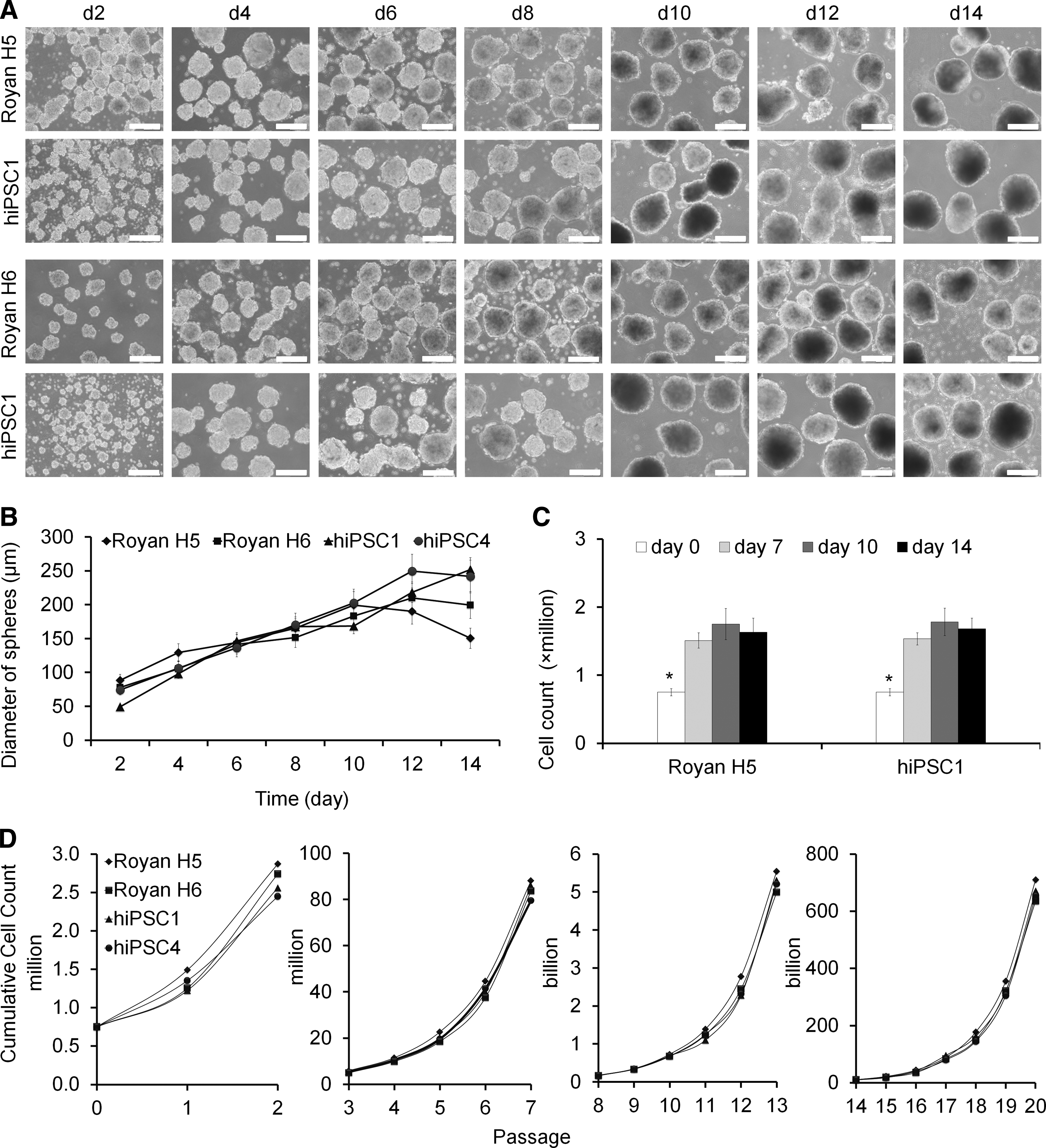
Aggregate growth kinetics, sphere diameters, and cumulative cell numbers in static suspension culture of hPSCs.
Characterization of all tested hPSC lines propagated in static suspension culture showed pluripotency markers after a minimum of 20 passages (Supplementary Results section and Appendix Fig. A1A, B). The expression of markers for spontaneous differentiation into the three germ layers—such as PAX6 and TAU for ectoderm; BRACHYURY and GATA binding protein 4 (GATA4) for mesoderm; and alphafetoprotein (AFP), Forkhead box A2 (FOXA2), and ALB for endoderm—was verified by RT-PCR (Appendix Fig. A1C).
Next, obtained cells from the static suspension culture were frozen and evaluated for viability after post-thawing at 0–3 h intervals and expression of pluripotency markers. Cell counts and viability values revealed that freezing/thawing process significantly decreased viability of the cultures after reviving (up to 20%; Appendix Fig. A2A). However, the revived cells were ALP positive; over 90% of the cells expressed nuclear markers OCT4 and NANOG, or the surface markers SSEA3 and TRA-1-60 (Appendix Results section and Appendix Fig. A2B, C).
Xeno-free expansion of hPSCs in dynamic suspension stirred bioreactor
The aforementioned results showed that all tested lines successfully propagated in the static suspension HFF-CM culture protocol and maintained their self-renewal capabilities. Accordingly, we selected this static culturing strategy as a starting platform for the dynamic suspension culture in a stirred bioreactor and bioprocess development for large-scale expansion of hPSCs.
Single-cell inoculated dynamic culture and impact of enzymatic dissociation medium type on hPSC expansion rate
Dynamic suspension cultures were conducted in 100 mL siliconized stirred bioreactors using Royan H5 and hiPSC1. To initiate the dynamic cultures, 1×10⁷ dissociated cells from the static cultures were transferred to 50 mL HFF-CM supplemented with bFGF and ROCKi in the stirred bioreactor. Cell aggregates were treated with ROCKi 1 h prior to enzymatic dissociation. The bioreactor was agitated at 35 rpm (increased to 40 rpm after 24 h) and medium refreshing began after 48 h of culture with the same medium and no ROCKi. After 24–48 h, small cell aggregates (40–80 μm) were observed in the bioreactor for both cell lines; aggregate sizes gradually increased due to cell division during the 10 days of culture (140–200 μm). After a minimum of four passages under the same conditions, the maximum FI in cell number was 3 for Royan H5 and 2.5 for hiPSC1. This was a slight improvement in cell proliferation in the dynamic culture compared with the static suspension culture (1.5–2 FI). In addition, enzymatic treatment of the cell aggregates with Accumax and Accutase resulted in higher cell viability (∼95%) and proliferation compared with TrypLE dissociation medium (Fig. 3; p<0.05). However, the optimum incubation time in the enzyme solution differed for different cell types of similar aggregate sizes, which indicated that different cell lines showed different resistance to enzymatic dissociation. This was possibly due to different E-cadherin expression levels (6–7 min for Royan H5 and Royan H6; 11–12 min for hiPSC1 and hiPSC4). 15 After 10 days, expansion rates in the single-cell inoculated dynamic culture increased to 4 FI for Royan H5 and 3 FI for hiPSC1 with the use of Accumax dissociation medium.

Impact of type of enzymatic dissociation medium on hPSC proliferation in single-cell inoculated dynamic suspension culture. Aggregates from suspension culture were treated with Rho kinase inhibitor 1 h prior to dissociation with Accumax™, Accutase™, and TrypLE™ dissociation medium. Enzymatic treatment was terminated by the addition of HFF-CM supplemented with 20% KnockOut™ SR XenoFree serum (*p<0.05).
Effect of cell inoculation density on expansion rates and metabolic activity
To determine optimum cell inoculation density for a single-cell inoculation of the dynamic suspension culture, bioreactors that contained 100 mL of HFF-CM supplemented with GlutaMAX and bFGF were inoculated with four cell inoculum concentrations: 2×10⁵, 3×10⁵, 5×10⁵, and 1×10⁶ cells/mL. Cell growth was indirectly monitored by measuring metabolic activity of the culture (Table 1 and Fig. 4). The exact cell numbers in the bioreactor could not be directly estimated due to heterogeneous cell aggregate formation; it was impossible to obtain a homogenous sample from floating aggregates in suspension culture. Therefore, we alternatively determined glucose consumption and lactate production kinetics for an indirect estimation of cell activity in the suspension aggregate culture. This method has been proposed for indirect monitoring of growth in immobilized mammalian cells, adherent cell cultures—such as human mesenchymal stem cells—and Chinese hamster ovary cell cultures.23–26 Metabolic activity profiles of Royan H5 and hiPSC1 using different cell inoculation densities are presented in Figure 4. The initial metabolic activity was higher in higher cell inoculation densities (up to 48 h), but after 2–3 days of culture, the exponential growth and metabolite activity of both cell lines significantly decreased in the 5×10⁵ and 1×10⁶ cells/mL inoculation densities (Fig. 4). These results were confirmed by kinetic studies of glucose consumption and lactate production rates (Table 1). As shown in Table 1, inoculation with cell densities higher than 3×10⁵ cells/mL decreased the duration of exponential growth and metabolic activity phase; thus, the culture reached its maximum cell density earlier. The associated exponential glucose consumption and lactate production rates increased with higher initial cell inoculation densities. The maximum and minimum glucose consumption and lactate production rates were obtained in 1×10⁶ cells/mL and 2×10⁵ cells/mL inoculation destinies for both tested cell lines (Table 1). However, after 10 days of culture, the maximum FI was obtained at the 3×10⁵ cells/mL inoculation density, which reached to 6.5-fold for Royan H5 and 4-fold for hiPSC1. The higher expansion rates in the 3×10⁵ cells/mL inoculation were possibly correlated to the extended exponential growth compared with (1) higher cell inoculation density culture and/or (2) efficient cell aggregation (due to appropriate initial cell numbers to the culture medium volume ratio after a single-cell inoculation). Lower expansion rates at higher cell inoculation densities could be related to the ratio of unsatisfactory cell numbers to the culture medium volume ratio, which resulted in cell loss due to sudden nutrient consumption and metabolite production, poor aggregation, and subsequently lower FI. The maximum cell density obtained for Royan H5 was 1.95×10⁶ cells/mL (3×10⁵ cells/mL inoculation) and for hiPSC1, it was 1.8×10⁶ (1×10⁶ cells/mL inoculation). These results have indicated that cell expansion rates of tested hPSC lines could be 2- to 3.5-fold improved by scaling up from a static suspension to dynamic suspension culture under the demonstrated culture conditions.
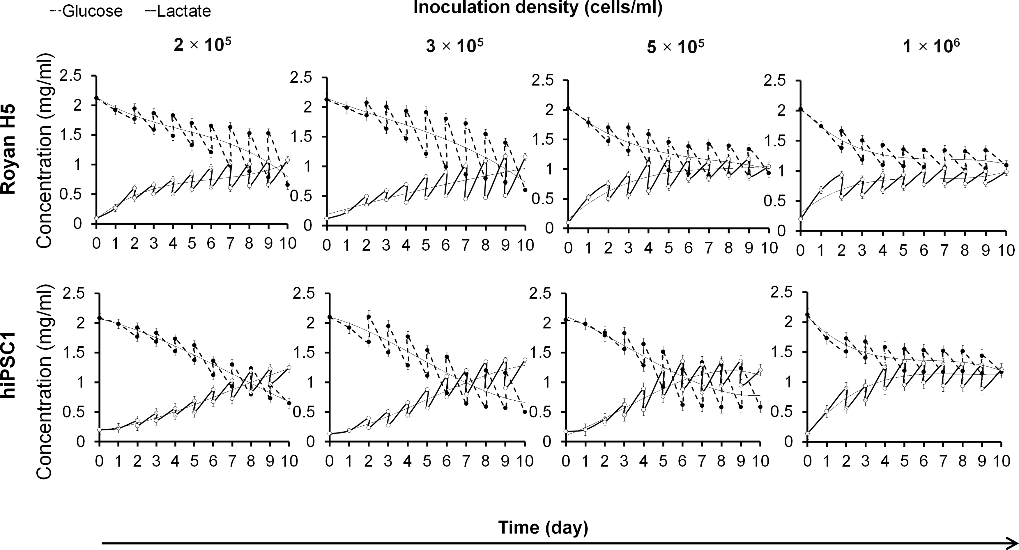
Glucose consumption and lactate production kinetics of single-cell inoculated suspension culture of hiPSCs at different cell inoculation densities in stirred bioreactor. Cells were seeded to the bioreactor at different cell inoculation densities (2×10⁵, 3×10⁵, 5×10⁵, and 1×10⁶ cells/mL). Metabolic activities were determined by high-performance liquid chromatography at 24-h intervals before and after medium refreshing.
Means with different superscript letters (a–c) within a column are significantly different (p<0.05).
hiPSCs, human induced pluripotent stem cells.
Mass production of size-controlled cell aggregates in suspension dynamic condition
After optimization of single-cell inoculation and passaging conditions, we employed different combinations of agitation rates (35–60 rpm) and medium viscosities (0.9 cp. [control, without shear protectant addition], 40 cp., and 70 cp.) by the addition of 0.1% and 0.2% CMC (shear protectant) to generate homogenous size-controlled aggregates of hPSCs in a dynamic suspension culture. The effects of these combinations on cell proliferation, viability, and aggregate size were extensively investigated. Proliferation and viability of both cell lines significantly decreased at agitation rates higher than 40 rpm (p<0.05; Fig. 5A). This was possibly correlated to excessive shear stress and consequent cell death during and after aggregation at higher mixing rates. The maximum FI approximated 6.5 for Royan H5 and 4 for hiPSC1 at 40 rpm agitation. We tested the effect of shear protectant addition on growth parameters at the 40 rpm agitation rate (Fig. 5A). As shown in Figure 5, a significant decrease in cell proliferation and viability of both cell lines was observed after addition of 0.2% CMC to the culture medium in 70 cp. medium viscosity. This negative effect could be related to high osmotic stress and possible CMC toxicity at this concentration level. The FI of Royan H5 decreased from 6.5 to 4 and hiPSC1 decreased from 4 to 2.5 in 70 cp. medium viscosity. Next, we studied the kinetics of Royan H5 and hiPSC1 aggregate sizes in different mixing rates and medium viscosity combinations. Our intent was to generate more homogenous aggregates without adverse effects on proliferation and increased risk of differentiation (Fig. 5B). As shown in Figure 5B, different aggregate sizes (150–310 μm) were generated by the use of different agitation rates and medium viscosity combinations. The mean aggregate size decreased at higher mixing rates and medium viscosities. To efficiently undergo large-scale production of size-controlled homogenous cell aggregates, we studied aggregate size kinetics during the culture period. The diverse effects of high agitation rates and shear protectant concentrations on cell viability and proliferation were investigated. We found that the combination of 50 rpm agitation rate and 40 cp. medium viscosity generated more homogeneous size-controlled aggregates (190–215 μm) without significant adverse effects on cell proliferation for both cell lines. We studied aggregate size kinetics of Royan H5 and hiPSC1 during 10 days of culture in both normal (no shear protectant addition, 40 rpm agitation) and viscous medium (40 cp., 50 rpm agitation; Fig. 6A). For both cell lines, aggregates were more homogeneous and smaller in diameter size in viscous medium compared with control medium. It was demonstrated that the sudden increase of aggregate size in normal medium (after 6 days of culture) was largely controlled in viscous medium while maintaining cell proliferation efficiency (Fig. 6B, C). The kinetics of aggregate area distribution also showed a similar trend. The range of cell aggregate areas was 30%–40% lower in viscous media compared with control medium (Fig. 6D, E).

Expansion, viability, and aggregate diameter of hPSCs cultured in dynamic suspension at different agitation rates and medium viscosities.
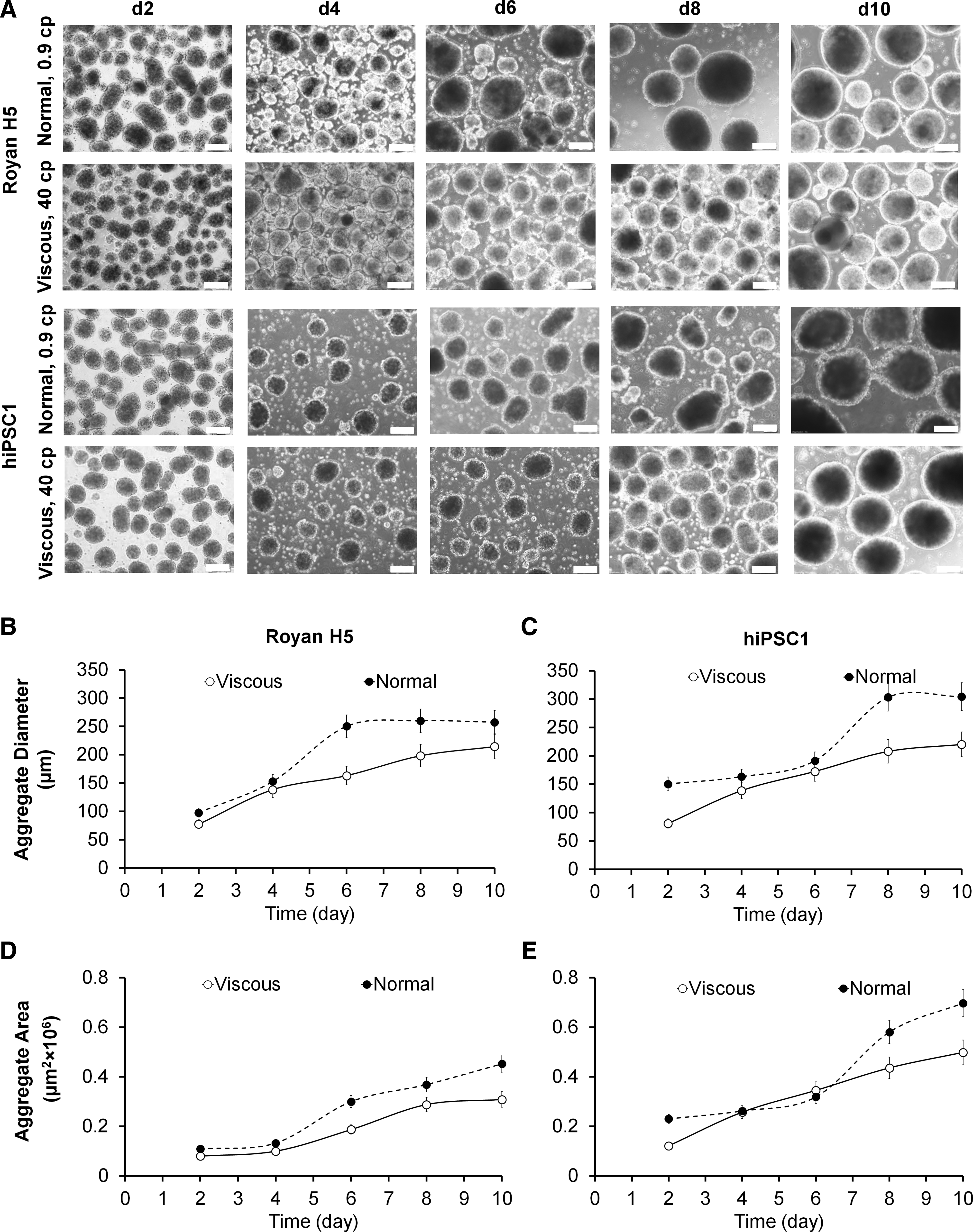
Kinetics of hPSC aggregate morphology, diameter, and area proliferated under optimized normal and viscous culture conditions in a dynamic suspension bioreactor.
Impact of oxygen concentration on cell proliferation in dynamic suspension culture
In the next step, we examined the effect of oxygen concentration on cell expansion as a key bioprocess parameter in the dynamic suspension culture of hPSC aggregates. We investigated the impact of three different oxygen concentration modes: hypoxia (5%–6% O2), uncontrolled (10%–13% O2), and normoxic conditions (18%–21% O2) on cell expansion and metabolic activity of Royan H5 and hiPSC1. The expansion rate significantly improved by controlling oxygen concentrations in the culture environment (Fig. 7A). Incubation of bioreactors under normoxic conditions resulted in higher expansion rates compared with uncontrolled and hypoxia conditions. The expansion rates of Royan H5 reached 8.2 FI and hiPSC1 increased to 7.5 FI after 10 days of incubation in normoxic conditions, which showed a 20% improvement for Royan H5 and 60% improvement for hiPSC1 compared with uncontrolled conditions. This was corroborated by a comparison of metabolic performances of both cell lines in uncontrolled and normoxic conditions (Fig. 7B). As shown in the figures, higher oxygen concentrations in normoxic conditions reduced the lag phase of the culture and extended the exponential growth phase of both cell lines to 9–10 days. Incubation of bioreactors under normoxic conditions resulted in efficient cell aggregation after single-cell inoculation and consequently resulted in higher expansion rates in the dynamic suspension culture.
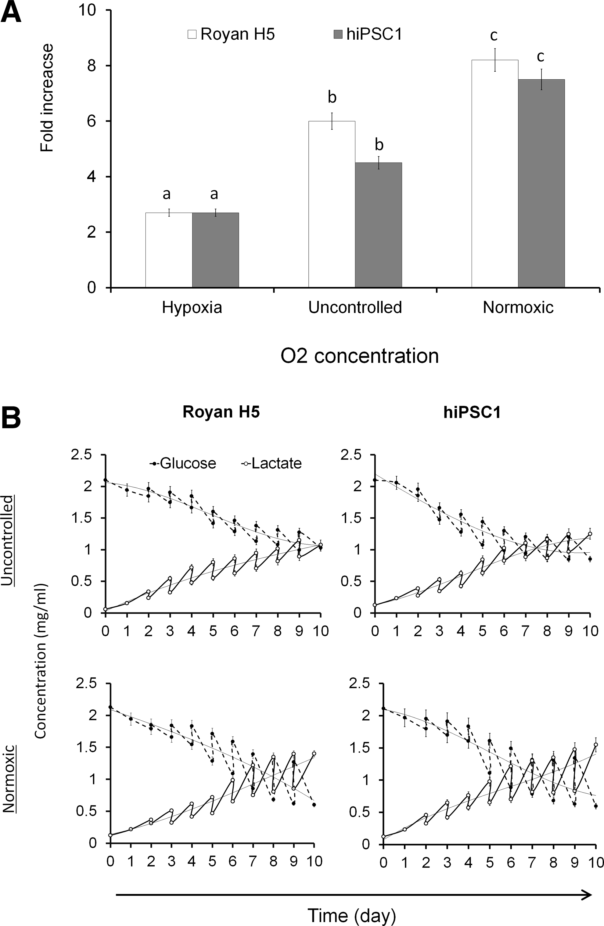
The effect of different oxygen concentration modes on expansion and metabolic activity of hPSCs cultured in a dynamic suspension bioreactor.
Cell aggregation kinetics in dynamic suspension culture
After optimization of passaging and hydrodynamic culture conditions, the aggregation and growth kinetics of both cell lines were monitored after single-cell inoculation and later during the first day of culture. We studied aggregation kinetics to understand the mechanism of aggregation and determined optimum conditions for agitation and medium refreshment to minimize cell loss after a single-cell inoculation. Aggregation monitoring showed that single cells of Royan H5 and hiPSC1 began to assemble in small cell aggregates after 4 h of inoculation. Afterward, cell aggregates grew; the majority of single cells attached to the formed aggregates after 10 h (Appendix Fig. A3). According to a kinetic study of cell numbers and viability, about 20% of the inoculated cells were lost during the first day of culture (Fig. 8). Metabolic activity measurements demonstrated that the exponential metabolic activity of both cell lines began after 4–6 h of inoculation, at the time of aggregate formation. We refreshed the medium (50% of the culture volume) after 48 h, when most viable single cells had attached to the formed aggregates. To improve the efficacy of aggregate formation and minimize cell damage due to high shear stress, agitation rates were started at 35 rpm and gradually increased to the maximum intended agitation rate during the first day of culture.
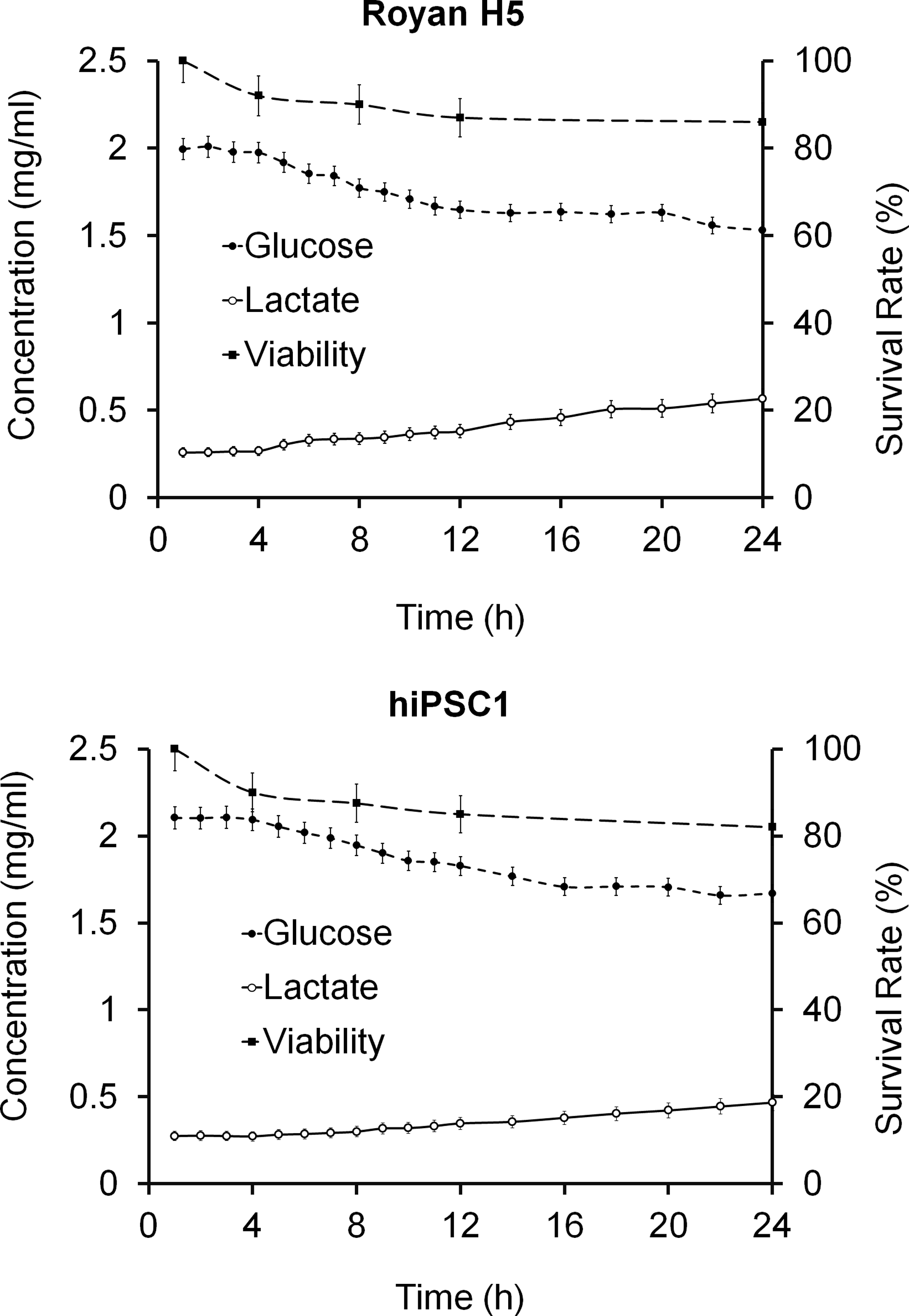
The survival rate and metabolic activity of Royan H5 and hiPSC1 cell lines cultured in a dynamic suspension bioreactor (inoculated at 3×10⁵ cells/mL density) determined by trypan blue and high-performance liquid chromatography, respectively. About 20% of the inoculated cells were lost during the first day of culture. Metabolic activity measurements showed that the exponential metabolic activity of both cell lines began after 4–6 h of inoculation, at the time of aggregate formation.
Characterization of hESCs and hiPSCs propagated in xeno-free dynamic suspension culture
We cultured all 4 tested cell lines by using the optimized dynamic suspension culture protocol that was characterized after a minimum of 10 passages in the bioreactor. As shown in Figure 9A, 2-day spheroids were plated on Matrigel-coated dishes for 5 days. These spheroids formed colonies typical of undifferentiated hESCs and hiPSCs with clear borders that consisted of compact cells with high nuclear:cytoplasm ratios. In addition, colonies obtained from all four tested suspension expanded lines were ALP positive (Fig. 9A). The spontaneous differentiation expression of the three germ layers was shown by RT-PCR (Fig. 9B). Pluripotency markers of cultured hESCs and hiPSCs in HFF-CM were probed by flow cytometry (Fig. 9C and D). Flow cytometry analysis of the hPSCs showed that most cells were double-positive for the nuclear markers OCT4 and Nanog (96%) and surface markers TRA-1-60 and SSEA3 (98%). Finally, we evaluated the in vitro differentiation potential of hESC and hiPSC lines cultured in xeno-free dynamic suspension by replacing CM with non-CM, following prolonged expansion. These results showed that all four hPSC lines tested can be successfully scaled up from static to dynamic suspension culture without compromising their self-renewal capabilities. Thus, we have shown that the developed scale-up platform can be used for mass production of homogenous size-controlled pluripotent hPSC aggregates in stirred suspension bioreactors under xeno-free conditions.
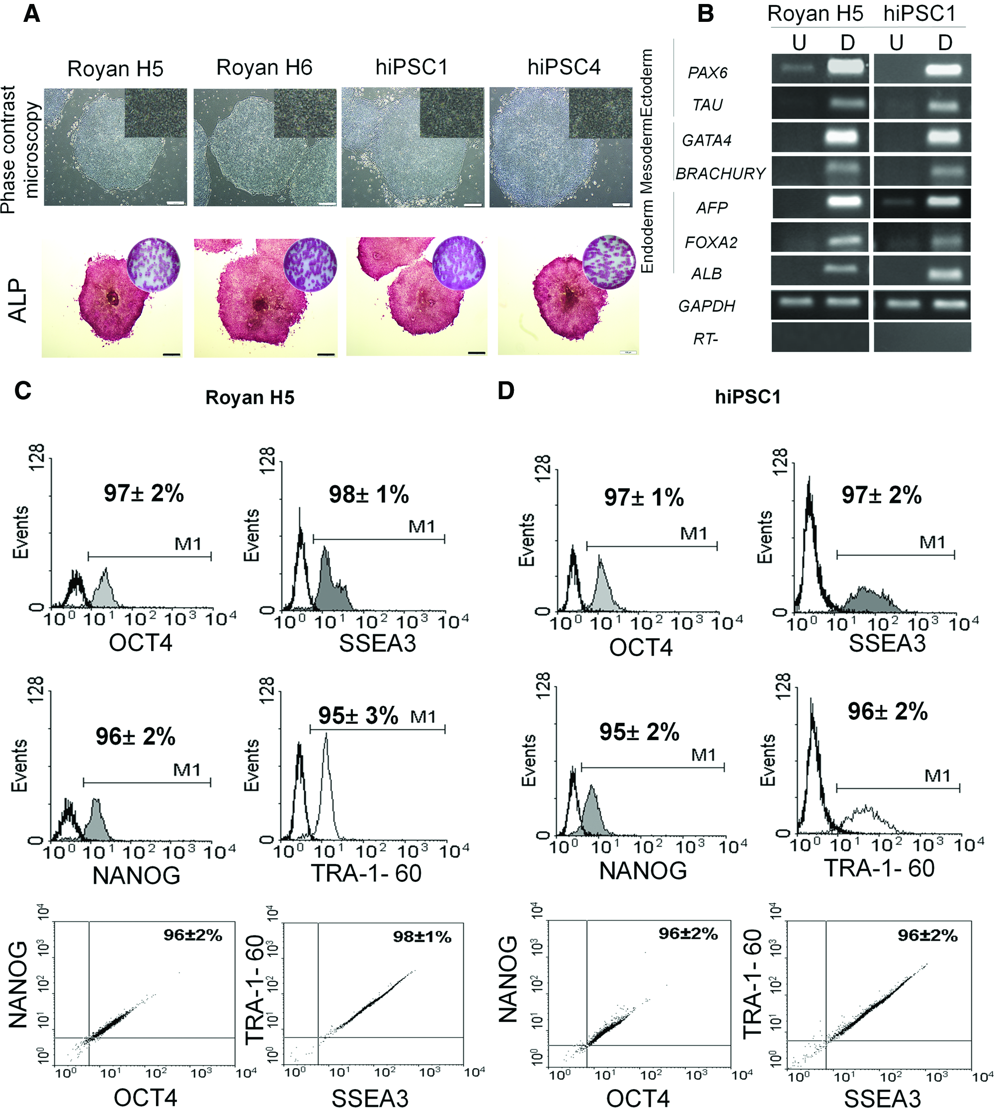
Characterization of hPSCs expanded in a xeno-free dynamic suspension culture under optimized culture conditions.
Scale-up platform for dynamic suspension culture
After stepwise optimization of key bioprocess parameters for mass production of hPSCs in the dynamic suspension culture, we explored a simple and fast scale-up platform for minimizing adaptation passages required when transforming adherent hESC and hiPSC cultures to static suspensions, followed by dynamic suspension cultures. We began with one frozen vial each of Royan H5 and hiPSC1 cell lines that contained 1×10⁶ cells/mL of freezing media (90% KnockOut™ SR+10% DMSO). After one passage in the adherent culture and three to four passages in the static suspension culture, the total number of cells reached 1×10⁷ cells for both cell lines. Then, all characterized cells were harvested and inoculated into a bioreactor with 50 mL HFF-CM and incubated at optimized culture conditions. After two passages in dynamic suspension culture, all cells (1.6×10⁸) were harvested and cryopreserved by the demonstrated freezing protocol. In the next step, we directly transferred the frozen cells (obtained from the dynamic suspension culture) to the bioreactor filled with 50 mL HFF-CM. We observed that both cell lines proliferated successfully and showed twofold expansion rates after 10 days. The expansion rates increased to 6.5 for Royan H5 and 5 for hiPSC1 after two to three passages while both cell lines were ALP positive and maintained self-renewal capabilities as demonstrated by flow cytometry analysis of OCT4 and NANOG pluripotency markers (Fig. 10). Finally, to explore the reproducibility of the developed dynamic suspension culture protocol for other hPSC lines, we cultivated Royan H6 and hiPSC4 using the same culturing strategy optimized for Royan H5 and hiPSC1 cell lines. Results showed that expansion rates of Royan H6 and hiPSC4 also improved by scaling up from a static suspension culture to dynamic suspension culture while maintaining pluripotency potential (6.5 FI for Royan H6 and 5 FI for hiPSC4).

Characterization of hPSCs cultured by direct transfer of suspension-adapted cells from frozen vial to dynamic culture in the bioreactor without adaptation passages in statistic suspension culture. Expression of ALP by Royan H5 and hiPSC1 after a minimum of five passages in dynamic culture in a bioreactor. The box within ALP shows the entire staining plate. Scale bars: 100 μm. Double expression of OCT4 and NANOG by flow cytometry with the same culturing strategy for both cell lines. Color images available online at
Discussion
In the present study, we developed a novel robust scale-up platform for mass production of homogeneous size-controlled hPSCs in dynamic suspension culture under xeno-free conditions. This has been achieved by identification of key bioprocess parameters involved in the suspension culture of these unique cells and by improvement in culture conditions through stepwise optimization of the most critical steps. A comparison of this study with other works that have focused on the suspension cultures of hESCs and hiPSCs as three-dimensional aggregates in a spinner bioreactor is presented in Table 2.
hESC, human embryonic stem cells; hPSCs, human pluripotent stem cells; bFGF, basic fibroblast growth factor; ROCKi, Rho kinase inhibitor; DMEM, Dulbecco's Modified Eagle Medium; ND, not determined.
The most critical biological and technical challenges of previous attempts aimed at the scale-up of hESC and hiPSC suspension cultures were as follows: (1) the adherent nature of these cells and necessity for time-consuming preadaptation passages in adherent and static suspension cultures before the transformation to a dynamic suspension culture11–13,15; (2) xeno-free and defined culture conditions that could support both proliferation and maintenance of self-renewal capabilities 10 ; (3) significant cell loss during each passage when using the single inoculation strategy2,10,15; (4) heterogeneous aggregate formation that resulted in the generation of heterogeneous cells with different pluripotency potentials11,13; (5) the negative effect of hydrodynamic stresses in dynamic culture conditions on cell proliferation and increased risk of spontaneous differentiation and karyotype abnormality2,13,15; and finally, (6) line-to-line variability in response to culture conditions.10,11,13,15,27 Most previously demonstrated protocols did not extensively study the growth and metabolic kinetics of the tested cell lines in proposed culture conditions in addition to the large-scale expansion in stirred suspension bioreactors (Table 2). Here, we have attempted to address the majority of the earlier mentioned challenges with the intent to design a novel bioprocess for large-scale expansion of highly pluripotent hPSCs. First, we screened eight different groups of xeno-free conditioned and/or defined hESC media combined with ROCKi treatment for their effectiveness in supporting proliferation and self-renewal of four hPSC lines (two hESC and two hiPSC lines) in suspension culture. We introduced the combination of DMEM/F12-CM supplemented with GlutaMAX and bFGF (100 ng/mL), and ROCKi treatment for maintenance of cell lines in a static suspension culture that was then scaled up to a dynamic suspension culture in a bioreactor. Thus, this medium could be used as an alternative medium to commercially defined mTeSR medium because of its higher efficacy and cost effectiveness. The main disadvantage of this CM is its nondefined status and possible lot-to-lot variability in composition, but recent feeder-free hESC medium screening studies have reported similar lot-to-lot variability in performance for commercial mTeSR medium. 28 Over the past few years, high-throughput screening of different molecules and extracellular matrices to develop a defined culture medium has gained increased attention, but most works have focused on relatively expensive molecules and components, such as recombinant laminin, fibronectin, synthetic peptides, or xylose derivatives.2,18,28,29 Therefore, the development of a cost-effective and robust defined medium for the suspension culture of hPSCs is still in progress for large-scale production of the cells under good manufacturing practice (GMP) conditions.
Next we showed that significant cell loss, which has been reported in previous protocols (up to 60%), could be minimized by the use of mild enzymatic dissociation optimized for each cell line, optimized ROCKi treatment, and determination of optimum cell density for the single-cell inoculation of each culture medium. The importance of cell inoculum density as a critical scale-up parameter for expansion of different human and animal cell lines has been demonstrated in many related studies.30–32 However, we did not find any detailed information on optimizing inoculation densities for hPSC cultures. Here, we found that expansion rates could be largely improved by choosing the right inoculation density for each cell line and culture medium. Practically, inoculation at very low cell densities would result in significant cell loss due to the lack of efficient cell-to-cell interactions. On the other hand, inoculation at very high cell densities can cause sudden depletion of nutrients in the medium and subsequent rapid production of metabolic waste, which adversely affects cell growth and expansion rates. Heterogeneous cell aggregate formation and subsequent limited diffusion in large aggregates is another disadvantage of very high cell inoculation densities, which can lead to necrosis in the center of large aggregates.
The generation of size-controlled aggregates is a very important parameter that should be taken into consideration in bioprocess development. Homogenous aggregate formation will facilitate and expedite the scale-up process due to the more effective enzymatic dissociation and subsequently lower cell loss after each passaging, higher expansion rates, and finally higher pluripotency and differentiation potentials.33–35 Here, we have shown that size-controlled aggregates could be generated by using precise control of the agitation rates and addition of shear protectants. Our proposed method is more scalable than other technologies developed for cell aggregate size control, such as microwell-mediated control, hanging drops, and microprinting technologies.33–35 In addition, we have demonstrated that the hydrodynamic culture condition of a bioreactor is a very important parameter for hPSC culture scale-up because of the very high sensitivity of hPSCs to shear stress. Importantly, increasing the agitation rates to improve nutrient delivery and homogenizing culture environment and subsequent aggregate formation should be well balanced in conjunction with limiting shear stress in dynamic conditions to minimize adverse effect on cell proliferation and pluripotency. To date, there have not been any practical investigations into shear effects on growth kinetics in hESC suspension cultures but the topic has been reviewed recently. 36
In the next step, we focused on the effect of oxygen concentration as a critical bioprocess parameter in suspension dynamic culture of hPSCs. We demonstrated that the expansion rates could be largely improved in normoxic conditions (up to 60% improvement in expansion rates). As culture volumes and cell densities increase, the surface area to volume ratios decrease and surface aeration may no longer be sufficient to ensure oxygen transfer to cells within aggregates. Thus, increasing the oxygen levels in the culture environment could lead to better proliferation and higher expansion rates. Therefore, the development of novel strategies to improve oxygen transfer to cells, such as membrane aeration and controlling dissolved oxygen concentration, is essential for successful scale-up of the hPSC culture.32,33,37 Finally, we have developed a fast and straightforward scale-up platform that eliminates the labor-intensive preadaptation passages required in previous suspension culture reports. In addition, all tested cell lines were successfully propagated using this culturing strategy while maintaining their self-renewal capabilities. This could be a great advantage over currently demonstrated suspension protocols that can simultaneously facilitate and expedite the production of a clinically relevant number of cells in a minimal culture time. For example, 1.9×10⁹ undifferentiated hPSCs could be generated within 30 days in four spinner flasks that contain 200 mL of HFF-CM with the use of this culturing strategy and scale-up platform (Fig. 11).

Schematic presentation of xeno-free bioprocess platform for mass production of hPSCs as carrier-free cell aggregates in a stirred suspension bioreactor. By using the proposed scale-up platform and optimized single-cell inoculated suspension culture, a clinically relevant cell number of hPSCs (∼2×10⁹ cells) were produced in four spinner flasks that contained a total of 800 mL HFF-CM over a 30-day period.
Conclusions
An optimized bioprocess for mass production of hPSCs under fully controlled, defined, and xeno-free conditions is necessary for cell manufacturing and cell therapy facilities that operate under GMP conditions. Suspension culture of hPSCs has facilitated the development of scalable culture platforms and production of clinically relevant number of cells in fully controlled bioreactors. However, more basic and applied research is needed prior to widespread commercial application of the demonstrated protocols. Exploring cell lines that show good adaptation to a suspension culture; development of cost-effective xeno-free defined culture media for suspension culture; indirect and nondestructive monitoring of key bioprocess parameters, such as cell growth kinetics, in an aggregate culture; an extensive study of the effect of bioreactor hydrodynamic conditions on hPSC aggregates; an automated straightforward platform for large-scale expansion and integrated differentiation; and finally, fundamentals of biological challenges in suspension culture are all excellent topics for future research.
Footnotes
Acknowledgments
This study was funded by grants provided from Royan Institute and the Iranian Council of Stem Cell Research and Technology. The authors also thank Seyedeh-Nafiseh Hassani, Mohammad Pakzad, Sepideh Mollamohammadi, and Adeleh Taei for their comments and technical assistance.
Disclosure Statement
None of the authors have any conflicts of interest to disclose and all authors support submission to this journal.