




Abstract
Human retinal progenitor cells (hRPCs), isolated from fetal retina, require extracellular matrix proteins such as fibronectin or laminin for successful attachment and self-renewal in vitro. Here we have shown that a novel synthetic vitronectin-mimicking surface supports self-renewal and multipotency of hRPCs in a chemically defined culture system. The morphology, adhesion, and proliferation of hRPC were equivalent on a novel vitronectin-mimicking surface (Synthemax) compared to a fibronectin-coated surface. When evaluated using real-time polymerase chain reaction, Western blotting, and flow cytometry, both surfaces maintained self-renewal of hRPCs, as shown by similar expression levels of Sox2, Nestin, cMyc, Klf4, and Pax6, with no change in integrin beta1 and integrin alpha5 expression. We suggest that the use of synthetic, xeno-free surfaces such as Synthemax will be useful for basic research studies, as well as development of translational strategies aimed at using stem cell transplantation to treat disease.
Introduction
One of the limiting factors for successful translation of this approach to the clinic is the ability to expand a cell line into a clinically relevant number of cells, while maintaining a nondifferentiated state. This requires establishing a line from a single source and expansion through multiple passages, with characterization both in vitro and in vivo. It is important to closely mimic the developing retinal microenvironment in a chemically defined cell culture system, including both soluble and insoluble factors.
There are two distinct approaches to culture central nervous system (CNS)-derived stem and progenitor cells: one utilizes neurosphere formation, allowing cells to grow as floating clonal aggregates,2,3 while another utilizes enzymatic dissociation and expansion of progenitors as a monolayer of attached cells.1,4,5 This dual approach has been exploited for both retinal precursors and progenitors, isolated from neonatal/fetal retina 6 or differentiated 7 from pluripotent stem cells. The benefits of monolayer expansion include higher reproducibility and homogeneity of the population. Also, since neurospheres vary in size, it is difficult to calculate proliferation and the doubling rate, critical parameters in cell expansion.
High concentrations of basic fibroblast growth factor (bFGF) and epidermal growth factor (EGF) can support self-renewal of hRPCs in a completely chemically defined system on fibronectin-coated surfaces 1 for several passages. As the fibronectin protein sequence is highly conserved, 8 trace amounts of full-length protein as well as short peptides in the cell suspension prepared for transplantation likely do not carry with it additional risks of host-versus-graft immunity. 9 However, lot-to-lot variations of purified fibronectin together with its high cost and the time-consuming process of coating vessels make the search for synthetic substrate a priority.
Several families of polymers have been investigated as a scaffold to culture retinal progenitors and precursors for transplantation. 10 Some of them inhibit proliferation, which is suitable for transplantation, but not for expansion, while others like poly (glycerol sebacate) 11 or poly (caprolactone) 12 require additional coating with fibronectin or laminin. Oxygen plasma treatment of polystyrene 13 and other polymers increases the hydrophilic properties of the surface, leading to an increase in the adhesion and growth of several cell types, such as fibroblasts, endotheliocytes, keratocytes, retinal pigment epithelium, mesenchymal stem cells, and others.14,15 However, CNS stem cells require fibronectin 16 or laminin17,18 as a substrate, and the same have been shown for retinal progenitor cells and precursors of different species, including mouse, 12 rat,19,20 pig, 5 and human.21,22
To our knowledge, no synthetic surfaces have been shown to be capable of maintaining the multipotency properties of retinal progenitors in chemically defined conditions.
Here we demonstrate the equivalency of a peptide-acrylate surface, enclosing an active vitronectin sequence (Corning Synthemax) and a fibronectin-coated surface for the self-renewal of hRPCs, as confirmed by similar adhesive and proliferative properties and the expression of specific stemness and eye-field markers.
Methods
Cell isolation and culture
All work with human tissue was considered as exempt from IRB approval. hRPC were isolated from human fetal neural retina at 16 weeks of gestational age as previously described. 21 Briefly, whole neuroretina was peeled from the retinal pigment epithelia layer, minced, and digested with collagenase I (Sigma-Aldrich). Cells and cell clusters were plated in flasks (Costar) coated with fibronectin (100 μg/mL for 30 min; Akron) in Ultraculture Media (Lonza), supplemented with 2 mM L-glutamine (Invitrogen), 20 ng/mL rh bFGF (Peprotech), and 10 ng/mL rh EGF (Peprotech). hRPC were passaged at 80% confluency using TrypZean (Sigma-Aldrich) with addition of benzonase (EMD Chemicals) and Defined Trypsin Inhibitor (Invitrogen). At each passage cell number and viability was estimated with Trypan blue (Sigma-Aldrich) staining using a hemacytometer with cells plated at a density of 10,000/cm2. For all experiments described here, we used hRPCs at passage 5 (12 population doublings in culture from time of isolation).
Adhesion
We performed two types of tests to assess the adhesive properties of the synthetic surface (Fig. 1). In the first set of tests, we plated 100,000 hRPCs into each well of a six-well plate (Costar tissue-culture treated and coated with fibronectin or Synthemax). The plates were then incubated for 1, 4, 6, 10, 20, or 30 min (37°C, 3% O2, 5% CO2, 100% humidity) and washed once with HBSS (with Ca++ and Mg++). We then randomly chose images (three fields of view for each condition, ∼50–150 cells within the field) and counted the number of cells attached using ImageJ (NIH). Experiments were performed three times for three different isolations of hRPCs, with all results combined and compared using a t-test with Bonferroni correction.
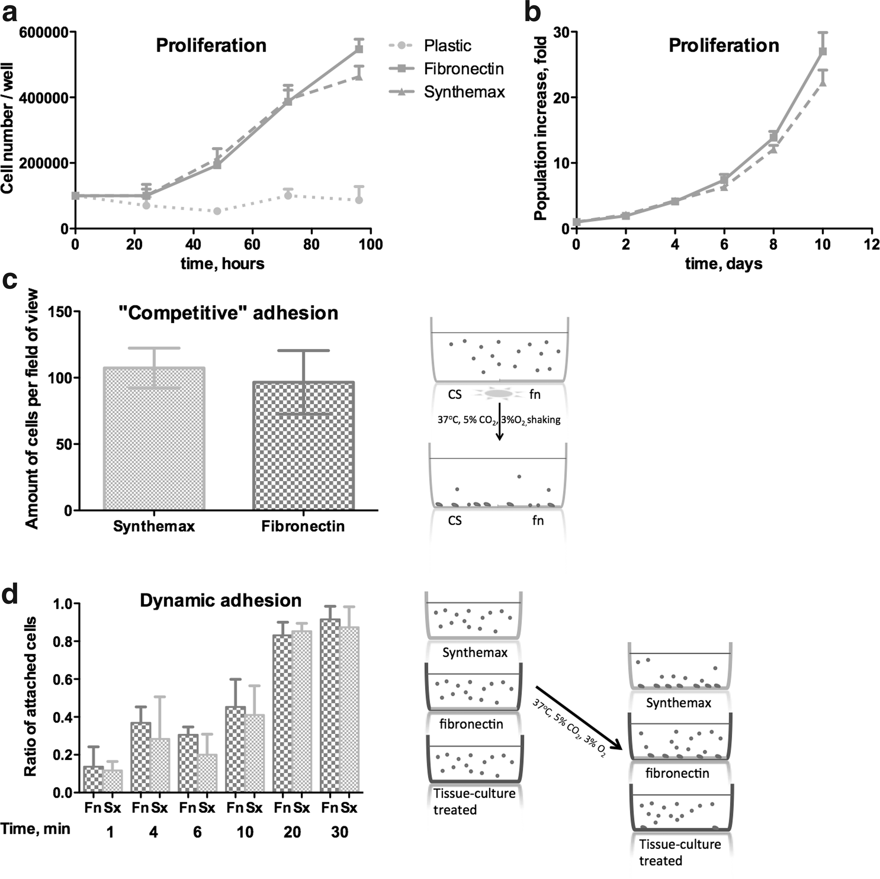
Synthetic surface mediates adhesion and proliferation of hRPC. Synthetic surface has adhesive properties, similar to fibronectin-coated plastic. We observe similar proliferative rate
The second set of experiments was designed to compare the adhesive properties of synthetic surface with those seen on fibronectin-coated tissue culture plastic. To do so, we coated one half of each well of a Synthemax plate with fibronectin (100 μg/mL solution for 30 min): to prevent leakage of fibronectin to the vitronectin-mimicking side, a hydrophobic line was made using a PAP-pen (Invitrogen). Wells were then rinsed with distilled water twice, and 100,000 cells were plated into each well. Plates were placed on top of a rotatory shaker in the incubator (37°C, 3% O2, 5% CO2, 100% humidity). After 30 min plates were washed with HBSS (with Ca++ and Mg++) and the number of attached cells determined as described above. Each experiment was performed three times for three different isolations of hRPCs, and again the results were combined and compared in this case by a paired t-test.
Proliferation
To compare proliferation of hRPCs on three different surfaces, 100,000 hRPCs were plated in media into each well of a six-well plate (tissue-culture-treated alone, and coated with fibronectin or Synthemax), and then incubated (37°C, 3% O2, 5% CO2, 100% humidity) for 24, 48, 72, or 96 h (after 48 h in culture media was replaced with fresh), harvested using TrypZean, benzonase, and DTI. Cell number and viability was estimated using Trypan Blue and a hemacytometer. Experiments were performed three times, with all results combined and compared using a t-test with Bonferroni correction.
To investigate the ability of the synthetic surface to maintain long-term proliferation of hRPCs, we plated hRPCs on fibronectin or Synthemax at the conditions described above. Cells were passaged every 2 days, and cell number was estimated with a hemacytometer. Experiments were performed three times, with all results combined and compared using a t-test with Bonferroni correction.
Flow cytometry
To perform flow cytometry analysis, hRPCs were plated on fibronectin-coated plastic or Synthemax; incubated for 48 h; harvested using TrypZean, benzonase, and DTI; washed in HBSS (without Ca++ or Mg++); and fixed in 4% paraformaldehyde (Sigma-Aldrich) for 20 min at 4°C. hRPCs were then washed, permeabilized in 0.2% Triton-X100 (Sigma-Aldrich) for 30 min at room temperature, blocked in 10% goat serum (Invitrogen) with 5% bovine serum albumin (BSA; Sigma-Aldrich) in phosphate buffered saline for 30 min at room temperature, washed, and stained with primary antibodies for 1 h at room temperature. We stained cells for stemness/eyfield markers (Sox2, Nestin, Pax6, Klf4) markers (see Supplementary Table S1; Supplementary Data are available online at
Western blotting
Protein was isolated from hRPCs cultured for 24 h on fibronectin-coated plastic or Synthemax using RIPA buffer with protease inhibitors (Sigma-Aldrich). Protein concentration was estimated using a BCA test (Thermo Scientific). Proteins were resolved under reducing conditions on 10% SDS-PAGE gel, transferred to PVDF membrane, blocked with 5% BSA in TBST, and stained with primary antibodies (GAPDH, Oct4, Sox2, Klf4, cMyc, Nestin, Integrin beta1, Integrin alpha5, TIMP2, MMP2) overnight at 4°C. After a triple wash in TBST, bands were identified with HRP-linked secondary antibodies (Amersham) (1:2500 in 5% BSA for 1 h at room temperature), and detected with an ECL HRP-substrate (Thermo scientific) using X-ray film (Thermo Scientific). The density of the bands was measured using ImageJ. The average expression level was calculated based on three experiments.
Real-time polymerase chain reaction
Total RNA was extracted from day 3 cell cultures on the vitronectin-mimicking surface and from cell cultured on fibronectin (RNeasy Mini kit; Qiagen) followed by column treatment with DNase I (Qiagen). Reverse transcription was performed with SuperScript III First-Strand Synthesis System (Invitrogen) and random primers (Sigma). Real-time quantitative polymerase chain reaction (PCR) was performed with Mastercycler ep Realplex2 (Eppendeorf North America) at 40 cycles with 100 ng of starting cDNA. SYBR green was used for amplification and data analyzed by delta CT method, Q1 program (Eppendorf North America). RNA was quantified with the delta CT method and normalized to GAPDH as an endogenous control. Each reaction was performed in triplicate.
Results
Adhesion and proliferation
The adhesive properties of the Synthemax surface did not differ quantitatively from those of fibronectin-coated tissue culture plastic (Fig. 1): in both cases more than 90% of the cells were attached after 30 min of incubation. However, in the control group (uncoated tissue-culture treated plastic) only single cells stuck to the surface (<5%), but did not spread: no processes or focal adhesion contacts were observed using phase-contrast microscopy (data not shown). Incubation of hRPCs on noncoated plastic for longer periods did allow adhesion, but they were unable to proliferate. Placing cells in “competing adhesion” conditions for 30 min similarly showed no difference in the number of attached cells between Synthemax and fibronectin-coated tissue culture plastic (Fig. 1c).
This attachment led to equal proliferative properties for 72 h of culture (Fig. 1a) with a population doubling time about 24 h. The only observed difference was the maximum density of hRPCs that can be achieved on the described surface and their behavior after reaching 95% confluency. At the cell density of 40,000 cells/cm2, confluency is about 80%, and 50,000 cells/cm2 gives 95% confluency. On fibronectin it is possible to reach 100,000 cells/cm2, although the hRPCs appear to decrease in size and start to form multiple layers. Synthemax has an upper limit of 60,000 cells/cm2, as the cells never overgrow. If cultured at this overconfluent condition for a longer period (>48h), hRPCs on fibronectin started to form clumps and then detach, while on Synthemax we never observed such behavior: hRPCs stopped proliferating and remained attached and spread. However, if passaged before reaching overconfluent conditions, hRPCs retain equal growth properties on fibronectin and synthetic surfaces.
Flow cytometry
We have not observed any difference in specific retinal progenitor marker expression between fibronectin or Synthemax groups (Fig. 2c). Stemness markers staining (Sox2, Klf4, Pax6, Nestin) showed no difference in hRPC population quality on these surfaces.

Synthetic vitronectin-mimicking surface maintains stemness properties of hRPC. By WB
Western blotting and real-time polymerase chain reaction
Results observed on flow cytometry were confirmed by Western blotting (Fig. 2a) and real-time polymerase chain reaction (Fig. 2c) for Sox2, Klf4, Oct4, Nestin, and cMyc. The expression level of beta1 as well as alpha5 subunits of integrin remains the same in both conditions. The level of TIMP2 and MMP9 in hRPCs did not differ between these surfaces.
Discussion
Clinical application of human stem and progenitor cells is highly dependent on development of a scalable system for expansion of a particular cell type. It should be highly reproducible, well standardized, and as defined as possible, including xeno-free conditions for all steps of manufacturing (isolation, expansion, and preparation of cells for grafting). Previous research was primarily focused on soluble factors such as media and supplements, with the best results for hRPCs achieved with the use of Ultraculture media, supplemented with rhEGF and rhbFGF (references). The utility of low-oxygen conditions for retinal progenitor culture and photoreceptor generation, previously well described for neural, mesenchymal, and pluripotent stem cells (refs), has also been confirmed. 23
Substrate components for this culture system have not been previously addressed. Two approaches have been described: one suggests growth of pig 2 and human 3 RPCs as neurospheres, while another utilizes different extracellular matrix-coated surfaces for attachment and expansion of RPCs as monolayer.5,21 Previously, we compared several recombinant and isolated proteins and peptides (fibronectin, laminin, collagen I, poly-D-lysine, poly-L-lysine) 1 as well as nondefined matrices (Matrigel; Geltrex) and found that fibronectin or laminin coating is sufficient for self-renewal of hRPC, while collagen, poly-lysine, and matrices, derived from Engelbreth-Holm-Swarm sarkoma do not mediate proper attachment and spreading. 1
Several polymer-based chemically defined culture surfaces have been described that are able to support the growth of pluripotent stem cells lines, such as poly (methyl vinyl ether-alt-maleic anhydride) (PMVE-alt-MA) 24 or commercially available NunclonVita or Synthemax 25 culture dishes. All other polymers described in the literature, including classic polystyrene and polypropylene, films, and scaffolds designed for transplantation, require additional coating with one or several of the integrin ligands mentioned above.
Here we have shown that the vitronectin-mimicking Synthemax surface is effective for maintaining multipotency of hRPCs in serum-free, xeno-free, chemically defined conditions. It has been previously described as an efficient and reproducible substrate for the culture of human embryonic stem cells 25 due to the synthetic vitronectin-mimicking, RGD-containing peptide 26 sequence within it. This arginyl-glycyl-aspartic acid motif, which is the ligand for integrin receptors, is more active within vitronectin 27 compared to other extracellular matrix proteins such as bone sialoprotein, laminin, or fibronectin.
Developing retinal neurons rely on integrin “outside-in” signaling, especially using the beta1 subfamily, 28 which requires vascular cell adhesion molecule or fibronectin for activation.17,29,30 The beta1 integrin subunit is required not only for proper migration and lamination of layers within the developing retina, but also for the cell survival.31,32 Inhibition of integrin beta 1 expression in the developing retina with antisense RNA resulted in the reduction of clone size generated from each precursor. 33 As we have shown here, expression of integrin beta1 subunit is significantly downregulated in hRPCs without proper ligands as substrates in the culture system, but does not differ between fibronectin or Synthemax surfaces.
We have not found any difference in the main transcription factors and other markers involved in multipotency and self-renewal of hRPC between fibronectin or Synthemax surface expansion conditions. However, the absence of ligands resulted in the rapid loss of Sox2, Klf4, and Oct4 in hRPCs. The equivalent proliferation rate on polymer compared to fibronectin-coated surfaces allows one to scale up the expansion of hRPC to sufficient quantities for clinical applications.
Conclusions
A novel synthetic vitronectin-mimicking surface supports self-renewal and multipotency of hRPCs in chemically defined culture system. This approach allows low-risk, cost-effective expansion of hRPCs for clinical applications and drug testing.
Footnotes
Acknowledgments
Authors thank Corning Inc. for culture plastic provided for the experiment.
Disclosure Statement
Authors declare no competing financial interests.
References
Supplementary Material
Please find the following supplemental material available below.
For Open Access articles published under a Creative Commons License, all supplemental material carries the same license as the article it is associated with.
For non-Open Access articles published, all supplemental material carries a non-exclusive license, and permission requests for re-use of supplemental material or any part of supplemental material shall be sent directly to the copyright owner as specified in the copyright notice associated with the article.
