
Abstract
A noninvasive quality monitoring of tissue-engineered constructs is a required component of any successful tissue-engineering technique. During a 2-week production period, ex vivo produced oral mucosa-equivalent constructs (EVPOMEs) may encounter adverse culturing conditions that might compromise their quality and render them ineffective. We demonstrate the application of near-infrared Raman spectroscopy to in vitro monitoring of EVPOMEs during their manufacturing process, with the ultimate goal of applying this technology in situ to monitor the grafted EVPOMEs. We identify Raman spectroscopic failure indicators for less-than optimal EVPOMEs that are stressed by higher temperature and exposure to higher than normal concentration of calcium ions. Raman spectra of EVPOMEs exposed to thermal and calcium stress showed correlation of the band height ratio of CH2 deformation to phenylalanine ring breathing modes, providing a Raman metric to distinguish between viable and nonviable constructs. We compared these results to histology and glucose consumption measurements, demonstrating that Raman spectroscopy is more sensitive and specific to changes in proteins' secondary structure not visible by H&E histology. We also exposed the EVPOMEs to rapamycin, a cell growth inhibitor and cell proliferation capacity preserver, and distinguished between EVPOMEs pretreated with 2 nM rapamycin and controls, using the ratio of the Amide III envelope to the phenylalanine band as an indicator.
Introduction
One of the challenges currently faced by tissue-engineered constructs is that most of these cell-based products require continuous monitoring to assess cell viability and metabolic function. 13 There is a number of bioanalytical techniques available to monitor the production of these constructs; however, these techniques are costly, invasive, and require sophisticated specimen preparation.14,15
In comparison, Raman spectroscopy offers a simple, noninvasive, and rapid method to monitor the quality of the tissue-engineered constructs during and at the end of their manufacturing process. Raman spectroscopy has been already effectively employed to provide biochemical information in cells and tissues16–19 and can also be used on tissue-engineered constructs.20–22 In this article, we demonstrate the utility of Raman spectroscopy for monitoring EVPOMEs during their fabrication in vitro. In particular, we identify Raman spectroscopic metrics, which serve as EVPOME failure indicators, as well as a metric that distinguishes EVPOMEs pretreated with rapamycin, a chemical that is capable of affecting cell behavior. 23
Rapamycin 24 possesses immunosuppressive, antifungal, and antitumor properties. It also functions as a cell-rejuvenating reagent and can preserve cell proliferation capacity and enhance cell longevity. 25 It is expected that rapamycin pretreatment of oral keratinocytes may increase EVPOME viability and alter its function.
Materials and Methods
Materials
The procedures for harvesting human oral mucosal tissue were approved by the University of Michigan Institutional Review Board. The methods for culturing primary human oral keratinocytes and EVPOMEs were detailed previously.3,11 In short, primary human oral keratinocytes were enzymatically dissociated from discarded oral mucosa tissues. The cell cultures were established in a chemically defined culture medium (EpiLife and EDGS; Invitrogen, Life Technologies) containing 0.06 mM calcium, 25 μg/mL gentamicin, and 0.375 μg/mL fungizone (Invitrogen, Life Technologies). To produce a single EVPOME, 2×10⁵ cells/cm2 were seeded on a 1-cm2 acellular cadaver dermis (AlloDerm®; LifeCell Corp.), which was presoaked in 5 μg/cm2 human type IV collagen (Sigma). Cells and AlloDerm substrates were immersed in a medium containing 1.2 mM calcium for 4 days and then raised to an air–liquid interphase for additional 7 days to allow cell stratification. Nonstressed EVPOMEs were produced at 37°C. Thermally stressed batches were maintained at 43°C overnight on day 9 postseeding for 24 h and switched back to 37°C for 24 h before analysis. Calcium-stressed EVPOMEs were cultured in a medium containing a higher-than-normal concentration of Ca2+ ions. For rapamycin studies, cells were grown in a medium containing 0.06 mM calcium and 2 nM rapamycin for 5 days before we used them to manufacture EVPOMEs. EVPOMEs for rapamycin experiments were cultured in 1.2 mM calcium either without rapamycin or in the presence of 2 nM rapamycin. At day 11 postseeding, EVPOME samples were collected, fixed with 10% formalin, and embedded in paraffin for histological examination. Specimens were cut into 5-μm sections and stained with hematoxylin and eosin. Histology samples were processed by The Dental School Histology Core Facility.
Typical cross-sections of AlloDerm substrates and EVPOMEs are shown in Figure 1. The thickness of AlloDerm and the keratinized layer formed on top varies from specimen to specimen. (The partial disruption of the cellular layer with thermal stress and a more gradual disruption of the cellular level with the increased concentration of calcium can be observed.) Since a 4×/0.2 NA objective was used in these experiments (see below), the spectral features of EVPOME reported here included contributions from both the AlloDerm substrate and the cellular layer above it.
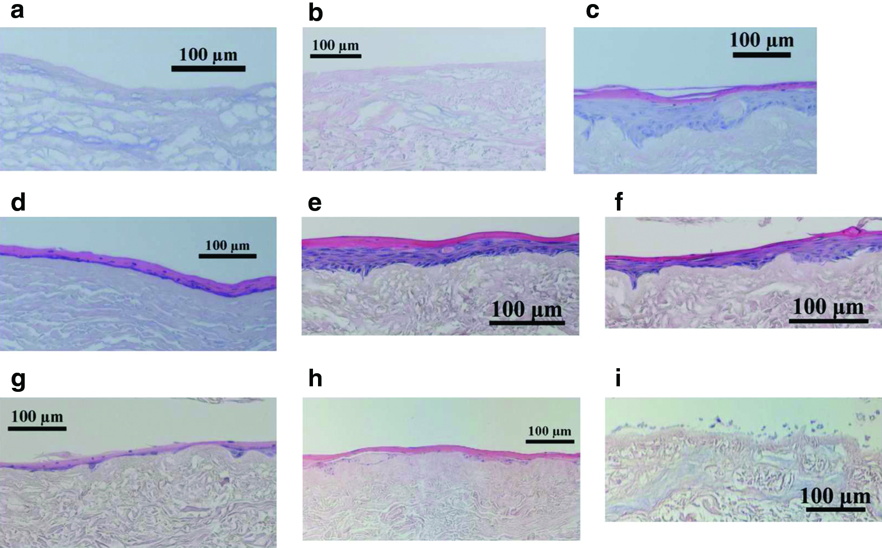
Images of cross-sections of specimens:
Raman spectroscopy
Raman spectra were obtained using a locally constructed Raman microscope. The system (previously described 26 ) is built around an epifluorescence microscope (Nikon E600). A line-focused 785-nm laser (Invictus; Kaiser Optical Systems, Inc.), focused through a 4X/0.2 NA objective, generated laser power of 120–130 mW at the specimen. The signal was collected through the same objective, transmitted toward, and focused into an imaging spectrograph (HoloSpec; Kaiser Optical Systems, Inc.). Raman spectra were collected through 25-μm and 50-μm slits, yielding an approximate spectral resolution of 4 and 8 cm−1, respectively. The spectra were recorded by a 1024×256 pixel deep-depletion charge-coupled device detector (Model DU 401-BR-DD; Andor Technology). The wavelength-dependent response of the detector and the wavelength scale of the spectrograph were calibrated using a HoloSpec Calibration Accessory (Kaiser Optical Systems, Inc.). Each spectrum was corrected for spectrograph image curvature, white-light corrected, dark-current subtracted, and baseline adjusted by a modified polyfit method. 27
In this study, we used a 4×/0.2 NA objective, which was intended to approximate the depth penetration of 0.22 NA multimode optical fibers that are commonly used in fiber-optic Raman probes. This was done in preparation to employing this technique to monitor the incorporation of EVPOMEs in situ into mice and eventually into human subjects.
Data collection
Measurements were made on multiple EVPOMEs and AlloDerm control batches (each batch corresponding to a single donor), with specimens from each batch studied on the same day under the same conditions. For stressed-versus-nonstressed experiments, the specimens from each batch included stressed (either thermal or Ca2+) and nonstressed EVPOMEs and AlloDerm controls. For rapamycin experiments, the batches included rapamycin-treated EVPOMEs and untreated EVPOME controls. EVPOME and AlloDerm specimens were mounted on sapphire flats. Spectral transects (∼100 μm in length) were collected at multiple locations on each specimen and averaged to give a single spectrum for the specimen. Each batch contained a total of 20–30 specimens of EVPOMEs and AlloDerm controls.
Data analysis
Raman bands were fitted by the Levenberg–Marquardt algorithm. Singular value decomposition (SVD) analysis 28 and fits were performed in Labview. T-tests were performed to evaluate the ability of Raman metrics to discriminate between the stressed and nonstressed status. Differences were considered significant at p<0.05. A threshold value was determined for the most significant Raman metrics to obtain better separation between stressed and nonstressed samples or between damaged and intact samples. Sensitivity and specificity were calculated according to a standard formula.
Results
In this study, both EVPOMEs and AlloDerm substrates without cells (controls) were subjected to two kinds of stress: (1) thermal by keeping the specimens at 43°C overnight on day 9, which induced temperature damage on cell growth and EVPOME development and (2) calcium by exposing specimens to various higher-than-normal concentrations of Ca2+ ions, which altered physiological development of the cells.
Thermal stress
The average Raman spectra of thermally stressed and nonstressed EVPOME and AlloDerm (collected from specimens shown in Fig. 1) are shown in Figure 2. Four major bands are seen: phenylalanine ring breathing mode at 1004 cm−1; amide III envelope, maximum at 1250 cm−1; CH2 deformation at 1450 cm−1; and amide I envelope, maximum at 1650 cm−1. The phenylalanine band is sharp and contains only one band, allowing for a relatively straightforward band fitting (see inset). The structure of the CH2 deformation band is more complex, and both amide I and amide III envelopes contain multiple overlapping unresolved bands. Since all of the spectra in Figure 2 were collected under similar conditions, the intensities of the bands could be compared without normalizing.

Raman spectra of thermally stressed and nonstressed EVPOMEs and AlloDerm. Phenylalanine at 1004 cm−1, broad amide III band at 1250 cm−1, CH2 at 1450 cm−1, and amide I at 1650 cm−1 bands are clearly visible. The inset shows the phenylalanine band. Color images available online at
The CH2/phenylalanine band ratios for thermally stressed and nonstressed EVPOMEs for three batches are shown in Figure 3a. We observed batch effects, which may be caused by factors such as variable thickness of AlloDerm substrates and different cellular layer quality because of small variations in execution of the production protocol and patient-specific variability in cell activity, as primary cell strains were used. While the individual CH2/phenylalanine band ratios were not the same for each batch, the ratio of stressed to nonstressed EVPOMEs for the CH2/phenylalanine band ratios was consistent. CH2/phenylalanine band ratios were always about 20% greater for thermally stressed than for nonstressed EVPOMEs (p=0.045).
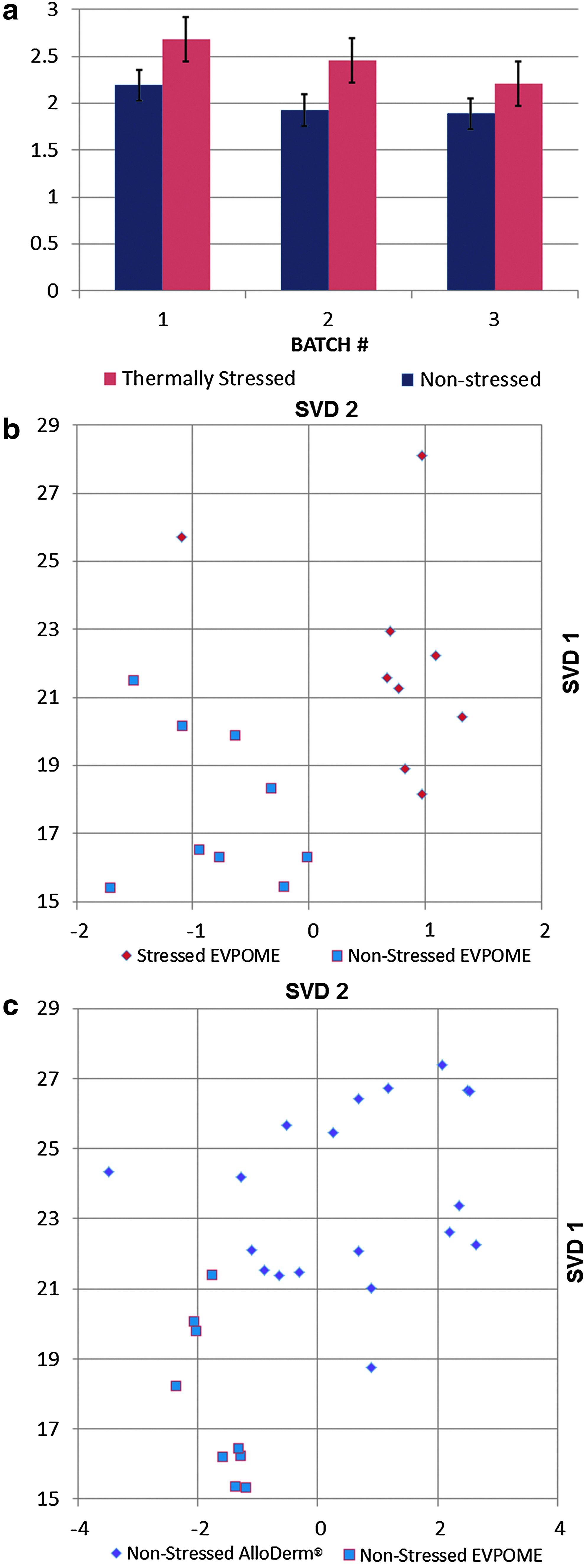
Thermal stressing of EVPOME.
Shape changes in the amide bands also occurred as a result of thermal stressing. Such changes along with band intensity information can be visualized using multivariate techniques. We have found that SVD applied to our data (see Fig. 3b) showed clear separation between stressed and nonstressed EVPOMEs. Furthermore, the SVD principal component plot comparing AlloDerm and EVPOMEs (Fig. 3c) distinguished between areas of EVPOME where the cell layer is severely disrupted and where it is intact.
Calcium stress
Raman spectra of EVPOMEs exposed to four different concentrations of Ca2+ ions are shown in Figure 4. The lowest concentration is considered physiologically normal, and thus the construct is assumed to be nonstressed. The Raman spectra were normalized to the phenylalanine band to help visualize the CH2/phenylalanine band intensity ratios for different concentrations. Major differences were seen in the amide I and amide III regions, especially for higher Ca2+ ion concentrations.
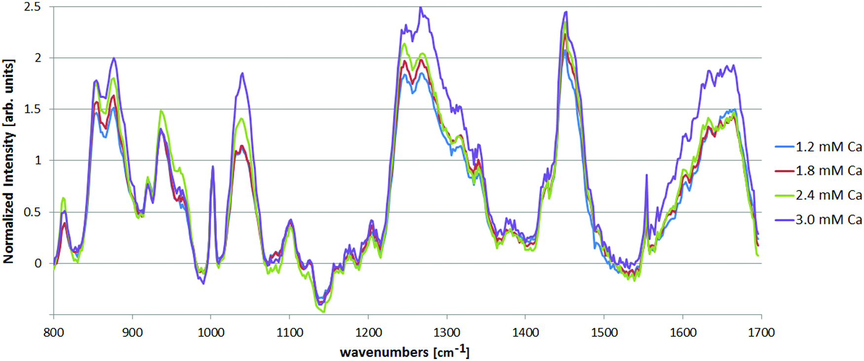
Normalized Raman spectra of EVPOMEs stressed by various concentrations of calcium ions. The spectra are normalized to phenylalanine for consistency and to allow fitting to a well-defined band. Color images available online at
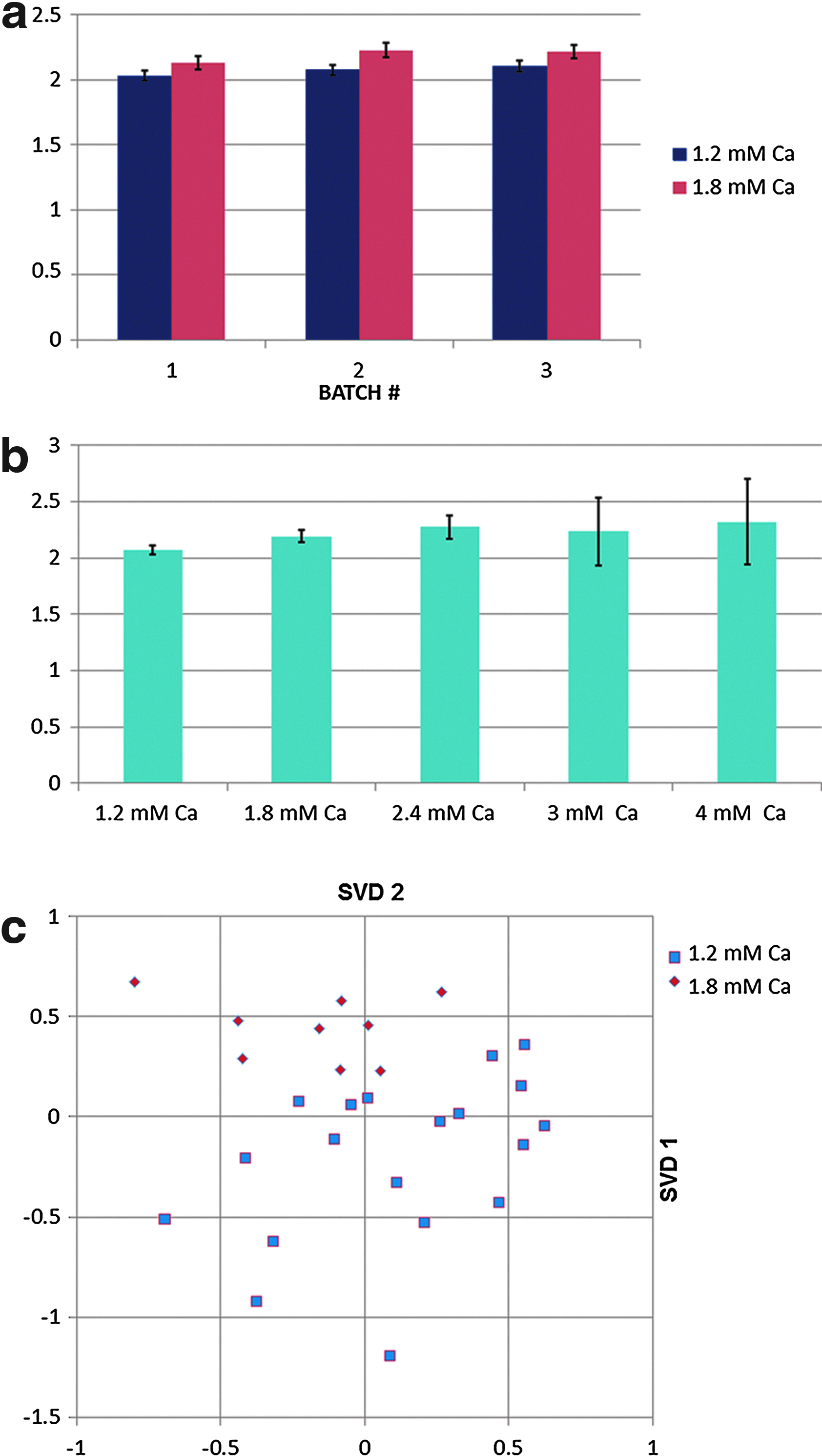
Batch effects were again observed (Fig. 5a). Figure 5b shows the CH2/phenylalanine band ratios for different concentrations averaged across batches. The ratio was always higher for stressed than for nonstressed specimens (p=0.03). At higher concentrations, the variation was greater (as indicated by larger error bars). The ratios of CH2 to phenylalanine bands were consistently higher for Ca2+-stressed specimens, although the difference between the stressed and the nonstressed (5%–7%) was not as large as in the case of thermal stressing (see Fig. 3a). Finally, Figure 5c shows that the results of SVD analysis showed separation of Ca2+-stressed and nonstressed EVPOME spectra.
Ca2+ ion stressing. The ratio of CH2 to phenylalanine band heights for
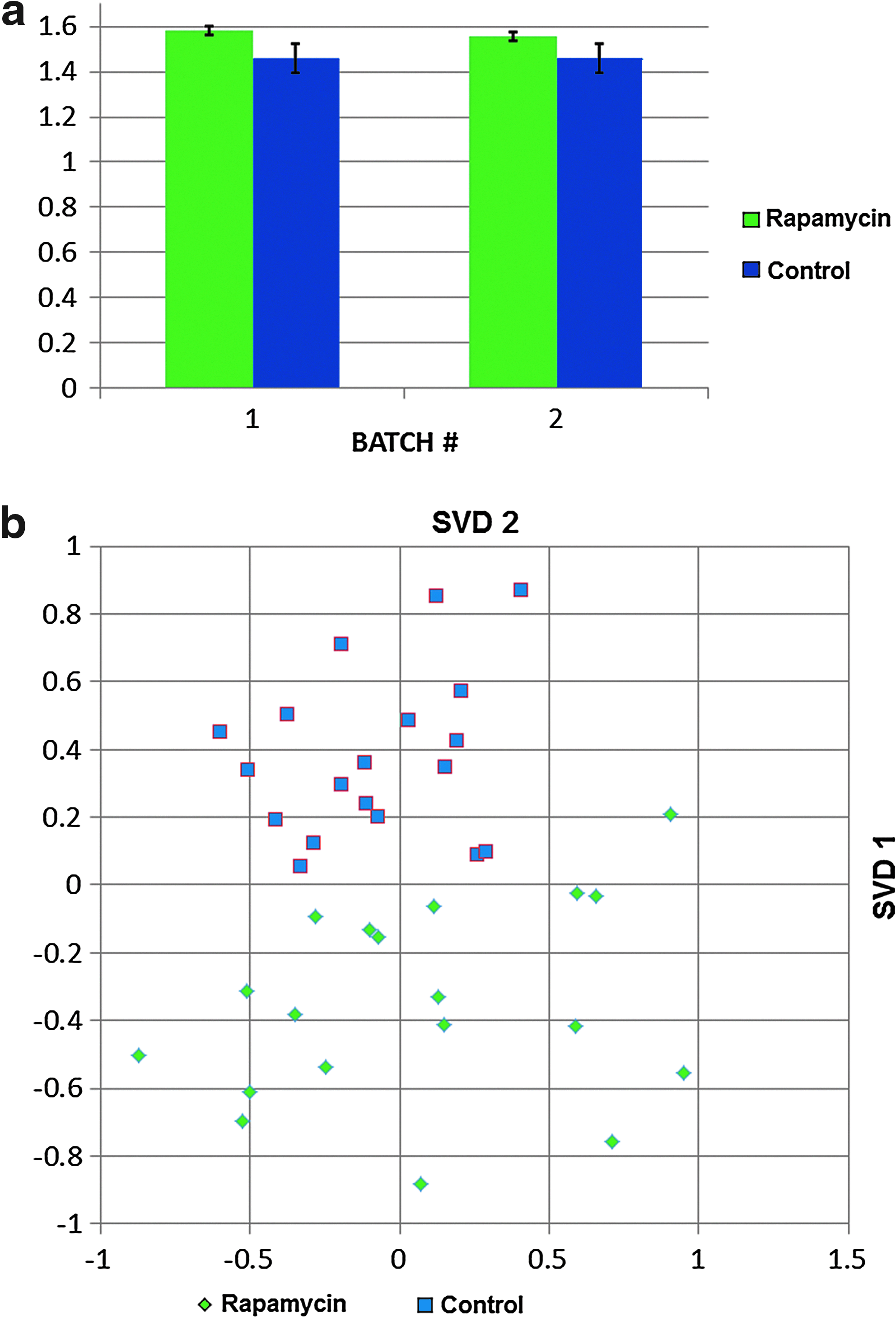
Rapamycin treatment
We also investigated Raman spectral changes resulting from pretreatment of the human keratinocytes with rapamycin. The averaged Raman spectrum of EVPOMEs pretreated with 2 nM of rapamycin was compared to the spectrum of control constructs without rapamycin pretreatment is shown in Figure 6. The ratio of the amide III band height to phenylalanine band height was always higher (p=0.02) for rapamycin-treated constructs than for controls (Fig. 7a), while the clearest separation was achieved via SVD analysis (Fig. 7b).

Normalized Raman spectra of EVPOME exposed to rapamycin (2 nM) versus control. Color images available online at
Histology and glucose consumption
In this study, the stress applied to each group of constructs was known in advance. In the general case, where constructs are manufactured for direct implantation, this information is not available, and the EVPOMEs are classified by histological examination of the H&E-stained cell layer. Figure 8a shows Ca2+- and thermally stressed specimens classified by the histological quality of the cellular layer. The specimens were classified as damaged or intact based on images of H&E-stained specimens, without knowledge of whether the specimens were stressed or nonstressed. Figure 8b shows the CH2/phenylalanine band height ratio for discriminating specimens pooled together across batches for damaged and intact cellular layer (classified based on H&E-stained images). Using the CH2/phenylalanine band height ratio and setting the threshold to 2.1 (the value that maximizes the sensitivity and specificity), intact and damaged specimens were classified with 82% sensitivity and 76% specificity. In comparison, plotting the CH2/phenylalanine band height ratio as a function of stress for both heat and calcium stressing (1.8 mM or higher Ca2+ concentration) and setting the threshold to 2.1 (see Fig. 8c), we obtained 75% sensitivity and 86% specificity for heat-stressed and 77% sensitivity and 63% specificity for Ca2+-stressed specimens.

Stress data for all EVPOME specimens:
Further, glucose consumption of cells is often used as a measure of overall viability, with low glucose consumption indicating low viability of cells. Figure 8d shows glucose consumption as a function of stress, indicating that the glucose consumption was poorly correlated with stress-induced cell damage for both thermal and Ca2+ stressing.
Discussion
It is clear that thermal stress caused structural changes in both EVPOMEs and AlloDerm. Thermally induced protein denaturing, which changes protein secondary structure, was visible in both amide bands. However, the intensity ratio of the CH2 deformation band to phenylalanine band was much easier to measure consistently, and is therefore proposed as a metric to establish whether or not a particular specimen had been exposed to stress.
Our analysis indicated that the CH2-to-phenylalanine band height ratio always increased with stress for both thermal and excess Ca2+ stress. The ratio consistently increased more with thermal stressing than with Ca2+ stressing. This finding was supported by histology, which indicated greater damage as a result of thermal stressing than Ca2+ stressing. The CH2-to-phenylalanine band height ratio tended to increase with the higher Ca2+ concentrations. By using this ratio alone, or together with the information from the other Raman bands, we can determine which constructs are most viable. Such EVPOMEs can then be used for implantation, reducing the rate of rejection due to the construct nonviability.
Further, by comparing the Raman spectra of EVPOMEs and acellular AlloDerm substrates, we were able to distinguish between the areas of EVPOMEs where the cell layer was severely disrupted. The disruption of a cell layer was at least partially responsible for the difference between stressed and nonstressed constructs.
We previously observed 21 that the intensity of the phenylalanine band decreased as a result of thermal stress, which was likely also caused by disruption of the keratin layer. The intensity of the phenylalanine band was only slightly different between stressed and nonstressed AlloDerm, a finding consistent with previously reported measurements. 20
The results of SVD analysis confirmed differences between stressed and nonstressed EVPOME spectra. As expected, the separation between Ca2+ stressed and nonstressed spectra was not as well defined as in the case of thermal stress, but in both cases, the stressed/nonstressed regions were clearly identifiable, confirming that additional information is contained in amide I, amide III, and other bands.
Raman spectral changes resulting from pretreatment of the human keratinocytes with rapamycin were also observed. In that case, the ratio of the amide III band height to phenylalanine band height was always higher for rapamycin-treated EVPOMEs, than for 1.2 mM Ca2+ controls.
It is unknown if our calcium ion or thermal stress protocols always result in some damage to the specimens. Either the EVPOMEs are not damaged despite being subjected to stress, or the damage is always present, but is not visible by histology. While the classification of EVPOMEs has been traditionally done by histological assessment or by measuring glucose consumption, Raman spectroscopy may provide an important noninvasive tool capable of providing complementary information to assess EVPOME cell viability.
Rapamycin treatment remains a special case because of the multiplicity of effects possible. While we have shown that Raman spectroscopy can distinguish rapamycin-treated from untreated specimens, we do not have enough information to propose specific mechanisms for the rapamycin-induced structural changes.
Conclusions
We have developed Raman spectroscopic criteria to noninvasively determine during the manufacturing process the viability of cells within the EVPOMEs. Band intensity ratios in well-defined bands can be used by themselves, or in the combination with shape changes in envelopes of overlapping bands. The clearest distinctions between stressed/nonstressed and rapamycin-treated EVPOMEs are obtained by SVD, which incorporates the greatest amount of information from their spectra.
We have also investigated the spectral changes resulting from the pretreatment of EVPOMEs with rapamycin and observed noticeable changes in the Raman spectra. We have determined the Raman metric that changes as a result of rapamycin pretreatment and show that with SVD, a clear difference between pretreated and control specimens is observable.
Footnotes
Acknowledgment
This work is supported in part by an NIH Grant R01 DE019431-01.
Disclosure Statement
No competing financial interests exist.