




Abstract
The three-dimensional culture of blood vessel wall cells now permits the construction of a human blood vessel mimic (BVM). Previous studies have used the human BVM as a tool to perform in vitro testing of medical devices and imaging instrumentation. The purpose of the current study was to enhance this technology through both the elimination of animal serum and the modification of scaffold properties in human BVM preparation. Additionally, BVMs were implanted with vascular stents to observe a potential cellular response to the devices in a serum-free environment. Serum-free culture of human adipose-derived stromal vascular fraction (SVF) cells was accomplished through sequential adaptation from a serum-supplemented medium. The adipose-derived SVF serves as a source of both human endothelium and human smooth muscle cells. Utilizing established pressure-sodding technologies, these cells were incorporated into the luminal surface of either expanded polytetrafluoroethylene (ePTFE) tubular scaffolds or electrospun poly(
Introduction
Recent work has shown the application of endothelial cell isolation, cellular interaction with biomaterial surfaces, bioreactor technology, and vascular tissue engineering toward the development and utilization of a three-dimensional (3D) in vitro blood vessel mimic (BVM).6–10 The BVM is based upon extensive studies evaluating the use of adipose-derived regenerative cells to create tissue-engineered vascular grafts for human therapeutic use.11–13 The BVM represents an in vitro tissue-engineered graft wherein cell-treated grafts are placed in a perfusion bioreactor to cultivate a cellular lining on the luminal surface. The purpose of the BVM is not for implantation, but rather to serve as a physiologically realistic, preclinical testing environment for evaluating and screening intravascular devices and for studying additional technologies such as vascular imaging modalities and drug delivery systems. The BVM system provides a more relevant overall cellular orientation and flow condition than common cell culture techniques and has the potential to provide cost–effective, efficient test results.
The BVM has previously been developed and applied utilizing animal serum-supplemented culture techniques. The use of animal serum for in vitro experimentation is associated with significant biological drawbacks, including poorly defined medium formulations and culture conditions, lot-to-lot composition variability, and risk of contamination.14–17 For this study, it was hypothesized that human BVMs can be engineered using SVF cells, expanded polytetrafluoroethylene (ePTFE) vascular grafts, and serum-free culture conditions. Additionally, to evaluate the effects that an alternative scaffold has on the development of serum-free BVMs, polymer scaffolds were created via electrospinning, a technique that allows for efficient, in-house production of materials that differ from ePTFE in composition and microstructure. Finally, BVMs were implanted with vascular stents to test the hypothesis that the cells have the ability to interact with intravascular devices in a serum-free environment. Converting the current BVM system to one that excludes serum is a step toward obtaining a total human model that can provide academic and industrial investigators the opportunity to perform controlled, consistent assessments of emerging stent designs via an in vitro tissue mimic rather than proceeding directly to animal studies.
Materials and Methods
Cells, reagents, and materials
Human SVF cells were isolated from adipose tissue as described in a following section, and HaCaT cells were received from Dr. Norbert Fusenig (German Cancer Research Center, Heidelberg, Germany). Collagenase was obtained from Worthington Biochemical Corp. (Lakewood, NJ), and bovine serum albumin (BSA) was purchased from Sigma-Aldrich (St. Louis, MO). Fetal bovine serum (FBS) was purchased from Gemini Bio-Products (West Sacramento, CA). Products acquired from Life Technologies (Carlsbad, CA) include Medium 199, Dulbecco's modified Eagle's medium (DMEM), HEPES buffer, and
Serum-free culture of human SVF cells
Human SVF cells were isolated from liposuction-derived adipose tissue.12,18,19 Human lipoaspirate was obtained from patients according to an approved University of Louisville IRB protocol. To isolate SVF cells, adipose tissue samples were digested at 37°C via application of animal-origin free collagenase (6 mg/mL in Dulbecco's divalent cation-free phosphate-buffered saline [DCF-PBS] containing 0.1% BSA), which was applied at a volume equal to the volume of adipose. The resulting suspension was centrifuged at 400 g for 4 min to separate stromal and vascular cells from the collagenase mixture and buoyant adipocytes. The SVF cell pellet was retrieved, washed in DCF-PBS, and resuspended in a complete medium. Primary cultures were initiated in 1% gelatin-coated flasks and washed to remove nonadherent cells. The adherent cell population was grown to confluence using a complete medium consisting of Medium 199, 20% FBS, 5 mM HEPES buffer, 2 mM
Preparation of surface modification medium
A medium conditioned by cultured HaCaT cells, a continuous human keratinocyte cell line, was utilized to modify the surfaces of the tubular scaffolds to augment cellular attachment. HaCaT cells release extracellular matrix proteins, including laminin-5, into the growth medium during culture, and previous research has shown that ePTFE modified with the protein-enriched medium promotes adherence and spreading of adipose-derived endothelial cells.22,23 Using similar techniques applied during SVF cell culture adaptation experiments, HaCaT cells were sequentially transitioned from the serum-supplemented medium consisting of high-glucose DMEM, 10% FBS, 5 mM HEPES buffer, and 2 mM
Preparation of BVM scaffolds
Expanded polytetrafluoroethylene
A commercially available ePTFE vascular graft was used as a cellular scaffold. The 4-mm I.D. grafts were cut into 4.5-cm lengths, mounted on barbed fittings, and autoclaved. The grafts were then denucleated with ethanol to remove air trapped in the interstices of the ePTFE, thus increasing the surface area for protein adsorption and subsequent cellular attachment. 24
Electrospun polymer scaffolds
PLCL, a polymer that has been mechanically evaluated previously for use in vascular grafts, 25 was utilized at an 80:20 (L-lactide:caprolactone) blend. The polymer was added to chloroform at 120 mg/mL and electrospun onto a rotating and transversing 4-mm O.D. mandrel. A syringe pump dispensing at a rate of 15 mL/h and a high-voltage power supply set to 20 kV were used in the electrospinning process. A total volume of 3 mL was dispensed per scaffold. After electrospinning, the scaffolds were cut into 4-cm lengths, mounted on barbed fittings, disinfected in 70% ethanol, and rinsed in sterile DCF-PBS.
BVM development in serum-free conditions
Prepared scaffolds were aseptically mounted into bioreactor chambers. In addition to the vessel chamber, which features a luminal inlet, a luminal outlet, and an extraluminal outlet, the bioreactor system consisted of a flow rate-controlled peristaltic pump (Watson-Marlow, Wilmington, MA) and a medium reservoir. Surface modification of scaffolds, using the HaCaT-conditioned medium described previously, was accomplished by circulating the medium throughout the bioreactor system and into the pores of the scaffold material. After surface modification, SVF cells, which had been transitioned to a serum-free medium, were harvested from culture flasks via application of TrypLE Express. Cells were pressure-sodded12,26 onto the inner surfaces of the scaffold by occluding the luminal flow path and injecting the cell suspension through a stopcock port located proximal to the vessel chamber. Pressure sodding forces the liquid part of the cell suspension through the pores of the scaffold material, while the cells themselves are filtered onto the scaffold surfaces. When cell sodding was complete, transmural flow of the perfusion medium was initiated at a low rate to allow for cellular attachment. The luminal path was then unclamped, and luminal flow was gradually increased until a flow rate of 15–22 mL/min was achieved. The BVMs were cultivated in this manner for 14–21 days at 37°C and 5% CO2, with the perfusion medium replaced regularly. The perfusion medium consisted of a basal Human Endothelial-SFM supplemented with Fungizone (29.5 ng/mL), penicillin (4.5 units/mL), and streptomycin (4.5 μg/mL).
Stent implantation
At the conclusion of 14 days of bioreactor cultivation, a group of electrospun BVMs was utilized for intravascular device implantation. Bare metal stents (4-mm expanded diameter) composed of a cobalt chromium alloy were deployed in the BVMs by inserting a sterile, stent-loaded angioplasty balloon catheter into the bioreactor system proximal to the vessel chamber and inflating the balloon to 5 atm for ∼30 s. To verify proper deployment of the devices, intravascular ultrasound imaging (Volcano S5 Imaging System, Volcano Eagle Eye Gold phased array coronary artery catheter; Volcano Corp., San Diego, CA) was used both before and after stent implantation. The BVMs were then flow conditioned with the serum-free medium for an added 7 days to allow for a cellular response to the devices. An additional electrospun BVM, which was designated as a control and did not receive a device, was flow conditioned for a total of 21 days.
Histology and immunohistochemistry
After bioreactor conditioning, nonstented BVMs were fixed under flow with either 10% neutral buffered formalin or HistoChoice, cut longitudinally and radially, paraffin embedded, and sectioned. Hematoxylin and eosin (H&E) staining was utilized to examine the basic tissue structure. Immunoperoxidase staining (hematoxylin counterstain) using antibodies against von Willebrand factor (vWF), α-smooth muscle actin, and vimentin was applied to detect the presence of endothelial cells, smooth muscle cells, and mesenchymal cells, respectively. Human aorta samples served as cell identification controls.
Scanning electron microscopy and 3D anaglyphs
High-magnification, high-resolution imaging of ePTFE, electrospun PLCL, and both stented and nonstented BVMs was performed using scanning electron microscopy (SEM, JSM-820 scanning electron microscope; JEOL Ltd., Tokyo, Japan). Before imaging, ePTFE and electrospun material were sputter-coated with a gold target. BVMs were treated with 3% glutaraldehyde fixative, cut longitudinally, and processed via dehydration, critical point drying, and sputter coating.
Two scanning electron micrographs were captured for both ePTFE and PLCL scaffolds. The second image was acquired at a 7° offset from the first image. The digital images were imported into Adobe Photoshop CS3, and 3D anaglyphs (red-cyan) of each material were constructed by combining the image pairs after assignment of the red channel to one image and the blue and green channels to the corresponding image.
Fluorescent nuclear staining
Electrospun BVMs implanted with stents were perfusion-fixed with 3% glutaraldehyde, cut longitudinally, and stained with nuclear-specific bisbenzimide (BBI) to determine whether or not cells from the BVM had interacted with the stents. Epifluorescence microscopy was used to visualize the nuclei of cells on stent surfaces.
Results
Serum-free cell culture
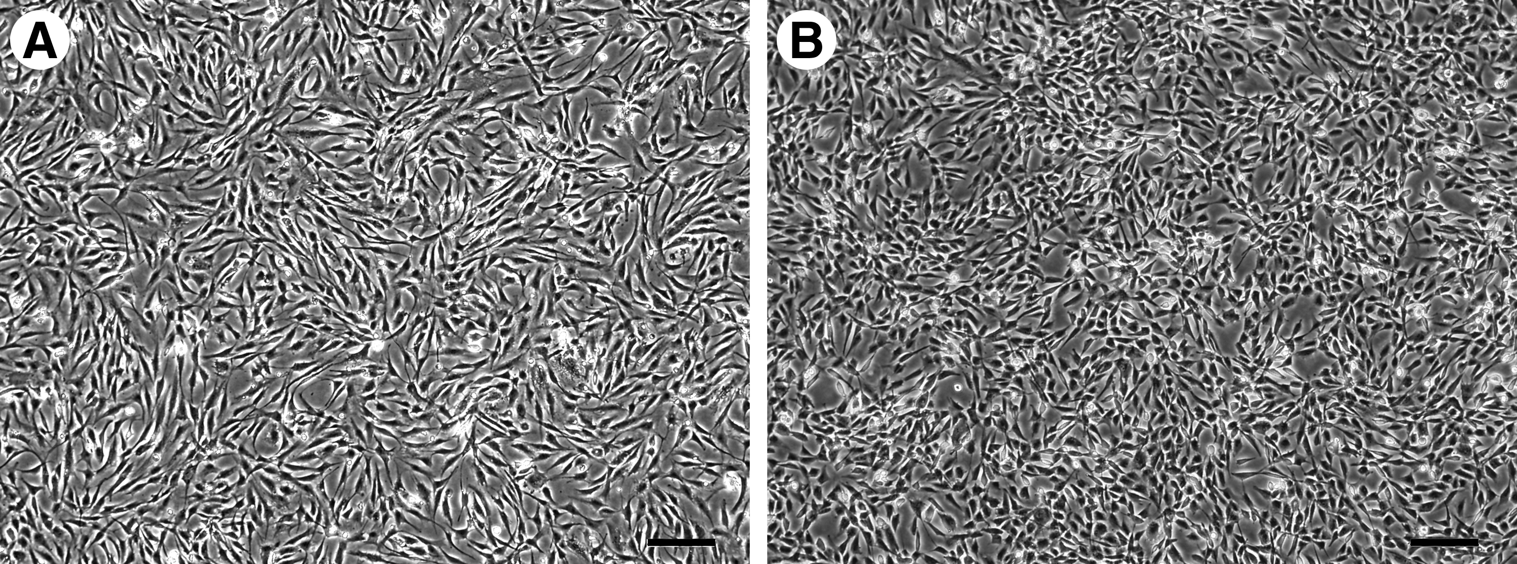
SVF cells sequentially adapted to the serum-free culture medium were examined regularly with an inverted phase-contrast microscope. In addition to cellular attachment, increasing cell density was observed throughout the conversion process, indicating that proliferation continued to occur under the changing culture conditions. Although slight morphological differences were present, SVF cells sustained in a 100% serum-free medium exhibited similar morphologies overall to those of SVF cells grown in the serum-supplemented medium (Fig. 1). Some regions of cellular aggregation were present in serum-free cultures. Morphological variations and cellular clumping could be attributed to the elimination of serum components, the addition of new components in the serum-free medium, the replacement of gelatin with human plasma fibronectin in augmenting cellular attachment, or a combination of these factors.
Phase-contrast micrographs of stromal vascular fraction cells cultured in
Histological and immunohistochemical evaluations
BVMs developed in the perfusion-based bioreactor system diagrammed in Figure 2 were analyzed using histological and immunohistochemical techniques.

Diagram of the blood vessel mimic (BVM) bioreactor system.
Expanded polytetrafluoroethylene BVM
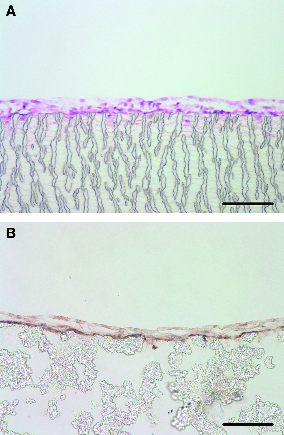
Examination of H&E-stained ePTFE BVM samples with bright-field microscopy verified a lining of intimal tissue within the construct (Fig. 3A). Both longitudinal and radial cross sections were used to study the general morphology of the lining, which consisted of multiple cellular layers. Tissue components showed very little penetration into the wall of the ePTFE scaffold. Immunohistochemical analysis of ePTFE BVM samples was performed to identify cellular phenotypes. Staining results revealed vimentin-positive cells throughout the tissue lining, indicating that cells of mesenchymal origin constitute the intimal layer within the BVM (Fig. 3B). Neither vWF nor α-smooth muscle actin antigens were detected.
Electrospun PLCL BVM
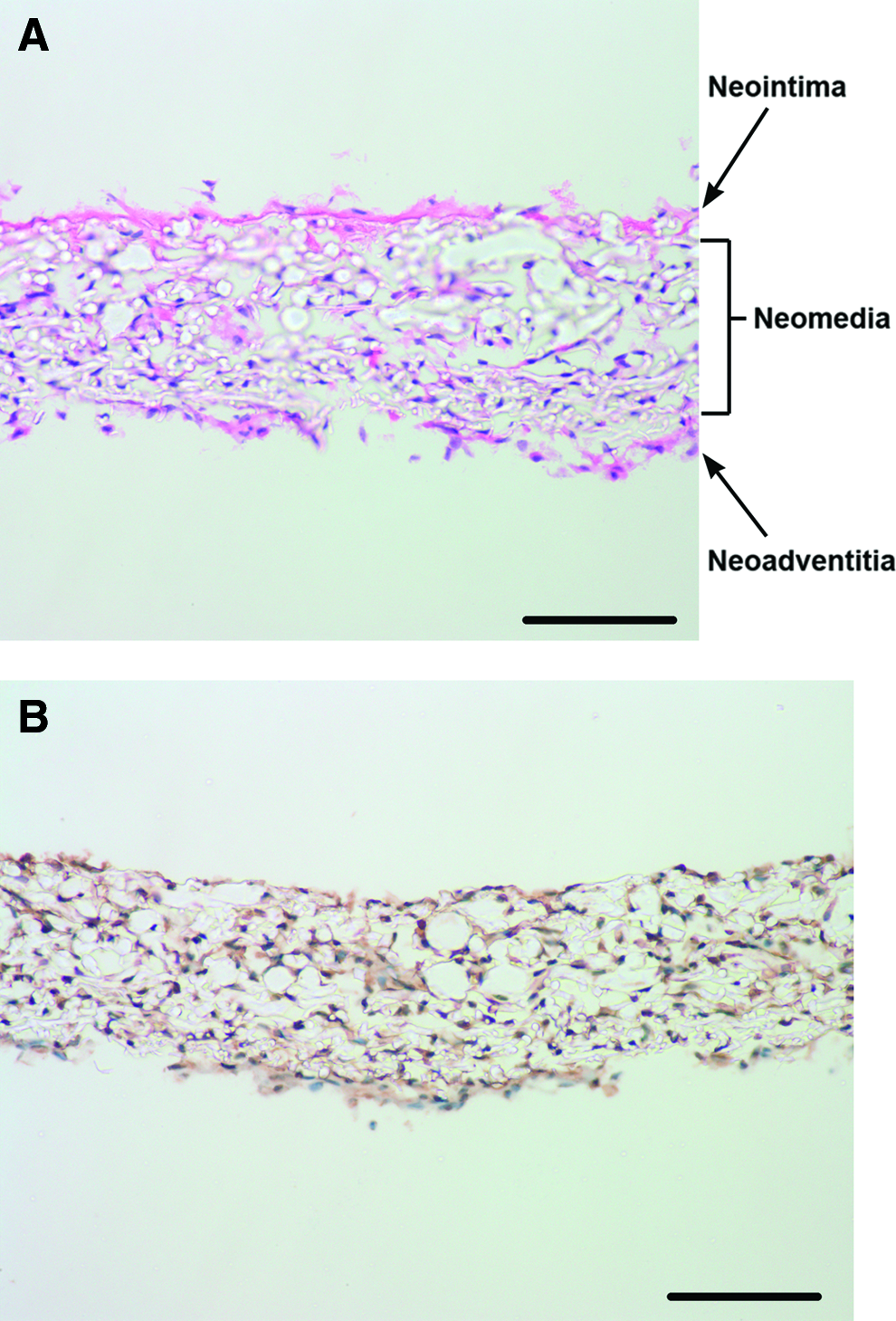
H&E staining of the nonstented electrospun BVM showed distinct structural differences from the ePTFE BVMs. Observation of longitudinal and radial cross sections revealed tissue present on the luminal surface (neointima), but also cellular infiltration throughout the walls of the polymer scaffold (neomedium) and tissue at the abluminal surface (neoadventitia) (Fig. 4A). The neointima consisted of monolayers of cells, as well as some localized regions of multi-layered cells. Immunohistochemistry was performed to characterize the cells within the electrospun BVM. Vimentin-positive mesenchymal cells were identified throughout all regions of the tissue-engineered construct (Fig. 4B). As was the case with the ePTFE BVMs, both vWF and α-smooth muscle actin stains were negative.
SEM and 3D anaglyphs
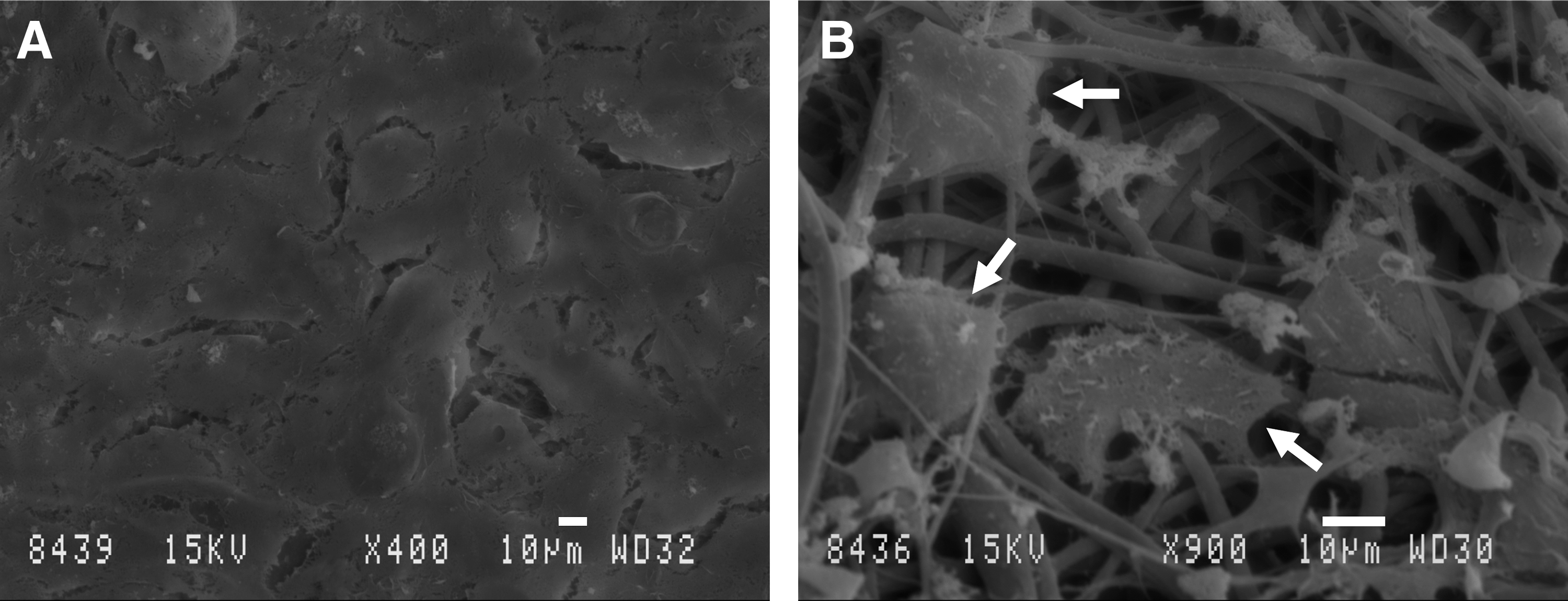
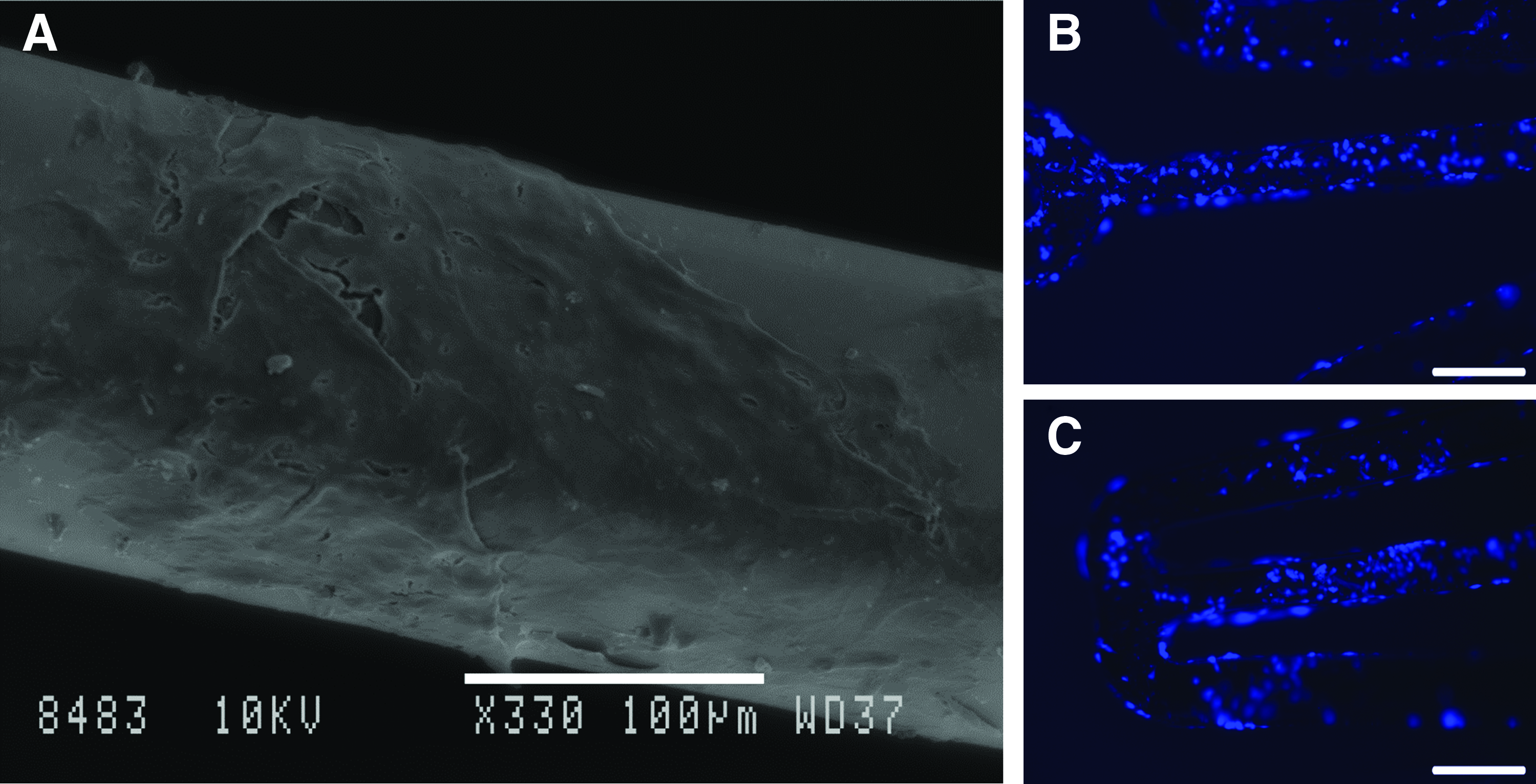
Analysis of the luminal and abluminal surfaces of the nonstented electrospun BVM via SEM verified the presence of cells at each location (Fig. 5A, B). The luminal cells of the BVM did not exhibit a specific alignment, such as the shear stress-induced alignment typical of the endothelium in blood vessels, but rather showed cobblestone morphologies. SEM was also utilized to compare ePTFE and electrospun PLCL surfaces (Fig. 6A, C). The porous characteristic of each material, which allowed for cells and scaffold to be combined via pressure sodding, is evident. Three-dimensional anaglyphs (Fig. 6B, D) generated from SEM images allow for a more informative visualization with respect to potential cellularization of the scaffolds. These 3D reconstructions detail the microstructural differences between the materials, specifically the solid nodes interconnected by thin fibers observed in ePTFE versus the nonwoven fiber mat produced by electrospinning. Finally, SEM imaging allowed for high-magnification visualization of the surfaces of stents that had been deployed in electrospun BVMs. Images revealed that while there were areas of stents that lacked cells, there were also regions of tissue growth over luminal and side surfaces of the devices (Fig. 7A). These results demonstrate that serum in not required for BVM cells to interact with implanted stents in a manner that may be characteristic of in vivo cellular healing activity.
In a serum-free electrospun BVM, scanning electron microscopy demonstrates

Two-dimensional scanning electron micrographs show
Fluorescent nuclear staining
Electrospun BVMs deployed with bare metal stents were stained with BBI, and en face images were acquired utilizing epifluorescence techniques. This type of analysis allowed for detection of cellular nuclei on stent surfaces. Results from this assessment confirmed the outcomes of SEM imaging. Cells were present along the luminal surfaces of the stent struts, indicating that a biological response to device implantation had begun in the BVMs (Fig. 7B, C).
Discussion
Preclinical testing of cardiovascular devices, imaging modalities, and therapeutics has been accomplished predominantly through the use of animal models. Although animal models are useful in that they provide a physiological environment that offers insight into biological processes and in vivo tissue responses to new devices, they are also associated with factors such as high costs, time consumption, and animal-to-human variability. Previous studies6,7 have supported the concept of employing BVMs as in vitro evaluation environments for intravascular devices such as stents. Incorporation of serum-free technologies into BVM development and utilization to eliminate the undesirable characteristics of animal serum and to create a more humanized model may lead to enhanced device evaluation and screening capabilities. This work has shown the transition of human SVF cells from serum-supplemented culture conditions to serum-free conditions and the subsequent bioreactor cultivation of a human cellular lining within a vascular graft, differences in development and structure of a serum-free BVM when incorporating SVF cells within a scaffold composed of electrospun fibers, and a human tissue response to implanted vascular stents in a serum-free environment.
The source of cells used for serum-free conversion and BVM construction was the SVF, which was isolated from human adipose tissue. The SVF is a desirable source of cells for creating BVMs, as it is readily available and consists of cell types such as microvessel endothelial cells, smooth muscle cells, and fibroblasts. The SVF cells showed viability and the capability to proliferate throughout adaptation to the serum-free medium, allowing the cells to be expanded to population sizes appropriate for sodding ePTFE vascular graft scaffolds. This study shows that human tissue-engineered tubular constructs can be created using serum-free methods, resulting in a 3D, biological environment that can be implanted with intravascular devices; however, it is limited in that endothelial cells were absent from the constructs. Luminal tissue within the grafts did not exhibit characteristics of functional endothelium, such as vWF expression, and consisted only of vimentin-positive undifferentiated mesenchymal cells or fibroblasts. In previous work, 6 utilization of cultured SVF cells, a similar bioreactor system, a 15 mL/min flow rate, and FBS-supplemented medium formulations resulted in BVMs that consisted of an intimal layer of endothelial cells within ePTFE vascular grafts, which supports the use of the BVM in modeling endothelialization. This suggests that incorporation of serum-free culture conditions into BVM development methods was specifically inadequate for the SVF cell population to maintain or to acquire endothelial phenotypes. SVF cells exhibit a high degree of plasticity 27 and could be more susceptible to dedifferentiation in the absence of serum. It is this plasticity however that allows this unique BVM to be viewed as a 3D system that could possibly be used to analyze the transition of mesenchymal cells from a less-differentiated state into endothelium and smooth muscle. Modifying the composition of the serum-free culture medium may be a viable option for directing SVF cell differentiation toward vascular phenotypes. Previous studies have shown the ability to induce the conversion adipose-derived cells into endothelial cells and smooth muscle cells based on the formulation of an in vitro culture medium.28,29 Additionally, adjusting fluid dynamic parameters of the bioreactor system to more closely resemble those of the native circulation is a potential approach to stimulating cell differentiation, as the current system was limited by subphysiological flow rates and pressure.
While the culture medium and bioreactor conditions are potential variables in altering serum-free BVM development, the current study addresses the utilization of a different type of scaffold. It was hypothesized that implementing a scaffold that differs in the microstructure from ePTFE may lead to a more physiological model of a blood vessel. The electrospinning method of scaffold production was selected given its prior use in generating porous fiber networks that show similarities to the protein fibers of the native extracellular matrix.30,31 As depicted by 3D SEM imaging, electrospinning PLCL resulted in tubular scaffolds of nonwoven fibers that provided an alternative physical environment for cellularization to that of ePTFE vascular grafts. Cells were able to infiltrate the electrospun scaffold wall in addition to forming a neointima. For subsequent research, specific scaffold properties such as porosity, wall thickness, and diffusion distance may be examined to identify the specific variables that facilitate such cellular invasion. Although the electrospun scaffold did not incite specific vascular phenotypes, cellular presence throughout the scaffold wall and abluminal surfaces has not been observed in BVMs created with ePTFE grafts and is evident that modifying scaffold properties can lead to variations in the morphological development of serum-free BVMs. This result was the basis for selecting electrospun BVMs for stent implantation in this study. By comprising multiple cellular layers, these BVMs are more anatomically similar to native blood vessels than the serum-free ePTFE BVMs and therefore are preferable for device testing. Additionally, the ability to form tissue layers throughout the electrospun BVM creates the opportunity to utilize defined cell types, such as arterial smooth muscle cells, in engineering physiologically realistic vascular components, such as a muscular neomedium. Increasing biomechanical stimulation may be particularly effective in influencing the development of tissue within electrospun BVMs, as synthesis of compliant scaffolds 25 may allow for circumferential stress and strain when exposed to pulsatile pressure.
In addition to applying electrospun scaffolds to BVM production, this work represents the first attempt at using a BVM for device assessment under serum-free conditions. Previous studies demonstrated that BVMs created using animal serum-supplemented culture methods and ePTFE vascular grafts exhibited intimal responses to bare metal and surface-modified stents.6,7 The results obtained from both SEM and BBI analysis during the current experiments show that cells within a tissue-engineered vessel mimic constructed using an electrospun fiber mesh responded to an implanted stent in the absence of serum. These findings suggest that BVMs, which are designed to provide an in vitro, the preclinical testing environment for intravascular devices, can be developed into multilayered tissue constructs under serum-free conditions and still be utilized to model interactions between tissue and devices. Future studies may involve in vitro intravascular device assessment under serum-free conditions using BVMs consisting of defined, large-vessel endothelial cells to provide a more relevant model of device endothelialization in an artery. Additionally, various types of stents, such as drug-eluting and protein-coated stents, could be tested to determine whether or not a serum-free BVM can exhibit differential tissue responses.
In conclusion, this work demonstrated the utilization of SVF cells, serum-free culture conditions, and bioreactor perfusion in engineering BVMs that differed structurally based on the nature of the scaffold. Additionally, it was shown that BVMs consisting of a matrix of electrospun fibers and multiple tissue layers have the capacity to demonstrate a cellular response to a deployed vascular stent in a serum-free environment. These results are significant as serum-free methods may offer an opportunity to precisely control and optimize vascular device testing conditions within in vitro BVMs. The ability to obtain reproducible and interpretable data is critical if the BVM is to be utilized as a validated model in the medical device industry and regulated by Good Manufacturing Practice and Good Laboratory Practice standards. Still, the outcomes of this study are limited in that development of BVMs using serum-free conditions did not result in an endothelial cell layer, which will be a requirement for accurate preclinical assessments of stent endothelialization. Furthermore, it is possible that implementation of serum-free device testing may mask important cellular responses that are dependent on the presence of serum. Therefore, moving forward, the effects of stent implantation in serum-free BVMs must be compared to in vivo biological responses to evaluate the use of this in vitro model as a suitable device-testing alternative.
Footnotes
Acknowledgments
The authors would like to acknowledge the Alternatives Research & Development Foundation for funding the entirety of this work as part of the 2010 Alternatives Research Grant Program.
Disclosure Statement
No competing financial interests exist.