
Abstract
In cartilage, tissue engineering bioreactors can create a controlled environment to study chondrocyte behavior under mechanical stimulation or produce chondrogenic grafts of clinically relevant size. Here we present a novel bioreactor that combines mechanical stimulation with a two compartment system through which nutrients can be supplied solely by diffusion from opposite sides of a tissue-engineered construct. This design is based on the hypothesis that creating gradients of nutrients, growth factors, and growth factor antagonists can aid in the generation of zonal tissue-engineered cartilage. Computational modeling predicted that the design facilitates the creation of a biologically relevant glucose gradient. This was confirmed by quantitative glucose measurements in cartilage explants. In this system, it is not only possible to create gradients of nutrients, but also of anabolic or catabolic factors. Therefore, the bioreactor design allows control over nutrient supply and mechanical stimulation useful for in vitro generation of cartilage constructs that can be used for the resurfacing of articulated joints or as a model for studying osteoarthritis disease progression.
Introduction
Cartilage can be considered as a simple tissue, as it only contains one cell type and only two major extracellular matrix components, proteoglycans and collagen type II. However, it is complex in the sense that it is a layered anisotropic structure, which is required to absorb strong mechanical forces, distribute load, and lubricate the joint.15,16 These functions arise, in part, from the zonal organization of the tissue. Collagen fibers, responsible for absorbing and distributing the load, are orientated parallel along the synovial surface and become perpendicularly orientated with depth anchoring in the subchondral bone plate. This results in a fountain-like structure with the purpose of distributing load and retrieving the tissue's original shape. 17 Glycosaminoglycans (GAGs) are distributed inhomogeneously as the articular surface is almost void of these molecules and their concentration increases with depth. 18 Water is attracted by the negatively charged proteoglycans. Upon deformation, water is expelled from the tissue, which flows back when the tissue relaxes. Although several bioreactor studies have demonstrated the ability to stimulate sulfated glycosamoniglycan (sGAG) deposition, a native distribution of sGAGs in tissue-engineered constructs has yet to be obtained. In addition, the fountain-like structure of collagen fibers has so far not been generated in vitro. As a result, it remains a challenge to engineer cartilaginous tissue in vitro that possesses the mechanical properties of native articular cartilage.
Several bioreactors for cartilage tissue engineering have been described. In these systems, nutrients are usually supplied through perfusion, creating shear stress, or static-like culture as in a culture dish. However, articular cartilage is not subjected to an external active flow and except for the perichondrium, it does not receive nutrients from the sides. It is situated between the synovial fluid and the subchondral bone plate, which physically separates the articular cartilage from the bone marrow. It has been postulated that cartilage is supplied with nutrients from the subchondral bone and from the synovial fluid.19,20 Applying this feature to in vitro culture was described by Chang et al., and can be mimicked in transwell cultures. However, no mechanical stimulation can be applied in this system. 21 Chondrocytes in their natural environment are subjected to mechanical load. Therefore, bioreactors for cartilage tissue engineering that are equipped with a compression module more closely resemble cartilage's in vivo environment. Load can either be applied by confined, 22 sliding, 23 or rotating compression. 24 Demarteau et al. showed that under perfusion and confined compression, sGAG metabolism was increased in cell-loaded polyethylene oxide terephthalate/poly butylene terephthalate block copolymer foam scaffolds, but this design did not originate in an inhomogenous sGAG distribution as in native cartilage. 22 Kock et al. showed production of collagen II next to a homogenous distribution of sGAGs under sliding indentation, but this did not result in fountain-like orientation of the collagen network. 23 The stimulatory effect of compression on the expression of zone-specific genes was shown by Grad et al. indicating the importance of mechanical stimulation in the regeneration of functional chondrogenic constructs in vitro.25,26
Here, we describe a bioreactor design in which nutrient supply from both the synovial and subchondral side can be mimicked. This configuration will facilitate the formation of gradients of nutrients, growth factors, and growth factor antagonists through the cartilage tissue. In addition, the bioreactor is complemented with options for confined compression. We hypothesize that the ability to create gradients of relevant factors combined with confined load will aid in the generation of neo-cartilage better resembling cartilage's natural organization and mechanical properties. In this study, we introduce the basic design of a dual compartment, compression-equipped novel bioreactor that can be used to engineer cartilage or osteochondral constructs in vitro.
Materials and Methods
Bioreactor design
The dynamic (Fig. 1A) and static (Fig. 1B) bioreactors were designed in Solidworks 2009, Edition SP4.1. The stainless steel housing of the dynamic bioreactor was produced by electrical discharge machining effectively creating a construct chamber of 1 cm3 (Fig. 1A). Removable round polymethylmethacrylate windows were installed at the front and back side of the housing and allowed for placement of the construct into the chamber. Windows contained a nitrile butadiene rubber O-ring ensuring a tight seal preventing leakage and infection.
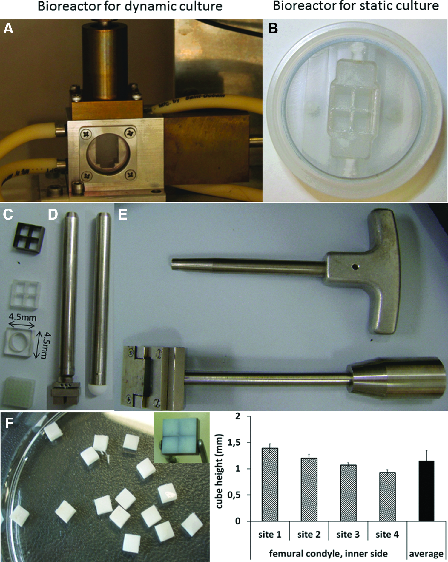
A novel bioreactor for articular cartilage explants.
The complete chamber has a height of 13 mm and a width and depth of 10 mm. Explants were placed in a 4-mm-high insert. Articular cartilage constructs were placed in a square insert (Fig. 1C). Osteochondral plugs were placed in a round insert (Fig. 1C). The inserts were placed in the reactor chamber on top of a 4.5-mm-high perforated polycarbonate plate (Fig. 1C, bottom) that allowed for diffusion of nutrients and metabolites. The square inserts were combined with a 4.5-mm-high perforated metal compression insert and the round inserts were combined with a 7-mm-diameter rounded Teflon head (Fig. 1D). Above and below the inserts are medium reservoirs with an approximate volume of 270 μL.
The culture medium within the channels of the perforated plates is refreshed by diffusion from the top and bottom medium reservoirs. The medium reservoirs are in turn refreshed by the laminar flow of a medium from four 10-mL syringes (Hamilton), which are driven by a syringe pump (kdScientific).
A stainless steel plunger is attached to the compression insert. This plunger applies top down compressional force on the tissue-engineered constructs by means of a separate steppenmotor. The exerted force is continuously monitored by a Futek pressure sensor (model LSB200, type FSH00105) and is configured and controlled via labVIEW software (National Instruments).
Additionally, we also designed a polycarbonate static bioreactor, which is identical to the dynamic bioreactor in the sense that samples of the same dimensions can be inserted and the volume of both medium reservoirs are identical to the dynamic bioreactor (Fig. 1B). The inlets were provided with a screw rod to enable the insertion of this bioreactor in a dynamic system. The device was placed in a custom-made four-well plate during culture. The static bioreactor lacked modalities for mechanical compression.
Material source
The knees of 10–12-month-old calves were collected from the local abattoir. Femoral condyles were sterily exposed by removing the muscles and patella.
For the articular cartilage explant model, cartilage slices were made with a custom-made slicer (Fig. 1E, bottom) to obtain slices of equal height. Since this slicer could not slice through the calcified zone of the cartilage, it was ensured that slices did not contain bone contamination. Cubes of 4.5 mm by 4.5 mm by 1–1.4 mm in height were punched from the slices with a four-frame stainless steel knife (Fig. 1C, top) and a puncher. The cubes were then transferred to the polycarbonate insert (Fig. 1C, second from the top) and loaded into the bioreactor chamber together with the metal compression and perforated bottom inserts. Explants were always cultured with the synovial side oriented upward and the subchondral side downward. The four explants were consistent in size. The thickness of the explants depended on the region of harvesting (Fig. 1F).
For the osteochondral model, 7-mm-diameter osteochondral plugs were punched from the condyles. These explants were about 6 mm in height to assure that cartilage and bone were properly attached after explantation. Afterward, bone was removed to adjust the explant's height to 3 mm and they were transferred to a polycarbonate insert (Fig. 1C, third from the top) and loaded into the chamber together with the Teflon compression and perforated bottom insert.
Computational modeling
Computational fluid dynamics of the fluid flow and diffusion in culture conditions were setup and solved in the MEMS module (microfluidics–flow with species transport–Incompressible-Navier Stokes) in Comsol Multiphysics version 3.5a software (Comsol).
The Navier–Stokes equation that was solved for incompressible fluid dynamics is as defined by Eq. 1:
where ρ is the fluid density (kg m3), u is the velocity field (m s−1), t is the time (s), η is the dynamic viscosity of the fluid (kg m−1 s−1), ∇ is the del operator, p is the pressure (Pa), and F represents other forces (gravity or centrifugal force), which in this case equals 0. It was assumed that a medium could not flow through the cartilage explants. The fluid flow was set at a velocity of 2.65×10⁻³ m/s (=0.5 mL/min).
Species transport was coupled to Navier–Stokes. Glucose and oxygen concentrations in the bioreactor chamber were modeled in steady state using the following assumptions (1) Walls in different conditions were considered rigid and impermeable, (2) no-slip boundary conditions were applied to surfaces, and (3) the glucose diffusion constant (Ð) in water was set to 9.0×10⁻¹⁰ m2/s and in cartilage was 3.0×10⁻¹⁰ m2/s.27,28 The initial concentration of glucose in the top compartment was 25 mM, representing high glucose Dulbecco's modified Eagle medium (DMEM), and was 0 mM in the bottom compartment, representing DMEM without glucose. The glucose consumption rate of chondrocytes in cartilage was considered to be 0.62×10⁻¹⁰ mol/(m3*s) (calculated from Ref. 29 ).
For the computation of oxygen gradients, we corrected for the difference in oxygen consumption in different layers. 30 Therefore, the explants were divided into ten different zones with the top two layers having a different consumption rate compared to the bottom eight layers. The rationale behind this division was described by Heywood et al., where oxygen consumption rates were measured in superficial and deep zone chondrocytes. Superficial zone cells were isolated from the top 20% of the tissue depth and deep zone cells from the remaining 80%. 30 Oxygen diffusion constant (Ð) in water was 3.05×10⁻⁹ m2/s and in cartilage was 2.2×10⁻⁹ m2/s. 31 The initial oxygen concentration in the top compartment was 0.254 mM, representing 100% air saturation, and 0.127 mM in the bottom compartment, representing 50% air saturation. The oxygen consumption rate in the top two zones was 3.53×10⁻⁶ mol/(m3*s) and in the bottom eight zones 6.758×10⁻⁶ mol/(m3*s). Consumption rates were calculated after Heywood et al. 30
It has to be noted that for glucose the consumption rate was assumed homogenous throughout the cartilage. However, as oxygen consumption rates are different in various layers, the same probably also holds for glucose consumption, but this is not supported by literature data.
Explants culture
Static culture
Cartilage explants were transferred to polycarbonate four-chamber (comparable to Fig. 1C) inserts and placed in the static bioreactor (Fig. 1B). A ring was used to separate the two medium compartments and the bioreactor was placed in a custom-made four-well plate. The top and bottom compartments were filled with the differentiation medium (DMEM, 100 U/mL penicillin/100 μg/mL streptomycin, 20 mM ascorbic acid, 40 μg/mL proline, 100 μg/mL sodium pyruvate, 1% insulin transferring, and selenium premix). In specific experiments, the medium in the top compartment was supplemented with 1.5 mg/mL hyaluronidase or collagenase. Volumes used were identical to those used in the dynamic bioreactor. Explants were cultured for 7 days without a medium change in a humidified atmosphere at 37°C.
Dynamic culture
Cartilage explants in the 4-chamber polycarbonate inserts were placed between a compression module insert (Fig. 1D) and a perforated cover plate in the bioreactor chamber (Fig. 1A). The reactor was connected to a syringe pump with syringes of 10 mL creating two separate medium compartments. Both compartments were filled with 30 mL of the differentiation medium saturated with 20% air. The concentration of glucose in the top compartment was 25 mM and in the bottom compartment was 0 mM. The medium flow was 0.5 mL/min. The culture was performed for 3 days at 37°C in an incubation unit, which is previously described. 12
Viability
Cell viability in the chondral and osteochondral model was assessed after 24 h of culture with three cycles of 1-h compression followed by 7 h of rest with a live/dead assay according to the manufacturer's protocol (Invitrogen). Live (green) and dead (red) cells were visualized with a separate FITC and Texas Red filters on a fluorescence microscope (Nikon Eclipse E600) and microphotographs were taken with Qcapture acquisition software. These pictures were overlaid in Photoshop and the areas of dead cells and the total area were analyzed with ImageJ.
Histology
For histology, explants were dissected top to bottom and fixed in 10% buffered formalin at 4°C overnight and decalcified in 12.5% ethylenediaminetetraacetic acid (EDTA) dissolved in H2O (pH=8.0) at 4°C for 4 weeks. The EDTA solution was refreshed every 7 days. Decalcified explants were embedded in Cryomatrix™ (Thermo Fisher Scientific). The cryomatrix blocks were sectioned in a cryotome into 10-μm-thick longitudinal sections, and then mounted onto Superfrost® Plus (Thermo Fisher Scientific) glass slides.
Safranin-O staining
Safranin-O staining was performed as previously described. 32 In short, sections were hydrated for 10 min in demi water and stained with Fast Green for 3 min, rinsed in 1% acetic acid and subsequently stained with Safranin-O for 5 min and dehydrated in 96% EtOH, 100% EtOH, and xylene for 2 min each. Sections were dried and mounted with a mounting medium.
Picrosirius Red
To visualize collagen fibers, sections were stained with the Picrosirius Red staining kit (BioSciences) according to the manufacturer's protocol. Shortly, sections were hydrated for 10 min in demi water, stained with hematoxylin for 8 min, and rinsed in distilled water followed by staining with Picrosirius Red. The stained sections were washed in 70% EtOH for 45 s and dehydrated in a graded ethanol series. Collagen fibrils were visualized using a Nikon polarization filter.
Microphotographs were taken using a light microscope (Nikon Eclipse E600) and Qcapture acquisition software. Image analysis was performed using ImageJ software.
Glucose analysis
After 3 days of dynamic culture, explants were physically separated into a top, middle, and bottom part, weighed and, after mincing, dissolved in 50 μL of phosphate-buffered saline. After 3 days of incubation at room temperature, the glucose concentration was analyzed using a Vitros DT60II medium analyzer (Ortho-Clinical Diagnostics), assuming that chondrocytes were inactive and not consuming glucose. Ten samples per zone were analyzed and the glucose content was corrected for the weight of the cartilage.
Statistical analysis
Statistical analysis was performed using a one-way ANOVA using SPSS version 19 followed by Tukey post hoc testing. Differences were considered statistically significant at p<0.05 and are indicated with an asterisk.
Results
Computational modeling of oxygen and glucose gradients
To predict whether our bioreactor design allowed for the formation of biologically relevant gradients of bioactive molecules, a computational model was made using glucose and oxygen as representative molecules. We modeled the design with the square cartilage only explants. Additionally, two different types of inlets were modeled, namely, a horizontal inlet and a diagonal inlet. The model predicted that with both inlets, biological relevant gradients were formed for both oxygen and glucose (Fig. 2). Interestingly, the two types of inlets created gradients that differed in concentration difference and distribution. The gradients generated by the horizontal inlets were larger than those created by the diagonal inlets. Thus, by a simple adaptation in the orientation of the inlets, the concentration gradients within the explant could be varied in a controlled manner.
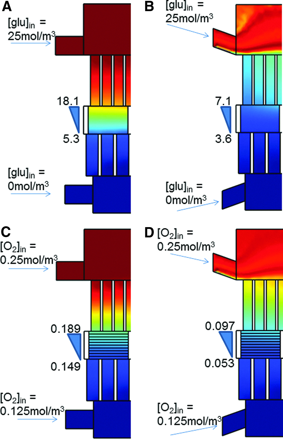
Computational modeling of the glucose and oxygen gradient formation within the explants in the dual flow bioreactor with two different designs. Prediction of the glucose gradient when horizontal
Experimental measurement of glucose gradient in explants
In the square cartilage explants without compression, some cell death was observed at the lateral sites of the explant likely due to sample processing.37,38 Hardly any cell death was observed in the top, in the center, or the bottom of the cartilage explant either in static or dynamic culture. Cartilage explants were successfully cultured for up to 7 days without loss of viability (data not shown). This showed that limitation of diffusion by confinement of the explant was not a limiting factor for chondrocyte survival. To validate the predicted gradient (Fig. 3A), cartilage explants were subsequently dynamically cultured for 3 days using a medium containing 25 mM of glucose in the top compartment and a medium containing 0 mM of glucose in the bottom compartment. Figure 3B shows the glucose concentration in the top, middle, and bottom layer of the explants after 3 days of culture. The concentration decreased with depth and the concentration in the top layer significantly differed from the one in the bottom layer. The decrease in the concentration from top to bottom suggests that a gradient can be formed during 3 days of culture. However, the experimental values did not fully comply with the computational model. This is most likely due to the fact that the diffusion coefficient of glucose through the tissue, in contrast to the model, is not homogenous, but differs from zone to zone. Another possibility could be that permeability of the tissue is higher than what is assumed in our model.33,34 Regardless, we validated the formation of a biologically relevant gradient using the dual-flow approach.
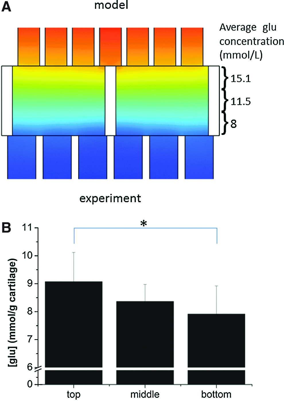
Predicted and experimental glucose concentrations in different zones of articular cartilage.
Gradient formation of cartilage degrading enzymes
We next assessed whether gradients in the explants could also be obtained by larger molecules like proteins. For this, we used enzymes involved in cartilage degradation as model compounds. Several enzymes involved in cartilage degradation enter the cartilage via the synovial fluid.35,36 In our models, we mimicked this by culturing explants using two physically separated medium compartments that are located on either side. The medium on the synovial side of the explant was supplemented with either hyaluronidase or collagenase. After 7 days of culture, explants were stained for sGAGs and collagens. Safranin-O and Picrosirius Red staining revealed a loss of sGAGs or collagen, respectively, at the synovial side of the cartilage as compared to a freshly isolated sample (Fig. 4A, C). Moreover, there was no loss at the lateral sides or the subchondral side indicating the absence of leakage. Quantification of the histological section showed a significant decrease in matrix components (Fig. 4B, D). In addition, no matrix degradation on the lateral sides of the explants could be observed. This strongly suggests that the system does not suffer from leakage ensuring that the additives from the top compartment can only reach the bottom compartment via diffusion through the tissue. Taken together, our bioreactor set up this system that allows for the creation of gradients of molecules of choice through cartilage explants.

Matrix degradation in a static dual compartment system.
Compression and cell viability
We next examined the effect of compression on chondrocyte viability. Square cartilage explants were compressed for 24 h (1-h compression, 7-h rest with 0.25 MPa compressive load [∼2.5% strain] at 0.33 Hz) and a live–dead assay was performed. In remarkable contrast to static or dynamic culture of cartilage explants, which showed negligible cell death in the top and bottom cell layers of the explant (data not shown), considerable cell death was observed after 24 h at the top and bottom of the explant (Fig. 5). Most cell death was noted at the top edges of the explant (Fig. 5, arrow), while cell death in the top central part of the explant was reduced (Fig. 5, asterisk). Chondrocytes in the middle of the explant did not show evidence of increased cell death. Cell death at the lateral sites was more pronounced than in static or dynamic culture.
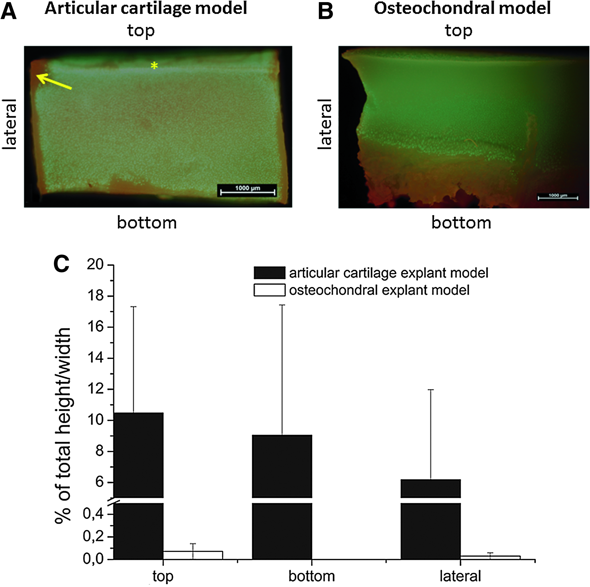
Design validation. Live/dead assays were performed on cartilage explants and on the cartilage in osteochondral explants after compression (24 h (1-h compression, 7-h rest) with 0.25 MPa compressive load (∼2.5% strain) at 0.33 Hz). In both constructs, cell death was only noted in layers at the periphery of the cartilage explants, but not in the center.
We reasoned that the square compression plate, which tight fitted in the cartilage inserts used for explant culture was responsible for the increased cell death, in particular, at the top lateral sites of the explant. This design was not compatible with tissue expansion and deformation upon compression likely resulting in high stresses at the borders of the explant resulting in the observed cell death. To test this, we redesigned the inset for explant culture in the bioreactor and the compression module. The new inset is designed for culturing cylindrical osteochondral plugs with a width of 7 mm. The top of the compression module was rounded and made out of teflon instead of metal. The rounded tip is compatible with tissue deformation at the lateral sides. Like the square cartilage explants, the cylindrical osteochondral plugs could be isolated and cultured in the bioreactor either statically or dynamically without noticeable cell death of chondrocytes anywhere in the explant for up to 2 weeks (data not shown). Interestingly, and in marked contrast to the previous design, 24 h of compression (1-h compression, 7-h rest with 0.25 MPa compressive load (∼2.5% strain) at 0.33 Hz) dramatically reduced the thickness of layers with cell death at the top, bottom, and lateral sites of the cartilage explants from an average 8% to less than 0.2% of the total construct height or width (Fig. 5). Also, the cell death at the top lateral sites was markedly reduced.
Discussion
This study reports on the development of a novel bioreactor for cartilage tissue engineering. Our design is unique due to the incorporation of both confined cyclic mechanical loading and nutrient supply via diffusion from both the top and bottom separately. This form of nutrient supply resembles the natural situation in native cartilage. The combination of both features in one bioreactor setup generates a system that more closely resembles the native environment of articular cartilage compared to previously reported bioreactors.22–24 We successfully succeeded in culturing cartilage explants and osteochondral plugs in our bioreactor for up to 2 weeks in static or dynamic flow without any significant cell death in the cartilage layers. In the square cartilage explants, some cell death was noted in the peripheral layers at the lateral sites of the construct. These likely arise as a consequence of cutting the cartilage.37,38 The data do indicate, however, that in our design diffusion of nutrients, oxygen, and waste products from top or bottom is sufficient to keep middle zone chondrocytes alive.
Computational modeling predicted that our design is suitable for the formation of gradients of small molecules like glucose and oxygen in cartilage explants. The formation of these gradients can be manipulated with simple modifications in the design by changing the position of the inlets for dynamic flow or by varying the nutrient concentrations above or below the construct. The design allows to explore the effect of gradients of a wide variety of molecules on healthy and tissue-engineered cartilage31,39 and to test the hypothesis that gradients of signaling molecules are important for reconstructing the cartilage's zonal architecture. For nutrients, like glucose, there is little known how the gradient in articular cartilage looks like. Since middle and deep zone chondrocytes rely on diffusion from either the synovial fluid or subchondral bone compartment, it is likely that such gradients exist. In line with this, Heywood et al. described differences in metabolic responses in the chondrocyte subpopulations, which may be involved in establishing specific zonal phenotypes.31,40 We performed two experiments to demonstrate the formation of a gradient in tissue explants cultured in our bioreactor by varying the concentration of compounds in the top and bottom compartment. First, we used glucose as a model compound for nutrients and added a concentration of 25 mM in the top compartment and 0 mM in the bottom compartment. Experimentally, a glucose gradient was detected across the cartilage explant. This experimental gradient did not exactly match the predicted one by computational modeling. In computational models, it is assumed that the glucose consumption rate in cartilage is homogenous, however, in reality it is not. 30 This is an important notion as it may also hold true for other nutrients as well as growth factors. For secreted soluble growth factors, the abundance, their zonal distribution, and the affinity and abundance of receptors for these factors will also play an important role. All these parameters, of which many are still unknown, will affect the diffusion of growth factors and nutrients and thus the nature of a gradient. Fundamental knowledge regarding diffusion coefficients of many nutrients and growth factors in cartilage is also lacking. All these factors likely contribute to the deviation in our modeled glucose gradient versus the experimentally determined values. It should be noted that the computational modeling and the experimental gradient across a cartilage explant were studied in the absence of mechanical load. Mechanical compression likely affects the diffusion of molecules in the cartilage as it results in water displacement. However, the true nature of the effects remains largely unknown. Our bioreactor can function as an enabling technology that can facilitate in depth studies of these topics.
Besides glucose, two proteins were used to study the formation of a gradient, namely, hyaluronidase and collagenase. These enzymes were added in the top compartment mimicking their presence in the synovial fluid as in OA.35,36 By studying the depletion of sGAGs and collagen from the tissue using histological staining, the formation of gradients could be easily studied. The results show that the absence of leakage at the lateral sites of the explant and the formation of a top down gradient. They demonstrate the feasibility of our design to build gradients of proteins in explants.
Next to culturing explants in an environment that mimics the natural situation in several key features, we hypothesize that creating gradients in a controlled way can contribute to the development of tissue-engineered grafts closely resembling the cartilage's natural architecture in vitro. Current research focuses on elucidating what concentration ranges stimulate the expression of zone-specific proteins like the superficial zone protein, cartilage intermediate layer protein, and cartilage oligomeric matrix protein. 41 Introduction of gradients may contribute to native matrix distribution in tissue-engineered cartilage. In addition, the effect of compression on these developing constructs can be studied in depth using this bioreactor.
Although rotating compression can best simulate the natural load of the joint, it is less controllable than confined or sliding compression, as different forces are exerted on the tissue (indentation and rotation) and each can have different effects on the tissue. Since load is an important feature in cartilage development, we complemented the two-compartment bioreactor with confined compression. When confined compression was applied to square cartilage explants using a tight fit perforated metal plate, substantial cell death was noted at the periphery of the explant particularly at the top lateral sites. This is likely explained by the design of the bioreactor's insets, which did not allow tissue deformation as a response upon load resulting in high stresses at the periphery of the construct and consequently cell death. This issue could be easily solved by adapting the inset for explant culture as well as the module used for confined compression. The new inset is compatible with the culture of cylindrical cartilage explants. Cylindrical osteochondral plugs can be easily isolated using standardized orthopedic equipment. For confined compression, a rounded module was used made out of Teflon. The mechanical properties of Teflon better resemble the mechanical properties of native cartilage compared to metal. In addition, the rounded surface of the tip is compatible with tissue expansion at the lateral sites upon load. Using this new design, negligible cell death was observed at the periphery of the cartilage explant. Besides the possibility for tissue expansion at the lateral sites upon load, the use of Teflon instead of metal for compression and the cylindrical shape of the explant may also contribute to improved cell survival upon load. Using this new design, it will be possible to culture chondral or even osteochondral explants or tissue-engineered constructs for prolonged periods of time in the bioreactor and study the role of compression.
Our system is advantageous over current mechanoperfusion bioreactor systems for cartilage tissue engineering as it is not based on convection, but on diffusion from physiologically relevant sides. This more closely mimics the natural environment of articular cartilage. This strategy allows us to take advantage of a two compartment system generating biologically relevant gradients in a tissue-engineered construct or explant culture. These gradients can be modulated by applying different concentrations of molecules of interest in each compartment. In addition, the gradient can be controlled by changing the orientation of the medium inlets.
Using simple modifications in our system, we can include other tissues such as bone and synovial tissue. This would further increase the resemblance of the microenvironment generated in our system to the native environment of cartilage. Moreover, with the addition of bone by explant culture of osteochondral plugs and/or synovium, it is able to function as a more accurate ex vivo model for degenerative cartilage diseases such as OA when compared to standard explants cultures. Particularly, it incorporates the idea that OA is not a single tissue disease, but a joint disease.42–45
In summary, we have developed a unique bioreactor system that captures two key features of the cartilage's natural environment. It has the ability to create gradients of nutrients and growth factors and apply confined cyclic compression. We believe that this will aid in development of an in vitro environment for cartilage homeostasis facilitating neo-cartilage formation. Therefore, our bioreactor holds the potential to grant a deeper insight in fundamental concepts of cartilage biology, model degenerative cartilage diseases like OA, and stimulates the development of novel therapeutics for such diseases.
Footnotes
Acknowledgments
This research forms part of the Project P2.02 OA control of the research program of the BioMedical Materials institute, cofunded by the Dutch Ministry of Economic Affairs, Agriculture and Innovation. The authors gratefully acknowledge the support of the TeRM Smart Mix Program of the Netherlands Ministry of Economic Affairs and the Netherlands Ministry of Education, Culture and Science.
Authors' Contributions
Study design: T.S., J.L., A.A., and M.K. Experimentation and data interpretation: T.S., J.L., F.D., and I.C. T.S. wrote the manuscript with the help of all authors. M.K. supervised the study.
Disclosure Statement
No competing financial interests exist.